MTX-23
Based on 1 publication(s) in Google Scholar
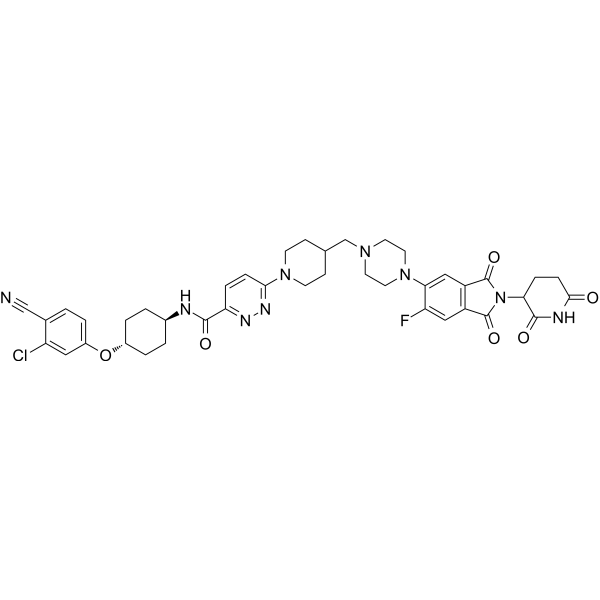
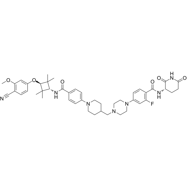
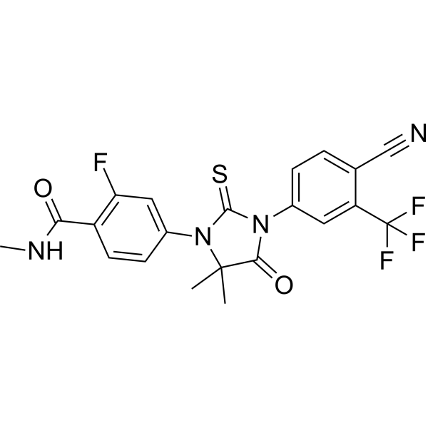
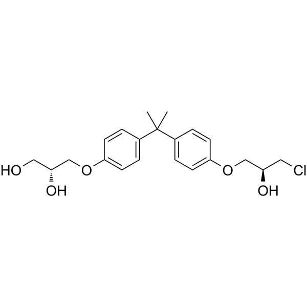
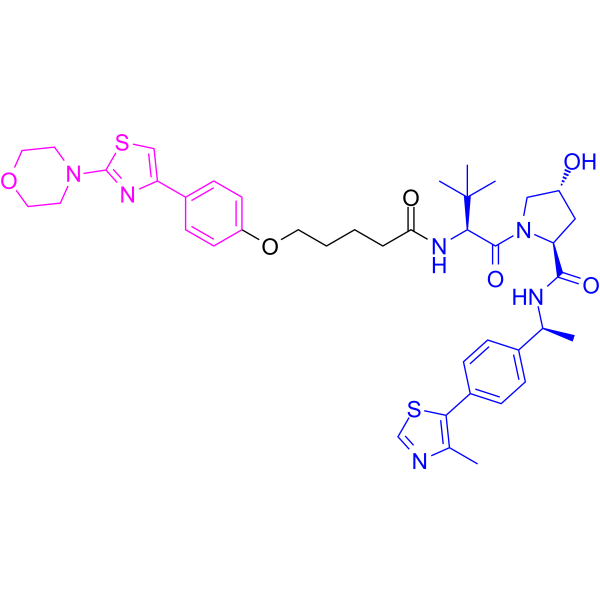
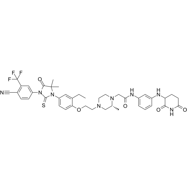
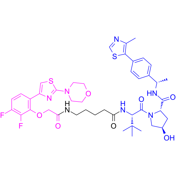
MTX-23 is a PROTAC degrader targeting androgen receptor splice variant 7 (AR-V7) and full-length androgen receptor (AR-FL), with DC50 values of 0.37 μM and 2 μM, respectively. MTX-23 binds to the DNA-binding domains of AR-V7 and AR-FL, recruits the VHL E3 ubiquitin ligase (E3 ubiquitin ligase) and induces their degradation. MTX-23 reduces the proliferation of prostate cancer cells, promotes their apoptosis (apoptosis), and also inhibits tumor growth in mouse models. MTX-23 can be used for the research of prostate cancer.
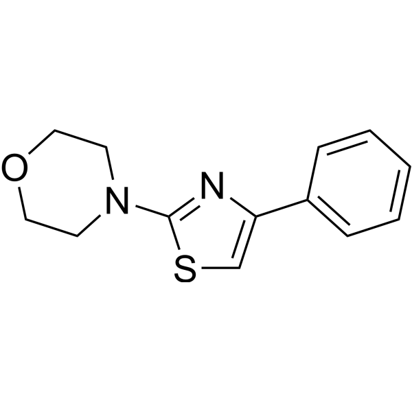
(Pink: AR-V7 ligand (HY-175939); Blue: VHL ligand (HY-112078); Black: linker).
Nos produits utilisent uniquement pour la recherche. Nous ne vendons pas aux patients.
- Pureté : 99.79%
- CAS No.: 2488296-74-6
- Formule: C43H53F2N7O7S2
- Masse moléculaire:882.05
-
Stockage:Powder -20°C, 3 years , 4°C, 2 years ; In solvent -80°C, 6 months , -20°C, 1 month
Publications Citing Use of MedChemExpress (MCE) MTX-23
MoreVoir tous les produits spécifiques à Isoform PROTACs
More
Activité biologique
Description
IC50 & Target
[1]|
AR-V7 0.37 μM (DC50) |
AR-FL 2 μM (DC50) |
In Vitro
MTX-23 degrades AR-V7 with a DC50 of 0.37 μmol/L and AR-FL with a DC50 of 2 μmol/L as measured by immunoblot analysis[1].
MTX-23 inhibits proliferation and increases apoptosis in androgen-responsive prostate cancer cells, with this antiproliferative effect dependent on the presence of AR-V7 and AR-FL[1].
MTX-23 inhibits proliferation in 12 human prostate cancer cell lines resistant to second-line antiandrogen therapies[1].
MedChemExpress (MCE) has not independently confirmed the accuracy of these methods. They are for reference only. Further protocols information, click here.
Chemical Information
-
CAS No. 2488296-74-6
-
Appearance Solid
-
Masse moléculaire 882.05
-
Formule C43H53F2N7O7S2
-
Color Off-white to light yellow
-
SMILES
O=C(N1[C@@H](C[C@H](C1)O)C(N[C@H](C2=CC=C(C3=C(N=CS3)C)C=C2)C)=O)[C@H](C(C)(C)C)NC(CCCCNC(COC4=C(C5=CSC(N6CCOCC6)=N5)C=CC(F)=C4F)=O)=O
-
Synonyms
PROTAC AR-V7 degrader-2
-
Livraison
Room temperature in continental US; may vary elsewhere.
-
Stockage
Powder -20°C 3 years 4°C 2 years In solvent -80°C 6 months -20°C 1 month
Publications (1)
-
Journal Impact Factor
-
Most Recent
-
Nature
2026 Apr;652(8110):763-773. PMID: 41882358
Solvant et solubilité
In Vitro:
DMSO : 100 mg/mL (113.37 mM; Need ultrasonic; Hygroscopic DMSO has a significant impact on the solubility of product, please use newly opened DMSO)
Please refer to the solubility information to select the appropriate solvent. Once prepared, please aliquot and store the solution to prevent product inactivation from repeated freeze-thaw cycles.
Storage method and period of stock solution: -80°C, 6 months; -20°C, 1 month. When stored at -80°C, please use it within 6 months. When stored at -20°C, please use it within 1 month.
Please refer to the solubility information to select the appropriate solvent. Once prepared, please aliquot and store the solution to prevent product inactivation from repeated freeze-thaw cycles.
Storage method and period of stock solution: -80°C, 6 months; -20°C, 1 month. When stored at -80°C, please use it within 6 months. When stored at -20°C, please use it within 1 month.
Concentration (start) × Volume (start) = Concentration (final) × Volume (final)
Protocole
-
Apoptosis
Apoptosis, also called programmed cell death, is generally characterized by distinct morphological characteristics.
-
TUNEL staining for apoptotic DNA fragmentation
TUNEL staining detects DNA strand breaks by using terminal deoxynucleotidyl transferase to add labeled nucleotides to exposed 3′-OH DNA termini, generating either microscopic staining in fixed cells or tissue sections, or fluorescence/cytometric signal in cell suspensions. TUNEL positivity reflects DNA fragmentation but should not be interpreted alone as definitive apoptosis, because TUNEL can also label necrotic, autolytic, mechanically damaged, or DNA-repair-associated DNA breaks.
-
Annexin V plus membrane-impermeant dye apoptosis staining
Annexin V-based apoptosis assays rely on the detection of phosphatidylserine (PS) externalization from the inner leaflet of the plasma membrane to the outer leaflet, an early biochemical hallmark of apoptosis. Fluorescently labeled Annexin V binds PS in a calcium-dependent manner, enabling identification of early apoptotic cells by flow cytometry or fluorescence microscopy. When combined with a membrane-impermeant DNA-binding dye (e. g. , propidium iodide), this approach allows discrimination between viable (Annexin V−/dye−), early apoptotic (Annexin V+/dye−), and late apoptotic or necrotic (Annexin V+/dye+) cell populations by assessing membrane integrity and PS exposure.
-
Research Protocol for Endocrine Diseases
Endocrine diseases often arise from disrupted hormone production, hormone signaling, or target-tissue responsiveness; for diabetes-focused endocrine disease models, insulin signaling regulates glucose uptake, hepatic glucose output, lipid metabolism, and β-cell compensation. Type 2 diabetes develops through interacting defects in insulin resistance, β-cell dysfunction, adipose inflammation, hepatic glucose overproduction, altered incretin signaling, and ectopic lipid metabolism. A major unresolved question is whether endocrine dysfunction is driven primarily by target-tissue insulin resistance, intrinsic β-cell failure, immune/inflammatory stress, or combined multi-organ failure that differs by disease stage.
-
Patient-Derived Xenograft (PDX)
Patient-derived xenograft (PDX) models are generated by engrafting primary human tumor tissue directly into immunodeficient mice, allowing in vivo propagation of patient tumor biology without initial in vitro adaptation. These models are used to preserve key histopathological and molecular characteristics of the original tumor and enable assessment of tumor growth dynamics and therapeutic response in a living organism. The biological readout is tumor engraftment and subsequent growth in the murine host, which reflects the ability of human tumor cells to survive, vascularize, and expand in an immunocompromised microenvironment.
-
Apoptosis Solutions
Apoptosis is a regulated, generally non-lytic cell-death pathway that removes unwanted, damaged, infected, or abnormal cells through coordinated morphological changes, caspase activation, DNA fragmentation, and membrane remodeling. The intrinsic apoptosis pathway is controlled mainly by mitochondrial outer membrane permeabilization, BCL-2 family proteins, cytochrome c release, apoptosome formation, caspase-9 activation, and downstream executioner caspase-3/7 activation. The extrinsic apoptosis pathway is initiated by death receptors such as Fas, TNFR, and TRAIL receptors, which recruit adaptor proteins and activate caspase-8 before engaging executioner caspases or mitochondrial amplification through BID cleavage. Apoptosis is linked to many phenotypes, including cancer cell killing, tissue homeostasis, immune regulation, neurodegeneration, infection response, and treatment-induced cytotoxicity; unresolved questions include how apoptosis interacts with necroptosis, pyroptosis, ferroptos
-
Patient-Derived Orthotopic Xenograft (PDOX)
Patient-derived orthotopic xenograft (PDOX) modeling implants fresh patient tumor tissue or patient-derived tumor cells into the anatomically corresponding organ or tissue site of immunodeficient mice, usually by surgical orthotopic implantation, to preserve patient tumor histology, local microenvironmental context, invasion, metastatic behavior, and treatment-response features better than subcutaneous implantation. PDOX readouts include tumor engraftment, orthotopic tumor growth, local invasion, metastasis, recurrence after resection, histologic similarity to the donor tumor, biomarker retention, molecular concordance, survival, and response or resistance to therapy. PDOX models are used for preclinical drug testing and individualized therapy evaluation, but engraftment success varies by tumor type and specimen quality.
-
Subcutaneous Cell-Line-Derived Xenograft
Subcutaneous cell-line-derived xenograft (CDX) models are established by implanting cultured human cancer cell lines into immunodeficient mice, where the injected cells form localized tumors that can be monitored in vivo as a measure of tumorigenic potential, growth kinetics, and treatment response. These models are widely used in oncology research because they allow reproducible tumor formation and enable comparative assessment of tumor growth between different cell lines or genetic manipulations in a controlled in vivo microenvironment. Subcutaneous implantation of cancer cells in immunodeficient mice is a standard approach for evaluating tumor growth behavior and therapeutic response across multiple cancer types, including prostate, esophageal, pancreatic, and colon cancer models.
-
Orthotopic Cell-Line Xenograft
Orthotopic cell-line xenograft models involve implantation of human cancer cell lines into the anatomically corresponding organ of immunodeficient mice to reproduce tumor growth within a native microenvironment, enabling more clinically relevant tumor behavior compared with subcutaneous models. These models are widely used because orthotopic placement better recapitulates tumor progression, including invasion and metastatic spread, which are often underrepresented in heterotopic implantation systems. Compared with conventional xenografts, orthotopic implantation is described as more technically complex but provides improved simulation of tumor-microenvironment interactions and metastatic behavior, making it particularly valuable for translational oncology research. Surgical orthotopic implantation approaches have been emphasized as enabling faithful reproduction of clinical cancer features, including metastasis and disease progression patterns that align with the tumor’s organ of origi
-
Cell Viability Determination by MTT Colorimetric Assay
The following protocol uses the MTT colorimetric assay as a classic literature-established method for assessing cell viability/metabolic activity in cultured mammalian cells. MTT[3-(4,5-dimethylthiazol-2-yl)-2,5-diphenyltetrazolium bromide] is reduced by metabolically active cells to a colored formazan product; the amount of formazan is quantified spectrophotometrically and provides an indirect measure of metabolically active viable cells. Importantly, MTT reduction reflects cellular oxidoreductase/metabolic activity rather than an absolute direct count of living cells, so changes in cellular metabolism can alter the signal independently of cell number.
Pureté et documentation
-
Fiche technique (276 KB)
-
SDS (393 KB)
- English - EN (393 KB)
- Français - FR (393 KB)
- Deutsch - DE (393 KB)
- Norwegian - NO (393 KB)
- Español - ES (393 KB)
- Swedish - SV (393 KB)
- Italian - IT (393 KB)
- Korean - KR (393 KB)
- Portuguese - PT (393 KB)
-
Instruction de manipulation (2659 KB)
Références
Complete Stock Solution Preparation Table
Please refer to the solubility information to select the appropriate solvent. Once prepared, please aliquot and store the solution to prevent product inactivation from repeated freeze-thaw cycles.
Storage method and period of stock solution: -80°C, 6 months; -20°C, 1 month. When stored at -80°C, please use it within 6 months. When stored at -20°C, please use it within 1 month.
| Optional Solvent | Concentration Solvent Mass | 1 mg | 5 mg | 10 mg | 25 mg |
|---|---|---|---|---|---|
| DMSO | 1 mM | 1.1337 mL | 5.6686 mL | 11.3372 mL | 28.3431 mL |
| 5 mM | 0.2267 mL | 1.1337 mL | 2.2674 mL | 5.6686 mL | |
| 10 mM | 0.1134 mL | 0.5669 mL | 1.1337 mL | 2.8343 mL | |
| 15 mM | 0.0756 mL | 0.3779 mL | 0.7558 mL | 1.8895 mL | |
| 20 mM | 0.0567 mL | 0.2834 mL | 0.5669 mL | 1.4172 mL | |
| 25 mM | 0.0453 mL | 0.2267 mL | 0.4535 mL | 1.1337 mL | |
| 30 mM | 0.0378 mL | 0.1890 mL | 0.3779 mL | 0.9448 mL | |
| 40 mM | 0.0283 mL | 0.1417 mL | 0.2834 mL | 0.7086 mL | |
| 50 mM | 0.0227 mL | 0.1134 mL | 0.2267 mL | 0.5669 mL | |
| 60 mM | 0.0189 mL | 0.0945 mL | 0.1890 mL | 0.4724 mL | |
| 80 mM | 0.0142 mL | 0.0709 mL | 0.1417 mL | 0.3543 mL | |
| 100 mM | 0.0113 mL | 0.0567 mL | 0.1134 mL | 0.2834 mL |
Keywords
- MTX-23
- 2488296-74-6
- PROTAC AR-V7 degrader-2
- MTX23
- MTX 23
- PROTACs
- Androgen Receptor
- Apoptosis
- AR-V7
- Androgen Receptor Full-Length
- Androgen Receptor Splice Variant-7
- prostate cancer cells
- mouse models
- Von Hippel-Lindau E3 ubiquitin ligase
- AR-FL
- androgen-responsive prostate cancer cells
- human prostate cancer cell lines
- castration-resistant prostate cancer
- Inhibitor
- inhibitor
- inhibit