



CB2-H
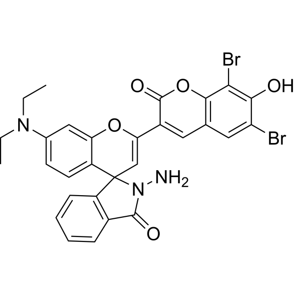
CB2-H is a dual-channel fluorescent probe for the simultaneous detection of HOCl and ONOO-. CB2-H enables the concurrent detection of HOCl and ONOO- at two independent channels without spectral cross-interference and can be applied for dual-channel fluorescence imaging of endogenously produced HOCl and ONOO- in living cells and zebrafish under different stimulants.
연구목적의 판매만을 진행합니다. 환자를 대상으로 한 판매는 하지 않습니다.
- 화학식: C29H23Br2N3O5
- 분자량:653.32
-
보관:
Please store the product under the recommended conditions in the Certificate of Analysis.

To place orders, for customer services and technical support, please contact: MedChemExpress USA
Tel: 609-228-6898 E-mail: [email protected] [email protected]


-
 Biological Activity
-
Chemical Information
- Protocol
- 순도&문서
- References
-
 Help & FAQs




Biological Activity
제품 설명
In Vitro
ONOO- can selectively oxidize the hydrazide group of CB2-H to afford the parent dye CB2 (Absmax/Emmax = 631/669 nm). In the case of HOCl, it undergoes an electrophilic attack on the benzopyran moiety of CB2-H to give a chlorinated product CB2-H-Cl, which inhibits the PET process within the probe and thus affords a turn-on fluorescence response at the coumarin channel (Absmax/Emmax = 407/468 nm)[1].
CB2-H (5 μM; 30 min) is capable of concurrently monitoring endogenously produced HOCl and ONOO- in living cells and zebrafish under different stimulants[1].
MedChemExpress (MCE) has not independently confirmed the accuracy of these methods. They are for reference only. Further protocols information, click here.


Chemical Information
-
분자량 653.32
-
화학식 C29H23Br2N3O5
-
SMILES
CCN(C1=CC=C2C(OC(C3=CC4=C(C(Br)=C(C(Br)=C4)O)OC3=O)=CC25C6=C(C(N5N)=O)C=CC=C6)=C1)CC
-
선적
Room temperature in continental US; may vary elsewhere.
-
보관
Please store the product under the recommended conditions in the Certificate of Analysis.

Protocol
-
Bioluminescent/Fluorescent Imaging Xenograft
Bioluminescent and fluorescent imaging xenograft models use tumor cells engineered to express optical reporters so tumor engraftment, growth, dissemination, and treatment response can be monitored longitudinally in living animals and validated ex vivo. Bioluminescence imaging usually measures luciferase activity after substrate administration and is commonly used as a surrogate for viable reporter-expressing tumor burden, while fluorescence imaging measures reporter or probe emission and can support tumor localization, ex vivo confirmation, or complementary multimodal analysis.


순도&문서
References

Calculators
Concentration (start) × Volume (start) = Concentration (final) × Volume (final)


CB2-H Related Classifications
HY-152103 Related Classifications
 Powered by Bioz