



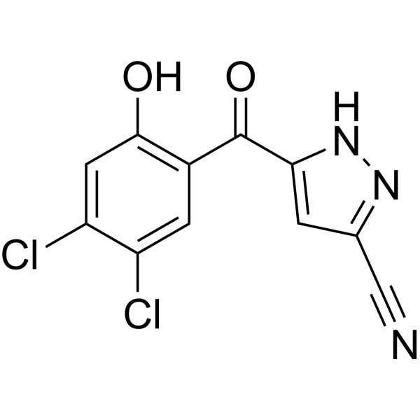
Antibacterial agent 83
Antibacterial agent 83 (compound 17h) displays potent antibacterial activity against various vancomycin-resistant Enterococcus faecalis (VRE) and methicillin-resistant Staphylococcus aureus (MRSA). Antibacterial agent 83 can significantly reduce the biofilm formation of MRSA and exhibited promising selectivity. Antibacterial agent 83 is metabolically stable in human liver microsomes.
Nur für Forschungszwecke. Wir verkaufen nicht an Patienten.
- CAS. Nr.: 2413865-92-4
- Formel: C11H5Cl2N3O2
- Molecular Weight:282.08
-
Speicherung:
Please store the product under the recommended conditions in the Certificate of Analysis.

To place orders, for customer services and technical support, please contact: MedChemExpress USA
Tel: 609-228-6898 E-mail: [email protected] [email protected]


-
 Biologische Aktivität
-
Chemical Information
- Protokoll
- Reinheit & Dokumentation
- Verweise
-
 Help & FAQs




Biologische Aktivität
Beschreibung
Cellular Effect
|
Cell Line

|
Type | Value | Description | References |
|---|---|---|---|---|
| L02 | IC50 |
10.7 μM
Compound: 17h
|
Cytotoxicity against human L02 cells after 72 hrs by CCK-8 assay
Cytotoxicity against human L02 cells after 72 hrs by CCK-8 assay
|
[PMID: 35398730] |
| L02 | IC50 |
3.02 μg/mL
Compound: 17h
|
Cytotoxicity against human L02 cells after 72 hrs by CCK-8 assay
Cytotoxicity against human L02 cells after 72 hrs by CCK-8 assay
|
[PMID: 35398730] |
In Vitro
Antibacterial agent 83 (compound 17h) shows antibacterial activity against Newmana, USA300b, and Mu50, with MIC of 0.25 μg/mL all[1].
Antibacterial agent 83 performs poor stability in the liver microsomes of rat, mouse and dog[1].
MedChemExpress (MCE) has not independently confirmed the accuracy of these methods. They are for reference only. Further protocols information, click here.


Chemical Information
-
CAS. Nr. 2413865-92-4
-
Molecular Weight 282.08
-
Formel C11H5Cl2N3O2
-
SMILES
N#CC1=NNC(C(C2=C(C=C(C(Cl)=C2)Cl)O)=O)=C1
-
Versand
Room temperature in continental US; may vary elsewhere.
-
Speicherung
Please store the product under the recommended conditions in the Certificate of Analysis.

Protokoll
-
Research Protocol for Infectious Diseases
Infectious-disease experiments test how pathogens interact with host barriers, innate immune receptors, inflammatory signaling, pathogen replication, and tissue injury; pattern-recognition receptors such as TLRs, RIG-I-like receptors, NOD-like receptors, and inflammasomes detect microbial molecules and activate NF-κB, interferon, and cytokine responses. The central hypothesis is that infection severity reflects the balance between pathogen burden and host response: protective inflammation restricts pathogen growth, whereas excessive or mislocalized inflammation contributes to tissue damage and disease phenotype. Unresolved questions include which host pathways are protective versus pathogenic, why some infection models fail to translate to human disease, and which combined readouts best predict clinically relevant infection outcomes.
-
Bacterial live/dead nucleic-acid viability staining
The LIVE/DEAD bacterial viability staining method is based on differential permeability of nucleic-acid-binding fluorescent dyes, most commonly SYTO 9 and propidium iodide (PI), which enables discrimination of bacterial populations with intact versus compromised cytoplasmic membranes. SYTO 9 penetrates both intact and damaged bacterial membranes and binds nucleic acids to produce green fluorescence, whereas propidium iodide penetrates only cells with compromised membranes and fluoresces red while also reducing SYTO 9 signal through competitive binding and fluorescence interactions. The resulting fluorescence pattern is interpreted as a proxy for membrane integrity, which is widely used as an indicator of bacterial viability in microscopy, flow cytometry, and spectroscopic platforms. However, mechanistic studies show that SYTO 9 and PI interactions involve displacement and fluorescence resonance energy transfer effects, which can influence signal interpretation depending on dye ratios a
-
Fungal Biofilm Culture
Fungal biofilm culture is an in vitro method for growing surface-attached fungal communities, most commonly Candida albicans, on abiotic substrates such as polystyrene wells, silicone elastomer, or polymethylmethacrylate; the assay models adhesion, proliferation, filamentation, extracellular-matrix-associated maturation, and dispersion. Biofilm output can be read by optical density at 600 nm for adherent biomass, XTT reduction for metabolic activity, CFU recovery for viable attached or dispersed cells, and microscopy for architecture.


Reinheit & Dokumentation
Verweise

Calculators
Konzentration (Stammlösung) × Volumen (Stammlösung) = Konzentration (Ziellösung) × Volumen (Ziellösung)


Antibacterial agent 83 Related Classifications
HY-143326 Related Classifications
 Powered by Bioz