



KMG-301AM
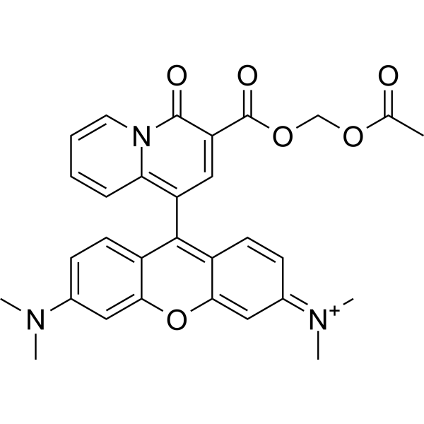
KMG-301AM is the acetoxy methyl esterified form of KMG-301. KMG-301AM successfully accumulates in mitochondria and then it is hydrolyzed to KMG-301. KMG-301 is an Mg2+-selective fluorescent probe functional in mitochondria in intact cells. Since the mitochondrial membrane is impermeable to KMG-301, it is not released upon depolarization of the mitochondrial membrane potential. KMG-301 can indicate changes in mitochondrial Mg2+ concentration and shows Mg2+ transport across the mitochondrial membrane in the early phases of a cellular model.
Nur für Forschungszwecke. Wir verkaufen nicht an Patienten.
- CAS. Nr.: 1372642-77-7
- Formel: C30H28N3O6+
- Molecular Weight:526.56
-
Speicherung:
4°C, protect from light
* In solvent : -80°C, 6 months; -20°C, 1 month (protect from light)

To place orders, for customer services and technical support, please contact: MedChemExpress USA
Tel: 609-228-6898 E-mail: [email protected] [email protected]


-
 Biologische Aktivität
-
Chemical Information
- Protokoll
- Reinheit & Dokumentation
- Verweise
-
 Help & FAQs




Biologische Aktivität
Beschreibung
In Vitro
Guidelines (Following is our recommended protocol. This protocol only provides a guideline, and should be modified according to your specific needs)[1].
1. For optical imaging, 20 μM KMG-301AM is applied to cells in Hanks' balanced salt solutions (HBSS) for 10 min on ice, so that hydrolysis of the acetoxymethyl ester by esterase present in the cytosol will be avoided.
HBSS contains (in mM): NaCl, 137; KCl, 5.4; CaCl2, 1.3; MgCl2, 0.5; MgSO4, 0.4; Na2HPO4, 0.3; KH2PO4, 0.4; NaHCO3, 4.2; D-glucose, 5.6; HEPES, 5 (pH adjusted to 7.4 with NaOH).
2. Then, the cells are washed twice with HBSS and incubated for 15 min at 37°C to allow for complete hydrolysis of the acetoxymethyl ester form in mitochondria.
3. If you want the simultaneous use of KMG-104AM (HY-128536) and KMG-301AM, cells are incubated with 5 μM KMG-104AM in HBSS for 30 min at 37°C, and then stained with KMG-301AM.
4. Isolated mitochondria are incubated with 20 μM KMG-301AM in Mitochondria Imaging Buffer (MIB) for 20 min at 37°C. Then, mitochondria are washed twice with MIB and further incubated for 15 min at 37°C.
MIB contains (in mM): KCl, 125; K2HPO4, 2; MgCl2, 1; HEPES, 5; EDTA, 0.02 (pH adjusted to 7.2 with KOH) or Mg2+-free MIB (without MgCl2)
5. Fluorescence imaging experiments are performed using a confocal laser scanning microscope system. KMG-301 is excited at 559 nm from a laser diode, and the signal was observed at 600-700 nm.
In the simultaneous measurement, KMG-104 and KMG-301 were excited at 488 nm from an Argon laser and 559 nm, respectively. Fluorescence was separated using a 560 nm dichroic mirror and observed at 500-545 nm and 600-700 nm, respectively.
MedChemExpress (MCE) has not independently confirmed the accuracy of these methods. They are for reference only.


Chemical Information
-
CAS. Nr. 1372642-77-7
-
Appearance Liquid
-
Molecular Weight 526.56
-
Formel C30H28N3O6+
-
Color Brown to red
-
SMILES
O=C1C(C(OCOC(C)=O)=O)=CC(C(C2=CC=C(N(C)C)C=C2O3)=C4C3=C/C(C=C4)=[N+](C)/C)=C5C=CC=CN51
-
Versand
Room temperature in continental US; may vary elsewhere.
-
Speicherung
4°C, protect from light
* In solvent : -80°C, 6 months; -20°C, 1 month (protect from light)


Reinheit & Dokumentation
-
Data Sheet (275 KB)
-
SDS (252 KB)
- English - EN (252 KB)
- Français - FR (252 KB)
- Deutsch - DE (252 KB)
- Norwegian - NO (252 KB)
- Español - ES (252 KB)
- Swedish - SV (252 KB)
- Italian - IT (252 KB)
- Korean - KR (252 KB)
- Portuguese - PT (252 KB)
-
Handling Instructions (2659 KB)
Verweise

Calculators
Konzentration (Stammlösung) × Volumen (Stammlösung) = Konzentration (Ziellösung) × Volumen (Ziellösung)


KMG-301AM Related Classifications
HY-126220 Related Classifications
 Powered by Bioz