



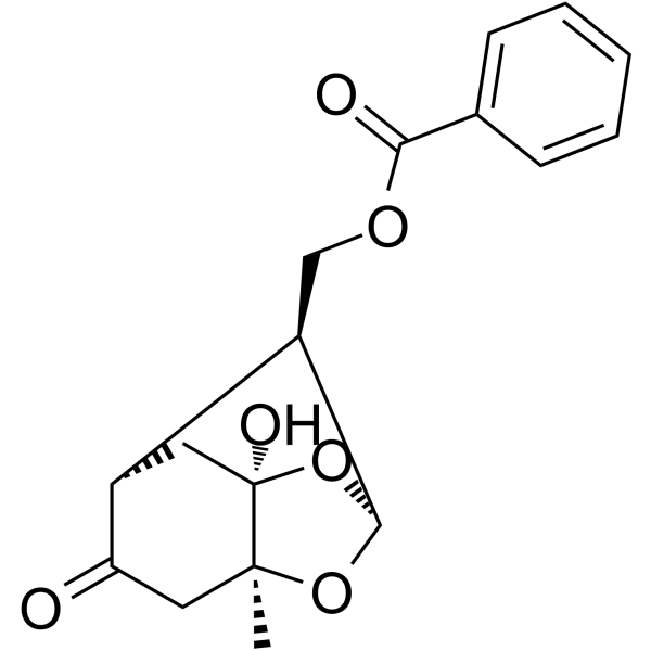
Paeoniflorigenone

Based on 1 Customer Validation
Paeoniflorigenone, isolated as an active ingredient from the root of moutan cortex, induces apoptosis selectively in the cancer cell lines and exhibits antiproliferative effect.
Nur für Forschungszwecke. Wir verkaufen nicht an Patienten.
- Reinheit : 98.50%
- CAS. Nr.: 80454-42-8
- Formel: C17H18O6
- Molecular Weight:318.32
-
Speicherung:
4°C, protect from light
* In solvent : -80°C, 6 months; -20°C, 1 month (protect from light)

To place orders, for customer services and technical support, please contact: MedChemExpress USA
Tel: 609-228-6898 E-mail: [email protected] [email protected]


-
 Biologische Aktivität
-
Chemical Information
- Protokoll
- Reinheit & Dokumentation
- Verweise
-
 Help & FAQs




Biologische Aktivität
Beschreibung
Cellular Effect
|
Cell Line

|
Type | Value | Description | References |
|---|---|---|---|---|
| RAW264.7 | IC50 |
6.87 μM
Compound: 25
|
Inhibition of LPS-induced NO production in mouse RAW264.7 cells after 24 hrs by Griess reaction
Inhibition of LPS-induced NO production in mouse RAW264.7 cells after 24 hrs by Griess reaction
|
[PMID: 23067550] |
In Vitro
Paeoniflorigenone (0-30 μM; 0-72 hours) significantly decreases the cell proliferation of tumor cell lines and inhibits the proliferation of normal cell lines[1].
Paeoniflorigenone (0-30 μM; 24-72 hours) significantly induces apoptotic DNA fragmentation selectively in tumor cells[1].
MedChemExpress (MCE) has not independently confirmed the accuracy of these methods. They are for reference only.
-
Cell Line:TIG-1, 3T3-L1, HL60, Jurkat and HeLa cells
-
Concentration:0-30 μM
-
Incubation Time:0-72 hours
-
Result:Significantly decreased the cell proliferation of tumor cell lines and inhibited the proliferation of normal cell lines.
-
Cell Line:TIG-1 and 3T3-L1 (normal cells), HL60, Jurkat, and HeLa cells (tumor cells)
-
Concentration:0, 1, 3, 10, 30 μM
-
Incubation Time:24 and 72 hours
-
Result:Induced DNA fragmentation in three tumor cell lines, but not in two normal cell lines.


Chemical Information
-
CAS. Nr. 80454-42-8
-
Appearance Oil
-
Molecular Weight 318.32
-
Formel C17H18O6
-
Color White to off-white
-
SMILES
O=C(C1=CC=CC=C1)OC[C@H]2[C@@H]3O[C@@]4(C[C@H]2C(C[C@@]4(O3)C)=O)O
-
Structure Classification
-
Initial Source
-
Versand
Room temperature in continental US; may vary elsewhere.
-
Speicherung
4°C, protect from light
* In solvent : -80°C, 6 months; -20°C, 1 month (protect from light)


Reinheit & Dokumentation
-
Data Sheet (276 KB)
-
SDS (252 KB)
- English - EN (252 KB)
- Français - FR (252 KB)
- Deutsch - DE (252 KB)
- Norwegian - NO (252 KB)
- Español - ES (252 KB)
- Swedish - SV (252 KB)
- Italian - IT (252 KB)
- Korean - KR (252 KB)
- Portuguese - PT (252 KB)
-
Handling Instructions (2659 KB)
Verweise

Calculators
Konzentration (Stammlösung) × Volumen (Stammlösung) = Konzentration (Ziellösung) × Volumen (Ziellösung)


Paeoniflorigenone Related Classifications
HY-N3119 Related Classifications
 Powered by Bioz