



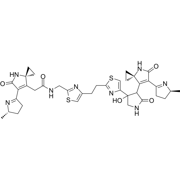
Colibactin 742
Colibactin 742 is a covalently binding DNA-damaging agent targeting DNA, with an IC50 of 5.2 μM against human cervical cancer cells (HeLa). Colibactin 742 covalently binds to DNA, forming interstrand crosslinks (ICLs), activating the Fanconi anemia DNA repair pathway, inducing γH2AX and FANCD2 foci formation and cell cycle arrest, while exacerbating mismatch repair deficiency (MMRd)-related mutations. Colibactin 742 can mimic the genotoxicity of natural Colibactin while avoiding its instability, and is mainly used in colorectal cancer (CRC) related research, including microbial tumorigenesis mechanisms, DNA damage repair pathways, and mutation signature analysis.
For research use only. We do not sell to patients.
- CAS No.: 2916559-62-9
- Formula: C37H42N8O5S2
- Molecular Weight:742.91
-
Storage:
Please store the product under the recommended conditions in the Certificate of Analysis.

To place orders, for customer services and technical support, please contact: MedChemExpress USA
Tel: 609-228-6898 E-mail: [email protected] [email protected]


-
 Biological Activity
-
Chemical Information
- Protocols
- Purity & Documentation
- References
-
 Help & FAQs




Biological Activity
Description
In Vitro
Colibactin 742 (100 nM-100 μM; 1.5 hours) induces stable DNA interstrand crosslinks (ICLs) using linearized pUC19 DNA as a substrate at both pH 5.0 and pH 7.0, and its activity is superior to Colibactin 770 (3a) at pH 5.0[1].
Colibactin 742 (1-100 μM; 4 hours treatment followed by 20 hours incubation) increases the number of FANCD2 and γH2AX foci-positive cells by 4 times at 11 μM in HeLa cells, and induces cell death at >33 μM[1].
Colibactin 742 (24 nM-100 μM; 72 hours) is cytotoxic to HeLa cells with an IC50 of 5.2 μM[1].
Colibactin 742 (10 μM, 100 μM; 4 hours, 12 hours) activates the p53 signaling pathway and induces cell cycle arrest, senescence, ER stress, and the IL-1 inflammatory pathway in FHC cells, upregulating the expression of genes such as CDKN1A, BTG2, and FAS[2].
Colibactin 742 (20 μM; 48 hours/10 times, with 48-96 hour intervals) upregulates BRCA1 and Fanconi in HCT 116 cells." Anemia, mismatch repair (MMR), and IFNL1-related pathway gene expression increase T>N ??single-base substitutions (SBS) and short insertion-deletion (Indel) mutations[2].
MedChemExpress (MCE) has not independently confirmed the accuracy of these methods. They are for reference only. Further protocols information, click here.
-
Cell Line:HeLa cells, Human Colonic Organoids (HDCs), HCT 116 cells
-
Concentration:HeLa: 24 nM - 100 μM; HCT 116: 20 μM (chronic treatment)
-
Incubation Time:48 h, 72 h
-
Result:Colibactin 742 exhibited cytotoxicity against HeLa cells with an IC50 of 5.2 μM after 72 hours of incubation.
Significantly reduced the viability and diameter of HDCs after 48 hours of incubation.
Chronic exposure to 20 μM, decreased the colony formation ability of HCT 116 cells in soft-agar assays.
In Vivo
MedChemExpress (MCE) has not independently confirmed the accuracy of these methods. They are for reference only.
-
Animal Model:Galleria mellonella larvae (average weight 250 mg) normal model[2]
-
Dosage:7.42 μg per larva (approximately 30 mg/kg)
-
Administration:Intrahemocoelic injection
-
Result:Caused increased cuticular melanization of the larvae compared with Colibactin 746 (inactive analog) 24 hours post-injection. Oral gavage of Colibactin 742 induced a higher level of DNA damage in the intestinal epithelium of the larvae, which was confirmed by the alkaline comet assay showing increased DNA strand breaks.


Chemical Information
-
CAS No. 2916559-62-9
-
Molecular Weight 742.91
-
Formula C37H42N8O5S2
-
SMILES
O=C(NCC1=NC(CCC2=NC(C3(O)CNC(C3C4=C(C5=N[C@@H](C)CC5)C(N[C@]46CC6)=O)=O)=CS2)=CS1)CC([C@@]7(CC7)N8)=C(C9=N[C@@H](C)CC9)C8=O
-
Shipping
Room temperature in continental US; may vary elsewhere.
-
Storage
Please store the product under the recommended conditions in the Certificate of Analysis.

Protocols
-
Cell Cytotoxicity Assay
Cytotoxicity assays are usually based on the assessment of cell membrane damage, which can also be indirectly detected by measuring cell viability. Detection methods include MTT assay, CKK-8 assay, LDH assay and ATP assay, etc.
-
Flow cytometric DNA-content cell-cycle staining
Flow cytometric DNA-content cell-cycle staining measures the fluorescence intensity of DNA-bound fluorochromes in single cells or nuclei to estimate DNA content distributions, allowing assignment of populations to G0/G1, S, and G2/M phases by DNA histogram deconvolution. Propidium iodide (PI) intercalates into DNA, and PI fluorescence is proportional to cellular DNA content when staining is performed under conditions that make DNA accessible and minimize non-DNA signal. Cells with G2/M DNA content are expected to show approximately twice the fluorescence intensity of G0/G1 cells, while S-phase cells occupy intermediate fluorescence values. PI-based DNA-content analysis can also detect cells with fractional DNA content, often reported as sub-G1, when DNA fragmentation and extraction during staining reduce retained DNA signal in apoptotic cells. DAPI is an alternative DNA fluorochrome for univariate DNA-content analysis, while bivariate approaches combining DNA content with proliferation
-
BrdU Incorporation Assay
Bromodeoxyuridine (BrdU) incorporation assay is based on the principle that BrdU, a thymidine analog, is incorporated into newly synthesized DNA during the S phase of the cell cycle, thereby serving as a marker of DNA replication and cellular proliferation. Incorporated BrdU can be detected using anti-BrdU antibodies following DNA denaturation, enabling visualization or quantification of proliferating cells through immunochemical detection methods such as immunofluorescence or immunohistochemistry.
-
Protocol for Cell Cycle
Cell-cycle analysis by flow cytometry measures DNA content in single cells to estimate the fraction of cells in G0/G1, S, and G2/M phases. Propidium iodide intercalates into DNA, and after RNA removal with RNase, fluorescence intensity reflects cellular DNA content: 2N cells are assigned to G0/G1, cells between 2N and 4N to S phase, and 4N cells to G2/M. DNA-content analysis alone cannot reliably separate G0 from G1 or G2 from M. Ki-67 can distinguish quiescent G0 cells from cycling cells, EdU or BrdU incorporation marks active DNA synthesis in S phase, and phospho-histone H3 staining identifies mitotic cells within the 4N population.
-
Genotoxicity/Mutagenicity Study
The bacterial reverse mutation assay detects point mutations that restore amino-acid prototrophy in auxotrophic Salmonella typhimurium or Escherichia coli tester strains; after exposure to a test article, mutagenic activity is read out as an increased number of revertant colonies on minimal agar compared with the vehicle control. The assay uses tester strains with different mutation targets so that base-substitution and frameshift mutagens can be detected, and testing is performed with and without exogenous mammalian metabolic activation because some chemicals require biotransformation to become mutagenic.


Purity & Documentation
-
Data Sheet (275 KB)
-
SDS (252 KB)
- Français - FR (252 KB)
- Deutsch - DE (252 KB)
- Norwegian - NO (252 KB)
- Español - ES (252 KB)
- Swedish - SV (252 KB)
- Italian - IT (252 KB)
- Korean - KR (252 KB)
- Portuguese - PT (252 KB)
-
Handling Instructions (2659 KB)
References
[1]. Wernke KM, et al. Probing Microbiome Genotoxicity: A Stable Colibactin Provides Insight into Structure-Activity Relationships and Facilitates Mechanism of Action Studies. J Am Chem Soc. 2021 Sep 29;143(38):15824-15833. [Content Brief]
[2]. Dougherty MW, et al. The microbial genotoxin colibactin exacerbates mismatch repair mutations in colorectal tumors. Neoplasia. 2023 Sep;43:100918. [Content Brief]
[3]. Agrawal R, et al. Colibactin Exerts Androgen-dependent and -independent Effects on Prostate Cancer. Eur Urol Oncol. 2025 Jun;8(3):716-730. [Content Brief]

Calculators
Concentration (start) × Volume (start) = Concentration (final) × Volume (final)


Colibactin 742 Related Classifications
HY-139621 Related Classifications
 Powered by Bioz