



Macrocarpal A

Based on 1 Customer Validation
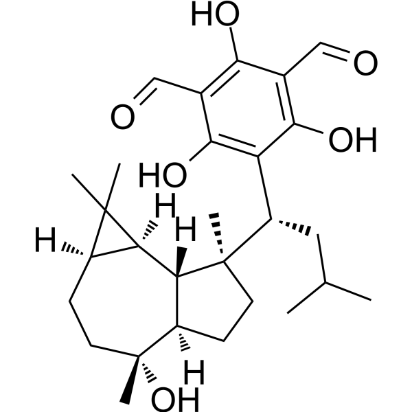
Macrocarpal A (10-epi-Eucarobustol F) is an antibacterial agent, which can be isolated from the leaves of Eucalyptus macrocarpa. Macrocarpal A inhibits the growth of Bacillus subtilis PCI219 (minimum inhibitory concentration below 0.2 µM) and Staphylococcus aureus FDA209P (minimum inhibitory concentration is 0.4 µM).
Nos produits utilisent uniquement pour la recherche. Nous ne vendons pas aux patients.
- Pureté : 99.71%
- CAS No.: 132951-90-7
- Formule: C28H40O6
- Masse moléculaire:472.61
-
Stockage:Powder -20°C, 3 years ; In solvent -80°C, 6 months , -20°C, 1 month

To place orders, for customer services and technical support, please contact: MedChemExpress USA
Tel: 609-228-6898 E-mail: [email protected] [email protected]


-
 Activité biologique
-
Chemical Information
- Protocole
- Pureté et documentation
- Références
-
 Help & FAQs




Activité biologique
Description
Cellular Effect
|
Cell Line

|
Type | Value | Description | References |
|---|---|---|---|---|
| A549 | IC50 |
<10 μM
Compound: Macrocarpal A
|
Cytotoxicity against human A549 cells assessed as reduction in cell viability incubated for 72 hrs by SRB assay
Cytotoxicity against human A549 cells assessed as reduction in cell viability incubated for 72 hrs by SRB assay
|
[PMID: 27142786] |
| HL-60 | IC50 |
<10 μM
Compound: Macrocarpal A
|
Cytotoxicity against human HL60 cells assessed as reduction in cell viability after 72 hrs by MTT assay
Cytotoxicity against human HL60 cells assessed as reduction in cell viability after 72 hrs by MTT assay
|
[PMID: 27142786] |


Chemical Information
-
CAS No. 132951-90-7
-
Appearance Solid
-
Masse moléculaire 472.61
-
Formule C28H40O6
-
Color White to off-white
-
SMILES
CC(C)C[C@@H](C1=C(C(C=O)=C(C(C=O)=C1O)O)O)[C@@]2([C@@]3([H])[C@@]4([H])[C@](CC[C@@](O)([C@]3([H])CC2)C)([H])C4(C)C)C
-
Synonyms
10-epi-Eucarobustol F
-
Structure Classification
-
Initial Source
-
Livraison
Room temperature in continental US; may vary elsewhere.
-
Stockage
Powder -20°C 3 years In solvent -80°C 6 months -20°C 1 month

Protocole
-
Research Protocol for Infectious Diseases
Infectious-disease experiments test how pathogens interact with host barriers, innate immune receptors, inflammatory signaling, pathogen replication, and tissue injury; pattern-recognition receptors such as TLRs, RIG-I-like receptors, NOD-like receptors, and inflammasomes detect microbial molecules and activate NF-κB, interferon, and cytokine responses. The central hypothesis is that infection severity reflects the balance between pathogen burden and host response: protective inflammation restricts pathogen growth, whereas excessive or mislocalized inflammation contributes to tissue damage and disease phenotype. Unresolved questions include which host pathways are protective versus pathogenic, why some infection models fail to translate to human disease, and which combined readouts best predict clinically relevant infection outcomes.


Pureté et documentation
-
Fiche technique (278 KB)
-
SDS (254 KB)
- English - EN (254 KB)
- Français - FR (254 KB)
- Deutsch - DE (254 KB)
- Norwegian - NO (254 KB)
- Español - ES (254 KB)
- Swedish - SV (254 KB)
- Italian - IT (254 KB)
- Korean - KR (254 KB)
- Portuguese - PT (254 KB)
-
Instruction de manipulation (2659 KB)
Références

Calculators
Concentration (start) × Volume (start) = Concentration (final) × Volume (final)


Macrocarpal A Related Classifications
HY-N3346 Related Classifications
 Powered by Bioz