



Odesivimab
Based on 1 publication(s) in Google Scholar
Odesivimab is a human monoclonal antibody, targeting Ebola virus glycoprotein with a KD value of 7.74 nM for recombinant histidine-tagged Makona strain Ebola virus glycoprotein ectodomain protein. Odesivimab can be used in research of Ebola virus infection.
Nos produits utilisent uniquement pour la recherche. Nous ne vendons pas aux patients.
- Pureté : 99.64%
- CAS No.: 2135632-30-1
- Masse moléculaire:146.16 kDa
-
Stockage:
Please store the product under the recommended conditions in the Certificate of Analysis.


To place orders, for customer services and technical support, please contact: MedChemExpress USA
Tel: 609-228-6898 E-mail: [email protected] [email protected]


-
 Activité biologique
-
Chemical Information
- Protocole
- Pureté et documentation
- Références
-
 Help & FAQs





Publications Citing Use of MedChemExpress (MCE) Odesivimab
More

Activité biologique
Description
Isotype
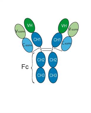
Human IgG1 kappa
Recommend Isotype Controls
Species Reactivity
Virus
IC50 & Target
Zaire Ebola virus
In Vitro
Odesivimab (REGN-3471) neutralizes pseudovirus particles in vitro. Odesivimab with Atoltivimab and Maftivimab has an IC50 value of 0.39 nM[1].
MedChemExpress (MCE) has not independently confirmed the accuracy of these methods. They are for reference only.
In Vivo
Gene ID
911829 [NCBI]
Accession
Conjugated
Unconjugated
Reconsititution
The product can be reconstituted/diluted with sterile PBS or saline.
Format
-
Human IgG1 kappa
Application
ELISA, FACS, Functional assay


Chemical Information
-
CAS No. 2135632-30-1
-
Appearance Liquid
-
Masse moléculaire 146.16 kDa
-
Color Colorless to light yellow
-
SMILES
[Odesivimab]
-
Synonyms
REGN3471
-
Livraison
Shipping with dry ice.
-
Formulation
Please refer to the lot-specific COA for specific buffer information.
-
Stockage
Please store the product under the recommended conditions in the Certificate of Analysis.


Publications (1)
-
Journal Impact Factor
-
Most Recent
-
Cell
Molecular characterization of Ebola virus glycoprotein V75A substitution in the 2018-2020 epidemic. [Abstract]2026 Feb 5;189(3):818-831.e19. PMID: 41576953


Pureté et documentation
-
Fiche technique (278 KB)
-
SDS (251 KB)
- English - EN (251 KB)
- Français - FR (251 KB)
- Deutsch - DE (251 KB)
- Norwegian - NO (251 KB)
- Español - ES (251 KB)
- Swedish - SV (251 KB)
- Italian - IT (251 KB)
- Korean - KR (251 KB)
- Portuguese - PT (251 KB)
-
Inhibitory Antibodies User Guide (603 KB)
Références

Calculators
Concentration (start) × Volume (start) = Concentration (final) × Volume (final)


Odesivimab Related Classifications
HY-P99764 Related Classifications
 Powered by Bioz