



Clodronate liposomes
Based on 11 publication(s) in Google Scholar
Clodronate liposomes, a mixture of Clodronate encapsulated by phospholipids, is a macrophage scavenger. Clodronate liposomes is engulfed by macrophages in the body. Under the action of lysosomal phosphatases in macrophages, Clodronate dissolved in liposomes will be gradually released and accumulated in cells. When a certain concentration is reached, macrophages will be irreversibly damaged and apoptosis will be induced.
For research use only. We do not sell to patients.
-
Storage:
Store at 4°C, do not freeze


To place orders, for customer services and technical support, please contact: MedChemExpress USA
Tel: 609-228-6898 E-mail: [email protected] [email protected]


-
 Biological Activity
-
Chemical Information
- Protocols
- Purity & Documentation
- References
-
 Help & FAQs




Publications Citing Use of MedChemExpress (MCE) Clodronate liposomes
More- Cancer Commun (Lond). 2025 Oct 13. [Abstract]
- Cancer Res. 2026 Mar 5. [Abstract]
- Cancer Res. 2025 Nov 6. [Abstract]
- Gut Microbes. 2026 Dec 31;18(1):2635820. [Abstract]
- Adv Sci (Weinh). 2026 Jul 3:e76411.
- J Control Release. 2025 Oct 28:114367. [Abstract]
- Phytomedicine. 2025 Oct:146:157117. [Abstract]
- Cell Mol Gastroenterol Hepatol. 2026 Mar 2:101765. [Abstract]
- Cell Oncol (Dordr). 2026 Mar 23;49(2):61. [Abstract]
- Biomedicines. 2026 May 18;14(5):1145. [Abstract]
- FASEB J. 2025 Aug 15;39(15):e70923. [Abstract]
 Customer Validation & Images

-
IF
-
In Vivo Efficacy Study
-
Histological Imaging/Staining


Biological Activity
Description
In Vivo
Clearing red pulp area of spleen and Kupffer macrophages:
Single dose: 200 μL/mouse (intravenous or intraperitoneal injection).
Long-term administration: The first dose is 150 μL/mouse, and then 100 μL/mouse every three days.
Depletion of alveolar macrophages
The best effect was obtained by intravenous injection (150-200 μL) combined with tracheal or intranasal administration (50 μL).
Depletion of brain microglia
Intraventricular cerebrospinal fluid injection, mice (10 μL), rats (50 μL).
MedChemExpress (MCE) has not independently confirmed the accuracy of these methods. They are for reference only.


Chemical Information
-
Appearance Liquid
-
Color Milky white to off-white
-
SMILES
[Clodronate liposomes]
-
Shipping
Room temperature in continental US; may vary elsewhere.
-
Storage
Store at 4°C, do not freeze


Publications (11)
-
Journal Impact Factor
-
Most Recent
-
Cancer Commun (Lond)
CD24 is a promising immunotherapeutic target for enhancing efficacy of third-generation EGFR-TKIs on EGFR-mutated lung cancer. [Abstract]2025 Oct 13. PMID: 41084191 -
Cancer Res
Metronomic Chemotherapy Induces Metabolic Reprogramming in Cancer Cells That Modulates Mature Regulatory Dendritic Cell Function to Stimulate Antitumor Immunity. [Abstract]2026 Mar 5. PMID: 41784618 -
Cancer Res
USP20-Driven Cholesterol Metabolism Links Inflammatory Signaling to Malignancy and Stromal Co-evolution in Pancreatic Cancer. [Abstract]2025 Nov 6. PMID: 41196022 -
Gut Microbes
Simulated microgravity induces cerebral dysfunction by disturbing protective microbiota-metabolite-microglia signaling across the gut‒brain ax is. [Abstract]2026 Dec 31;18(1):2635820. PMID: 41729099 -
-
J Control Release
Enhancing TIM-3 immunotherapy with epirubicin-loaded pH sensitive fusion membrane nanoparticles for effective glioblastoma treatment. [Abstract]2025 Oct 28:114367. PMID: 41167336 -
Phytomedicine
Luteolin Induces GPX4-dependent Ferroptosis and Enhances Immune Activation in Colon Cancer. [Abstract]2025 Oct:146:157117. PMID: 40812220 -
Cell Mol Gastroenterol Hepatol
Ubiquitin-Conjugating Enzyme E2O Primes Hepatocytes to Restore Immune Tolerance in Autoimmune Hepatitis via Inhibiting Y-Box Binding Protein 1/Interleukin-6 Axis. [Abstract]2026 Mar 2:101765. PMID: 41780884 -
Cell Oncol (Dordr)
Targeting senescence-like tumor-associated macrophages sensitizes chemotherapy in triple-negative breast cancer. [Abstract]2026 Mar 23;49(2):61. PMID: 41870812
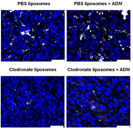
Clodronate liposomes purchased from MedChemExpress. Usage Cited in: Cell Oncol (Dordr). 2026 Mar 23;49(2):61. [Abstract]
Verification of macrophage deletion by Clodronate liposomes. Clodronate liposomes (40 mg/kg; i.p.) were administered twice weekly for two weeks before tumor inoculation and then weekly for four additional weeks. IF analysis of F4/80+ macrophages in the following groups: PBS-liposomes, ADM, Clodronateliposome, or ADM + Clodronate-liposome. Scale bar, 50 μm.
-
Biomedicines
HIF-1α Promotes Macrophage Extracellular Trap Formation and Exacerbates Acute Lung Injury in Neonatal Sepsis. [Abstract]2026 May 18;14(5):1145. PMID: 42193470 -
FASEB J
M2c Macrophages Mediate YAP1 to Promote Vascularized Bone Regeneration in Distraction Osteogenesis. [Abstract]2025 Aug 15;39(15):e70923. PMID: 40788163
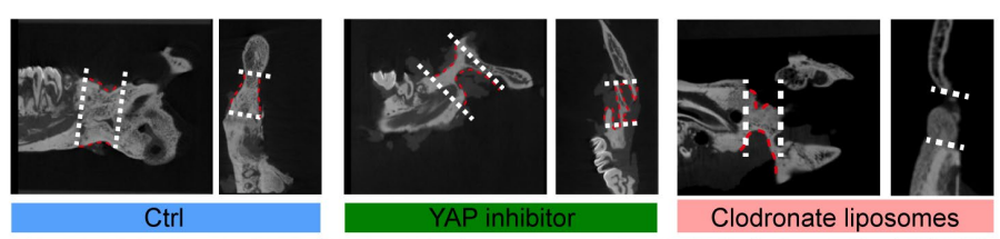
Clodronate liposomes purchased from MedChemExpress. Usage Cited in: FASEB J. 2025 Aug 15;39(15):e70923. [Abstract]
Representative micro‐CT images showing new bone formation in each group. Both the YAP1 inhibitor group and the Clodronate liposomes group exhibited impaired bone regeneration compared to the Ctrl group.
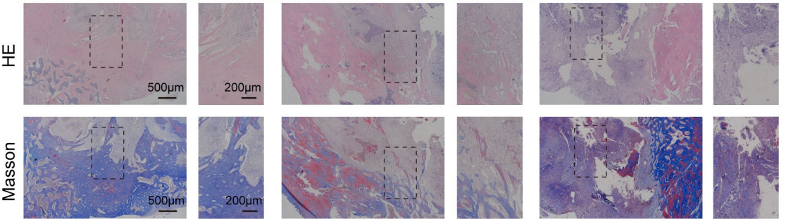
Clodronate liposomes purchased from MedChemExpress. Usage Cited in: FASEB J. 2025 Aug 15;39(15):e70923. [Abstract]
H&E staining and Masson's trichrome staining showed dense lamellar bone formation in the Ctrl group, while the YAP1 inhibitor and Clodronate liposomes groups exhibited sparse trabeculae, fibrous tissue infiltration, or immature callus.

Protocols
-
Apoptosis
Apoptosis, also called programmed cell death, is generally characterized by distinct morphological characteristics.
-
TUNEL staining for apoptotic DNA fragmentation
TUNEL staining detects DNA strand breaks by using terminal deoxynucleotidyl transferase to add labeled nucleotides to exposed 3′-OH DNA termini, generating either microscopic staining in fixed cells or tissue sections, or fluorescence/cytometric signal in cell suspensions. TUNEL positivity reflects DNA fragmentation but should not be interpreted alone as definitive apoptosis, because TUNEL can also label necrotic, autolytic, mechanically damaged, or DNA-repair-associated DNA breaks.
-
Annexin V plus membrane-impermeant dye apoptosis staining
Annexin V-based apoptosis assays rely on the detection of phosphatidylserine (PS) externalization from the inner leaflet of the plasma membrane to the outer leaflet, an early biochemical hallmark of apoptosis. Fluorescently labeled Annexin V binds PS in a calcium-dependent manner, enabling identification of early apoptotic cells by flow cytometry or fluorescence microscopy. When combined with a membrane-impermeant DNA-binding dye (e. g. , propidium iodide), this approach allows discrimination between viable (Annexin V−/dye−), early apoptotic (Annexin V+/dye−), and late apoptotic or necrotic (Annexin V+/dye+) cell populations by assessing membrane integrity and PS exposure.
-
Apoptosis Solutions
Apoptosis is a regulated, generally non-lytic cell-death pathway that removes unwanted, damaged, infected, or abnormal cells through coordinated morphological changes, caspase activation, DNA fragmentation, and membrane remodeling. The intrinsic apoptosis pathway is controlled mainly by mitochondrial outer membrane permeabilization, BCL-2 family proteins, cytochrome c release, apoptosome formation, caspase-9 activation, and downstream executioner caspase-3/7 activation. The extrinsic apoptosis pathway is initiated by death receptors such as Fas, TNFR, and TRAIL receptors, which recruit adaptor proteins and activate caspase-8 before engaging executioner caspases or mitochondrial amplification through BID cleavage. Apoptosis is linked to many phenotypes, including cancer cell killing, tissue homeostasis, immune regulation, neurodegeneration, infection response, and treatment-induced cytotoxicity; unresolved questions include how apoptosis interacts with necroptosis, pyroptosis, ferroptos
-
Lysosome and acidic-vesicle live-cell staining
Lysosome and acidic-vesicle live-cell staining detects acidic intracellular compartments by using membrane-permeant acidotropic probes that accumulate in low-pH vesicles, including lysosomes, late endosomes, autolysosomes, and acidic phagosomes. LysoTracker staining is commonly used as an intensity-based readout of acidic lysosomal compartment abundance or enlargement, while acridine orange produces green fluorescence in less concentrated compartments and red fluorescence after concentration-dependent accumulation in acidic vesicular organelles. Loss or reduction of acridine-orange red signal can be used as a readout of lysosomal membrane permeabilization or reduced acidic-vesicle integrity. This protocol is designed for live cultured cells and can be adapted for fluorescence microscopy, high-content imaging, plate-reader readout, or flow cytometry when the selected literature supports the readout. Because these dyes report acidotropic accumulation rather than lysosome identity alone,


Purity & Documentation
-
Data Sheet (273 KB)
-
SDS (251 KB)
- English - EN (251 KB)
- Français - FR (251 KB)
- Deutsch - DE (251 KB)
- Norwegian - NO (251 KB)
- Español - ES (251 KB)
- Swedish - SV (251 KB)
- Italian - IT (251 KB)
- Korean - KR (251 KB)
- Portuguese - PT (251 KB)
-
Handling Instructions (2659 KB)
References

Calculators
Concentration (start) × Volume (start) = Concentration (final) × Volume (final)


Clodronate liposomes Related Classifications
HY-172202 Related Classifications
 Powered by Bioz