



CO probe 1
Based on 1 publication(s) in Google Scholar
CO probe 1 (probe 2) is a highly efficient fluorescent CO probe (Ex=493 nm) with an allyl ether reaction site. In the presence of PdCl₂, CO reduces Pd2+ to Pd0, triggering a Tsuji-Trost reaction that removes the allyl protecting group, releases fluorescein, and generates a significant fluorescence signal. CO probe 1 has high selectivity, rapid response (fluorescence enhancement of 150 times within 20 minutes), and low cytotoxicity, and can be used for real-time imaging of CO in living cells. CO probe 1 may be used to study pathological mechanisms involving CO signaling regulation, such as inflammation, vascular disease, or cancer.
For research use only. We do not sell to patients.
- Purity : 99.88%
- CAS No.: 1043865-37-7
- Formula: C26H18Cl2O5
- Molecular Weight:481.32
-
Storage:
-20°C, protect from light
* In solvent : -80°C, 6 months; -20°C, 1 month (protect from light)


To place orders, for customer services and technical support, please contact: MedChemExpress USA
Tel: 609-228-6898 E-mail: [email protected] [email protected]


-
 Biological Activity
-
Chemical Information
-
Solvent & Solubility
- Protocols
- Purity & Documentation
- References
-
 Help & FAQs


-
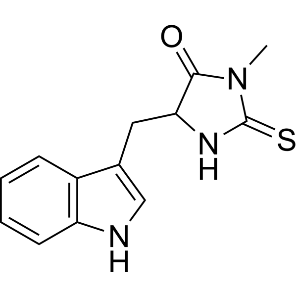
HY-15760
Necrostatin-1RIPK1 Inhibitor -
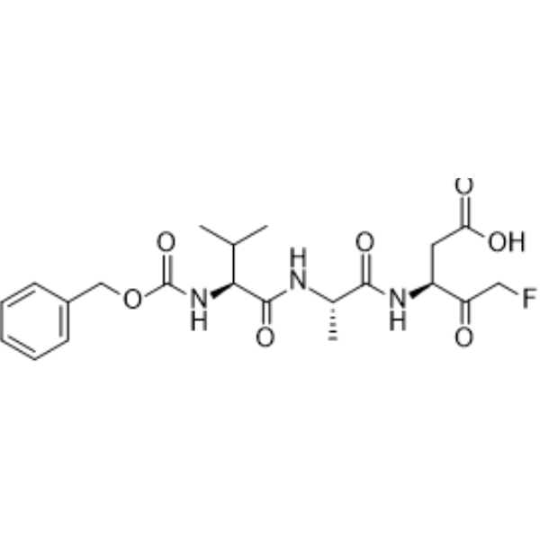
HY-16658B
Z-VAD-FMKPan-Caspase Inhibitor -
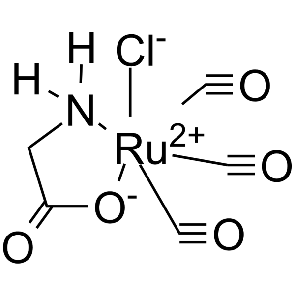
HY-100581
CORM-3CO releasing molecule -
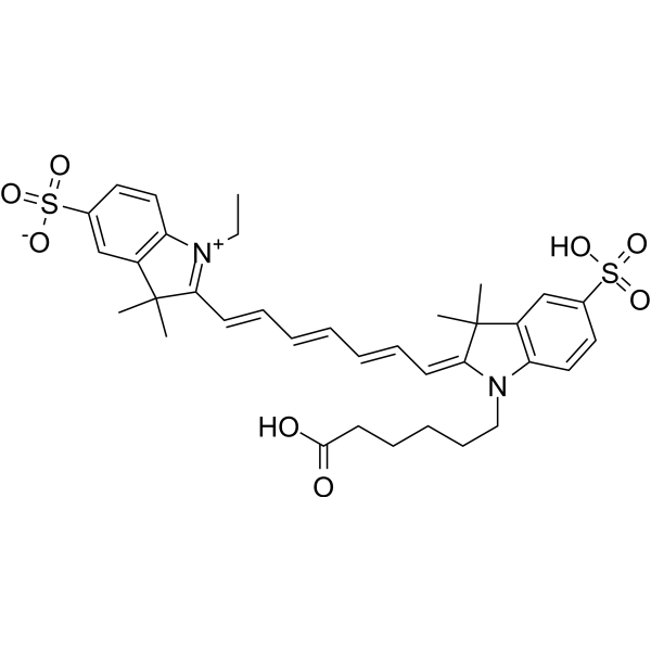
HY-D0825
CY7Fluorescent Dye -
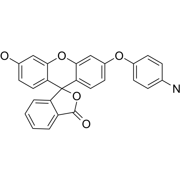
HY-119977
APFFluorescence Probe -
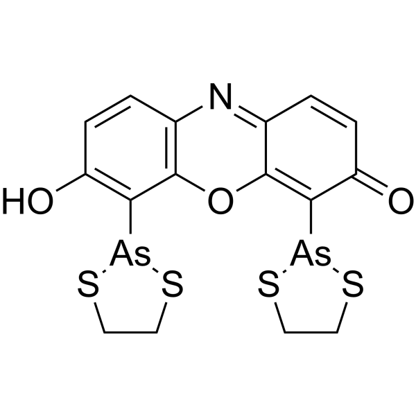
HY-130533
ReAsH-EDT2Protein Fluorescent Dye -

HY-123067
NBD-FructoseFluorescent Agent -
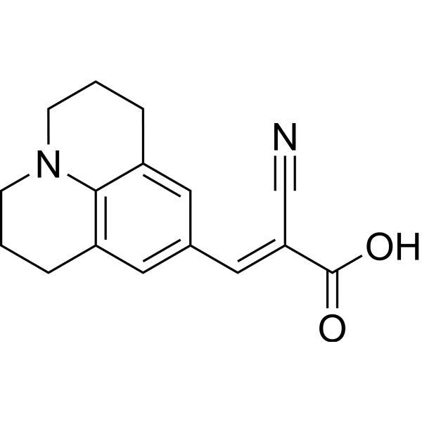
HY-D0054
9-(2-Carboxy-2-cyanovinyl)julolidineFluorescent Molecular Rotor -

HY-D2476
H2S Fluorescent probe 1Fluorescent Probe -

HY-158800
1-NBD-decanoyl-2-decanoyl-sn-glycerolDGK Fluorescent Substrate -
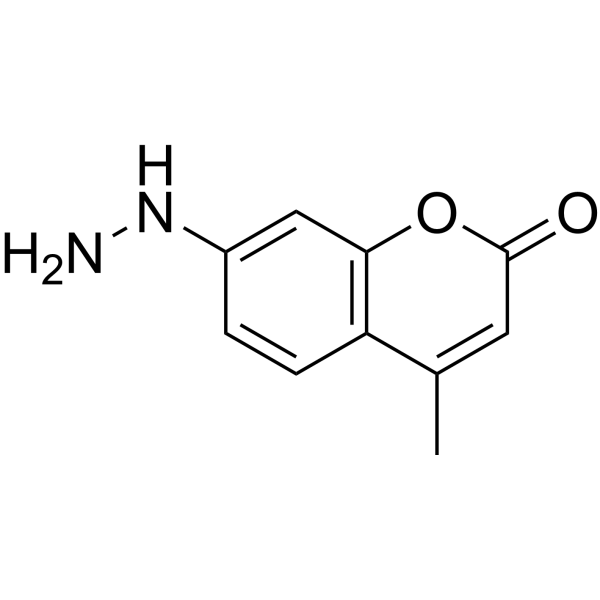
HY-124197
Coumarin hydrazineCarbonyl Fluorescent Probe -
HY-118213
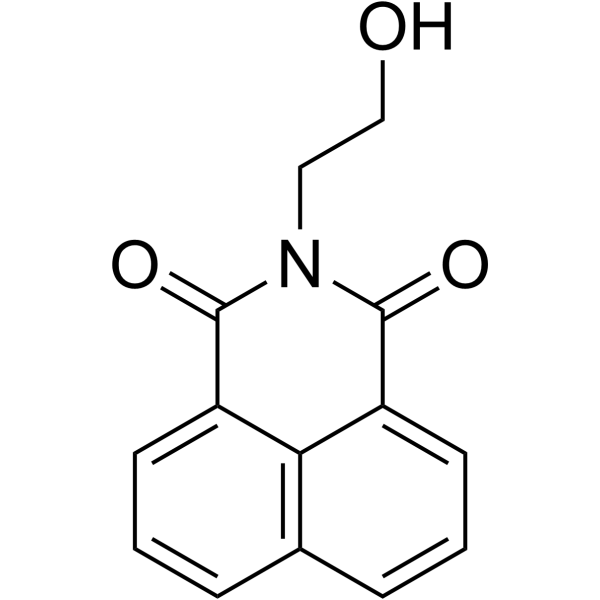
N-(2-Hydroxyethyl)-1,8-naphthalimideFluorochrome
Publications Citing Use of MedChemExpress (MCE) CO probe 1
More

Biological Activity
Description
In Vitro
Guidelines (Following is our recommended protocol. This protocol only provides a guideline, and should be modified according to your specific needs).
CO probe 1 CO imaging steps in living cells[1]:
1. Before the cell imaging experiment, cells were seeded in 24-well culture plates overnight.
2. Set up control group and fluorescence group. Control group: Living cells were incubated with CO probe 1 (1 μM) at 37 °C for 30 minutes. Fluorescence group: Living cells were incubated with a mixture of CO probe 1 and PdCl2 (1 μM each) at 37 °C for 30 minutes.
3. After the above two groups were incubated, they were washed three times with PBS and then imaged.
* For monitoring exogenous CO, HeLa cells were pretreated with CORM-3 (HY-100581) (1, 5, and 10 μM, respectively) at 37 °C for 30 min and then incubated with a mixture of CO probe 1 and PdCl2 (1 μM each) at 37 °C for 30 min. Cells were washed with PBS buffer before imaging.
* For monitoring CO stimulated by heme, the procedure was essentially the same as for imaging exogenous CO, but heme (100 μM) was used instead of CORM 3 (HY-100581) in the cell pretreatment.
MedChemExpress (MCE) has not independently confirmed the accuracy of these methods. They are for reference only. Further protocols information, click here.


Chemical Information
-
CAS No. 1043865-37-7
-
Appearance Solid
-
Molecular Weight 481.32
-
Formula C26H18Cl2O5
-
Color Light yellow to yellow
-
SMILES
O=C1OC2(C3=CC(Cl)=C(C=C3OC4=CC(OCC=C)=C(C=C42)Cl)OCC=C)C5=C1C=CC=C5
-
Shipping
Room temperature in continental US; may vary elsewhere.
-
Storage
-20°C, protect from light
* In solvent : -80°C, 6 months; -20°C, 1 month (protect from light)


Publications (1)
-
Journal Impact Factor
-
Most Recent


Solvent & Solubility
In Vitro:
DMSO : 25 mg/mL (51.94 mM; ultrasonic and warming and heat to 60°C; Hygroscopic DMSO has a significant impact on the solubility of product, please use newly opened DMSO)
Please refer to the solubility information to select the appropriate solvent. Once prepared, please aliquot and store the solution to prevent product inactivation from repeated freeze-thaw cycles.
Storage method and period of stock solution: -80°C, 6 months; -20°C, 1 month (protect from light). When stored at -80°C, please use it within 6 months. When stored at -20°C, please use it within 1 month.
Please refer to the solubility information to select the appropriate solvent. Once prepared, please aliquot and store the solution to prevent product inactivation from repeated freeze-thaw cycles.
Storage method and period of stock solution: -80°C, 6 months; -20°C, 1 month (protect from light). When stored at -80°C, please use it within 6 months. When stored at -20°C, please use it within 1 month.
Concentration (start) × Volume (start) = Concentration (final) × Volume (final)

Protocols
-
Cell Cytotoxicity Assay
Cytotoxicity assays are usually based on the assessment of cell membrane damage, which can also be indirectly detected by measuring cell viability. Detection methods include MTT assay, CKK-8 assay, LDH assay and ATP assay, etc.
-
Mammalian live/dead viability and cytotoxicity staining
Live/dead viability and cytotoxicity staining assays are based on the simultaneous detection of intracellular esterase activity in metabolically active (viable) cells and membrane integrity loss in non-viable cells. In commonly used dual-staining approaches, membrane-permeant fluorogenic substrates are converted by intracellular esterases into fluorescent products in live cells, while impermeant DNA-binding dyes selectively enter cells with compromised plasma membranes and label nucleic acids in dead or dying cells, enabling discrimination between viable and non-viable populations by fluorescence microscopy or flow cytometry.
-
Research Protocol for Cardiovascular Diseases
Cardiovascular disease can be modeled as maladaptive cardiac remodeling, where ischemic injury or pressure overload activates inflammatory signaling, fibroblast activation, extracellular-matrix deposition, cardiomyocyte hypertrophy, vascular remodeling, and progressive ventricular dysfunction. The TGF-β/SMAD axis is a central profibrotic pathway after myocardial injury and pressure overload, while innate immune and cytokine pathways regulate leukocyte recruitment, scar formation, and adverse remodeling. Key unresolved questions include which inflammatory signals are reparative versus harmful, when fibrosis is protective versus maladaptive, and whether pathway inhibition improves function without weakening necessary infarct healing or compensatory remodeling.
-
Research Protocol for Inflammation-related Diseases
The NLRP3 inflammasome is a cytosolic innate immune signaling platform that integrates priming signals and danger-signal activation to promote caspase-1 activation, maturation of IL-1β and IL-18, and gasdermin D-mediated pyroptotic cell death. The core experimental logic is to determine whether inflammatory disease phenotypes are driven by increased NLRP3 expression, ASC-containing inflammasome assembly, caspase-1 cleavage, GSDMD cleavage, and extracellular release of IL-1β/IL-18 rather than by nonspecific cell injury alone. The pathway is strongly linked to inflammation-related disease phenotypes because monosodium urate crystals activate NALP3/NLRP3 inflammasome signaling in gout-like crystal inflammation, cholesterol crystals activate NLRP3 inflammasomes in atherogenesis models, and DSS-induced intestinal inflammation has been reported to involve NLRP3 inflammasome activity. However, experimental colitis studies also show context-dependent protective effects of NLRP3 inflammasome co
-
Cell Viability Determination by MTT Colorimetric Assay
The following protocol uses the MTT colorimetric assay as a classic literature-established method for assessing cell viability/metabolic activity in cultured mammalian cells. MTT[3-(4,5-dimethylthiazol-2-yl)-2,5-diphenyltetrazolium bromide] is reduced by metabolically active cells to a colored formazan product; the amount of formazan is quantified spectrophotometrically and provides an indirect measure of metabolically active viable cells. Importantly, MTT reduction reflects cellular oxidoreductase/metabolic activity rather than an absolute direct count of living cells, so changes in cellular metabolism can alter the signal independently of cell number.


Purity & Documentation
-
Data Sheet (274 KB)
-
SDS (252 KB)
- English - EN (252 KB)
- Français - FR (252 KB)
- Deutsch - DE (252 KB)
- Norwegian - NO (252 KB)
- Español - ES (252 KB)
- Swedish - SV (252 KB)
- Italian - IT (252 KB)
- Korean - KR (252 KB)
- Portuguese - PT (252 KB)
-
Handling Instructions (2659 KB)
References


Complete Stock Solution Preparation Table
Please refer to the solubility information to select the appropriate solvent. Once prepared, please aliquot and store the solution to prevent product inactivation from repeated freeze-thaw cycles.
Storage method and period of stock solution: -80°C, 6 months; -20°C, 1 month (protect from light). When stored at -80°C, please use it within 6 months. When stored at -20°C, please use it within 1 month.
| Optional Solvent | Concentration Solvent Mass | 1 mg | 5 mg | 10 mg | 25 mg |
|---|---|---|---|---|---|
| DMSO | 1 mM | 2.0776 mL | 10.3881 mL | 20.7762 mL | 51.9405 mL |
| 5 mM | 0.4155 mL | 2.0776 mL | 4.1552 mL | 10.3881 mL | |
| 10 mM | 0.2078 mL | 1.0388 mL | 2.0776 mL | 5.1940 mL | |
| 15 mM | 0.1385 mL | 0.6925 mL | 1.3851 mL | 3.4627 mL | |
| 20 mM | 0.1039 mL | 0.5194 mL | 1.0388 mL | 2.5970 mL | |
| 25 mM | 0.0831 mL | 0.4155 mL | 0.8310 mL | 2.0776 mL | |
| 30 mM | 0.0693 mL | 0.3463 mL | 0.6925 mL | 1.7313 mL | |
| 40 mM | 0.0519 mL | 0.2597 mL | 0.5194 mL | 1.2985 mL | |
| 50 mM | 0.0416 mL | 0.2078 mL | 0.4155 mL | 1.0388 mL |


CO probe 1 Related Classifications
HY-D1244 Related Classifications
 Powered by Bioz