



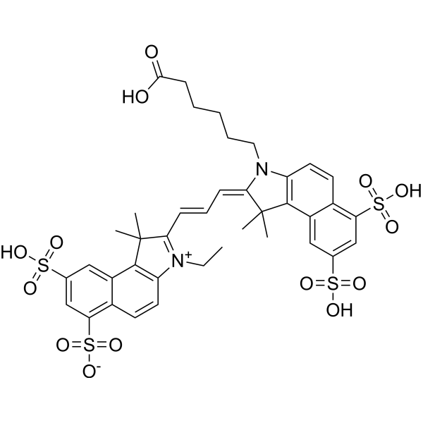
Cy3.5
Cy3.5 is a cyanine-based red fluorescent dye (Ex/Em ≈ 560-581/600-612 nm). After covalent conjugation with proteins, Cy3.5 exhibits extraordinary fluorescence enhancement with minimal quenching even at high labeling ratios. Cy3.5 can form a FRET dye pair with Cy5.5, which maintains stable FRET efficiency in various biochemical buffers and deoxygenated systems. Cy3.5 is applicable to studies related to fluorescent labeling of antibodies/proteins, single-molecule FRET imaging of double-stranded DNA and nucleosomes, SPION functionalization, in vivo tracing, and other relevant research.
연구목적의 판매만을 진행합니다. 환자를 대상으로 한 판매는 하지 않습니다.
- CAS No.: 1284240-77-2
- 화학식: C39H42N2O14S4
- 분자량:891.02
-
보관:
Please store the product under the recommended conditions in the Certificate of Analysis.

To place orders, for customer services and technical support, please contact: MedChemExpress USA
Tel: 609-228-6898 E-mail: [email protected] [email protected]


-
 Biological Activity
-
Chemical Information
- 순도&문서
- References
-
 Help & FAQs




Biological Activity
제품 설명
In Vitro
Operating Instructions (The following is a recommended experimental protocol for guidance only, and adjustments are required based on specific requirements)
1. Stock Solution Preparation
1.1 Solvent: DMSO.
1.2 Concentration Recommendations: Prepare a stock solution by dissolving a sufficient amount of Cy3.5 monofunctional dye (capable of labeling 1 mg protein) in 50 μL DMSO, followed by 2-fold serial gradient dilution in DMSO for labeling purposes; alternatively, prepare a 25 μM stock solution.
2. Working Solution Preparation
2.1 Diluents: 10 mM Tris-Cl (pH 8); imaging buffer; nucleosome buffer; 25 mM carbonate buffer (pH=9.3) containing amino-PVA coated SPION dispersion (2.5 mg Iron/mL).
2.2 Working Concentrations: 1 μM (double-stranded DNA); ~1 μM (nucleosomes).
2.3 Notes: Adjust the working solution concentration as needed; prepare and use immediately.
3. Staining Procedures
3.1 Protein Samples (goat IgG, bovine Ig, BSA)[1]:
3.1.1 Incubation Conditions: Incubate at room temperature for 30 min, and vortex to mix after 10 min and 20 min of incubation respectively.
3.1.2 Washing Step: Use gel filtration to separate Cy3.5 bound to proteins from unreacted dye in Buffer A (100 mM NaCl, 50 mM NaH2PO4, 1 mM EDTA, pH 7.5).
3.2 Nucleic Acid Samples (single-stranded DNA and double-stranded DNA)[2]:
3.2.1 Incubation Conditions:
3.2.1.1 Single-stranded DNA Labeling: Incubate with shaking at 30 °C overnight.
3.2.1.2 Double-stranded DNA Surface Immobilization: Incubate biotinylated double-stranded DNA in the imaging chamber for 5 min.
3.2.2 Washing Steps:
3.2.2.1 After Streptavidin Incubation for Double-stranded DNA: Wash with T50 buffer (10 mM Tris-HCl, pH 8, 50 mM NaCl) to remove excess streptavidin.
3.2.2.2 After Double-stranded DNA Immobilization: Wash with imaging buffer to remove excess double-stranded DNA.
3.3 Chromatin Samples (nucleosomes)[2]:
3.3.1 Incubation Conditions:
3.3.1.1 Histone Labeling: Label H2A K119C with Cy3.5 maleimide.
3.3.1.2 Nucleosome Surface Immobilization: Incubate biotinylated nucleosomes in the imaging chamber for 5 min.
3.3.2 Washing Steps:
3.3.2.1 After BSA Incubation for Nucleosomes: Wash with T50 buffer to remove excess BSA.
3.3.2.2 After Streptavidin Incubation for Nucleosomes: Wash with T50 buffer to remove excess streptavidin.
3.3.2.3 After Nucleosome Immobilization: Wash with nucleosome buffer to remove excess nucleosomes.
3.4 Amino-PVA Coated SPION Dispersion (for subsequent sheep tissue imaging)[3]:
3.4.1 Incubation Condition: Incubate for 1 hour.
3.4.2 Washing Step: Use a disposable PD-10 desalting column pre-equilibrated with 25 mM phosphate buffer to separate labeled SPIONs from excess unbound Cy3.5, and elute the labeled particles with 25 mM phosphate buffer.
4. Detection and Analysis
4.1 Instrument Types: Fluorescence spectrophotometer, total internal reflection fluorescence microscope, fluorescence microscope, laser confocal scanning microscope.
4.2 Excitation/Emission Wavelengths:
4.2.1 Excitation wavelengths: 560 nm, 561 nm, 581 nm; emission wavelength: 612/69 nm.
4.2.2 Fluorescence evaluation of sheep tissue samples uses an I3 filter, with an excitation range of 450-490 nm.
4.3 Result Analysis:
4.3.1 Protein Samples: Compared to free Cy3.5 in buffer, Cy3.5-labeled antibodies show enhanced fluorescence; at lower fluorophore/protein ratios, the fluorescence intensity of single Cy3.5 labels is higher; cumulative fluorescence increases with rising fluorophore/protein ratios and reaches a plateau, with only minimal quenching observed even at high ratios.
4.3.2 Nucleic Acid and Chromatin Samples: Cy3.5 maintains stable emission for approximately 9.3 min under continuous illumination, with rare blinking; the FRET efficiency between Cy3.5 and Cy5.5 on double-stranded DNA decreases as the distance between dyes increases, and remains stable in various biochemical buffers and deoxygenated systems; three distinct FRET peaks (0.64, 0.49, 0.31) correspond to Cy3.5-labeled nucleosomes with only proximal labeling, dual labeling, and only distal labeling, respectively.
4.3.3 Sheep Tissue Samples: Strong tissue background fluorescence is observed; the fluorescence signal of Cy3.5-functionalized SPIONs cannot be clearly distinguished from the background, and no distinct fluorescence localization or specific color change attributable to Cy3.5 is confirmed in systemic organs.
MedChemExpress (MCE) has not independently confirmed the accuracy of these methods. They are for reference only.


Chemical Information
-
CAS No. 1284240-77-2
-
분자량 891.02
-
화학식 C39H42N2O14S4
-
SMILES
O=S(C1=CC(S(=O)([O-])=O)=C2C(C3=C([N+](CC)=C(/C=C/C=C4N(CCCCCC(O)=O)C5=C(C6=CC(S(=O)(O)=O)=CC(S(=O)(O)=O)=C6C=C5)C/4(C)C)C3(C)C)C=C2)=C1)(O)=O
-
선적
Room temperature in continental US; may vary elsewhere.
-
보관
Please store the product under the recommended conditions in the Certificate of Analysis.


순도&문서
References
[1]. Gruber HJ, et al. Anomalous fluorescence enhancement of Cy3 and cy3.5 versus anomalous fluorescence loss of Cy5 and Cy7 upon covalent linking to IgG and noncovalent binding to avidin. Bioconjugate chemistry. 2000;11(5):696-704. [Content Brief]
[2]. Ghoneim M, et al. Single-Molecule Characterization of Cy3.5 -Cy5.5 Dye Pair for FRET Studies of Nucleic Acids and Nucleosomes. Journal of fluorescence. 2023 Mar;33(2):413-421. [Content Brief]

Calculators
Concentration (start) × Volume (start) = Concentration (final) × Volume (final)


Cy3.5 Related Classifications
HY-D0922 Related Classifications
 Powered by Bioz