



Cysmethynil
Based on 1 publication(s) in Google Scholar
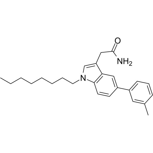
Cysmethynil is an Icmt inhibitor(IC50 = 2.4 μM). Cysmethynil inhibites RAS membrane binding and EGF signal transduction. Cysmethynil prevents the cells in the G1 phase and induces autophagy. Cysmethynil inhibits PC3 cells proliferation, has synergistic effect with Paclitaxel (HY-B0015) and Doxorubicin (HY-15142A). Cysmethynil has anti-tumor effects and can be used for solid tumor (such as prostate cancer et al.) research.
For research use only. We do not sell to patients.
- Purity : 99.07%
- CAS No.: 851636-83-4
- Formula: C25H32N2O
- Molecular Weight:376.53
-
Storage:Powder -20°C, 3 years , 4°C, 2 years ; In solvent -80°C, 6 months , -20°C, 1 month

To place orders, for customer services and technical support, please contact: MedChemExpress USA
Tel: 609-228-6898 E-mail: [email protected] [email protected]


-
 Biological Activity
-
Chemical Information
-
Solvent & Solubility
- Protocols
- Purity & Documentation
- References
-
 Help & FAQs


-
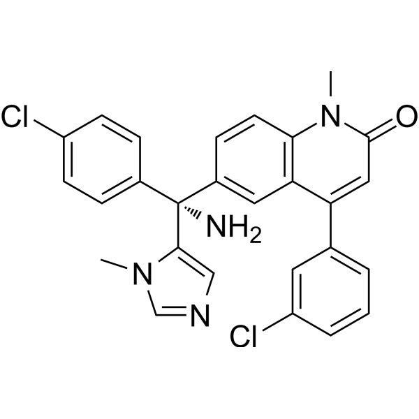
HY-10502
TipifarnibFTase Inhibitor -
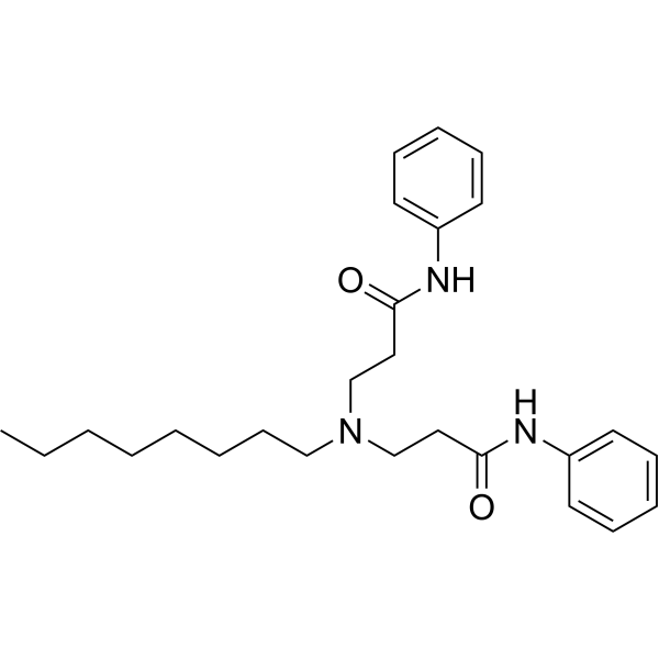
HY-155954
UCM-1336ICMT Inhibitor -
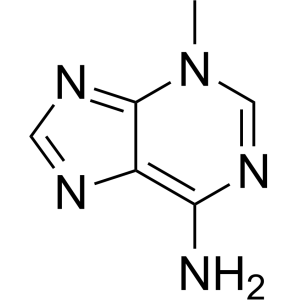
HY-19312
3-MethyladenineAutophagy/PI3K Inhibitor -
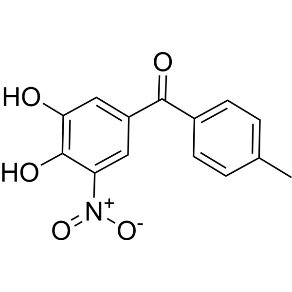
HY-17406
TolcaponeCOMT Inhibitor -
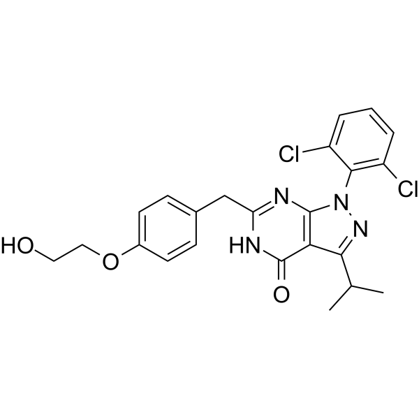
HY-112346
RGB-286147CDK/CRK Inhibitor -
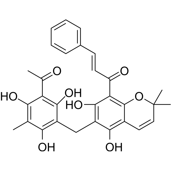
HY-18980
RottlerinPKC Inhibitor -

HY-19312R
3-Methyladenine (Standard)Autophagy/PI3K Inhibitor -
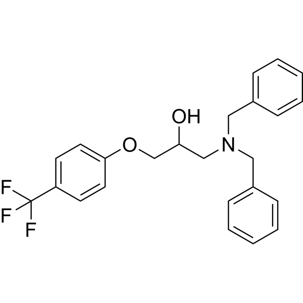
HY-134656
BC1618Fbxo48 Inhibitor -

HY-135533
Chromomycin A2Autophagy Inducer -
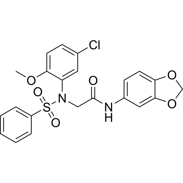
HY-111383
LX2343Beta-secretase Inhibitor -

HY-17406R
Tolcapone (Standard)COMT Inhibitor -
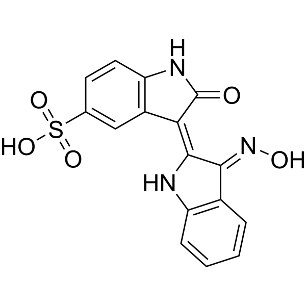
HY-111931
Indirubin-3'-monoxime-5-sulphonic acidCDK Inhibitor
Publications Citing Use of MedChemExpress (MCE) Cysmethynil
More

Biological Activity
Description
IC50 & Target
IC50 = 2.4 μM for Icmt
Cellular Effect
|
Cell Line

|
Type | Value | Description | References |
|---|---|---|---|---|
| HepG2 | IC50 |
19.3 μM
Compound: 1
|
Antiproliferative activity against human HepG2 cells after 72 hrs by MTT assay
Antiproliferative activity against human HepG2 cells after 72 hrs by MTT assay
|
[PMID: 23514631] |
| IMR-90 | IC50 |
29.2 μM
Compound: 1
|
Antiproliferative activity against human IMR90 cells after 72 hrs by MTT assay
Antiproliferative activity against human IMR90 cells after 72 hrs by MTT assay
|
[PMID: 23514631] |
| MDA-MB-231 | IC50 |
21.8 μM
Compound: 45292
|
Antiproliferative activity against human MDA-MB-231 cells after 72 hrs by colorimetric tetrazolium assay
Antiproliferative activity against human MDA-MB-231 cells after 72 hrs by colorimetric tetrazolium assay
|
[PMID: 20809634] |
| MDA-MB-231 | IC50 |
22.1 μM
Compound: 1
|
Antiproliferative activity against human MDA-MB-231 cells after 72 hrs by MTT assay
Antiproliferative activity against human MDA-MB-231 cells after 72 hrs by MTT assay
|
[PMID: 23514631] |
| PC-3 | IC50 |
21.3 μM
Compound: 1
|
Antiproliferative activity against human PC3 cells after 72 hrs by MTT assay
Antiproliferative activity against human PC3 cells after 72 hrs by MTT assay
|
[PMID: 23514631] |
| Sf9 | IC50 |
1.5 μM
Compound: 45292
|
Inhibition of recombinant ICMT expressed in Sf9 cells using [3H] AdoMet after 20 mins by scintillation counting
Inhibition of recombinant ICMT expressed in Sf9 cells using [3H] AdoMet after 20 mins by scintillation counting
|
[PMID: 20809634] |
| Sf9 | IC50 |
2.4 μM
Compound: cysmethynil
|
Inhibition of recombinant ICMT expressed in Sf9 cells using [3H]-AdoMet after 20 mins by scintillation counter
Inhibition of recombinant ICMT expressed in Sf9 cells using [3H]-AdoMet after 20 mins by scintillation counter
|
[PMID: 21661760] |
| Sf9 | IC50 |
1.9 μM
Compound: 1
|
Inhibition of recombinant Icmt (unknown origin) expressed in Sf9 cells assessed as transfer of methyl group from [3H]-S-adenosylmethionine to [3H]-biotin-S-farnesyl-L-cysteine after 30 mins
Inhibition of recombinant Icmt (unknown origin) expressed in Sf9 cells assessed as transfer of methyl group from [3H]-S-adenosylmethionine to [3H]-biotin-S-farnesyl-L-cysteine after 30 mins
|
[PMID: 23514631] |
| Sf9 | IC50 |
2.4 μM
Compound: 31
|
Inhibition of recombinant ICMT (unknown origin) expressed in sf9 cells using biotin S-farnesyl-L-cysteine as substrate
Inhibition of recombinant ICMT (unknown origin) expressed in sf9 cells using biotin S-farnesyl-L-cysteine as substrate
|
[PMID: 23566315] |
In Vitro
Cysmethynil (15 μM or 20 μM or 30 μM; 6 days) inhibits Icmt+/+ cells and Icmt-/-/ICMT cells proliferation, unaffects Icmt-/-cells growth [1].
Cysmethynil (20-30 μM; 1-6 days) inhibits PC3 cells proliferation and results in a dose- and time-dependent reduction in the number of viable PC3 cells[2].
MedChemExpress (MCE) has not independently confirmed the accuracy of these methods. They are for reference only. Further protocols information, click here.
-
Cell Line:PC3 cells[2]
-
Concentration:20-30 μM cysmethynil
-
Incubation Time:1-6 days
-
Result:Resulted in a dose- and time-dependent reduction in the number of viable PC3 cells.
In Vivo
Cysmethynil (20 mg/kg; intraperitoneal injection; three times a week; 2 weeks; alone or combined with Paclitaxel (HY-B0015)/ Doxorubicin (HY-15142A) ) is not toxic to mice and induces only moderate inhibition of tumor growth as single agent, the combination with Paclitaxel(HY-B0015)/ Doxorubicin(HY-15142A) results in significantly greater efficacy in inhibiting tumor growth. Cysmethynil sensitizes cervical cancer cells to chemotherapy agent in xenograft mouse model[3].
MedChemExpress (MCE) has not independently confirmed the accuracy of these methods. They are for reference only.
-
Animal Model:6 week-old SCID mice with SiHa cells in the mice flank [3]
-
Dosage:20 mg/kg (single; combination with Paclitaxel or Doxorubicin)
-
Administration:intraperitoneal injection; three times a week; 2 weeks
-
Result:Decreased in tumor size when treated with Cys alone, or Cysmethynil+Paclitaxel(HY-B0015)/Cysmethynil+ Doxorubicin(HY-15142A) .
-
Animal Model:SCID mice with PC3 cells in a xenograft model[2]
-
Dosage:0.1 mg/g or 0.2 mg/g Cysmethynil
-
Administration:intraperitoneal injection; every 48 h; 28 days
-
Result:Decreased in tumor size when treated with both doses of cysmethynil.


Chemical Information
-
CAS No. 851636-83-4
-
Appearance Solid
-
Molecular Weight 376.53
-
Formula C25H32N2O
-
Color White to light yellow
-
SMILES
O=C(N)CC1=CN(CCCCCCCC)C2=C1C=C(C3=CC=CC(C)=C3)C=C2
-
Shipping
Room temperature in continental US; may vary elsewhere.
-
Storage
Powder -20°C 3 years 4°C 2 years In solvent -80°C 6 months -20°C 1 month


Publications (1)
-
Journal Impact Factor
-
Most Recent
-
J Ovarian Res
Vitamin B6 promotes the activation of primordial follicles through the PI3K/Akt signaling pathway. [Abstract]2025 Dec 17;18(1):296. PMID: 41408298


Solvent & Solubility
In Vitro:
DMSO : 100 mg/mL (265.58 mM; Need ultrasonic; Hygroscopic DMSO has a significant impact on the solubility of product, please use newly opened DMSO)
Please refer to the solubility information to select the appropriate solvent. Once prepared, please aliquot and store the solution to prevent product inactivation from repeated freeze-thaw cycles.
Storage method and period of stock solution: -80°C, 6 months; -20°C, 1 month. When stored at -80°C, please use it within 6 months. When stored at -20°C, please use it within 1 month.
Please refer to the solubility information to select the appropriate solvent. Once prepared, please aliquot and store the solution to prevent product inactivation from repeated freeze-thaw cycles.
Storage method and period of stock solution: -80°C, 6 months; -20°C, 1 month. When stored at -80°C, please use it within 6 months. When stored at -20°C, please use it within 1 month.
Concentration (start) × Volume (start) = Concentration (final) × Volume (final)

Protocols
-
Cell Cytotoxicity Assay
Cytotoxicity assays are usually based on the assessment of cell membrane damage, which can also be indirectly detected by measuring cell viability. Detection methods include MTT assay, CKK-8 assay, LDH assay and ATP assay, etc.
-
Autophagy
Autophagy is a process in which eukaryotic cells use lysosomes to degrade their own cytoplasmic proteins and damaged organelles under the regulation of autophagy related gene (Atg). Microtubule-associated proteins light chain 3 (LC3) is recognized as autophagy marker, which transfers from cytoplasmic LC3 (LC3-I) to membrane type (LC3-II). LC3-II/I ratio could be detected by Western Blot and fluorescence microscopy.
-
Lysosome and acidic-vesicle live-cell staining
Lysosome and acidic-vesicle live-cell staining detects acidic intracellular compartments by using membrane-permeant acidotropic probes that accumulate in low-pH vesicles, including lysosomes, late endosomes, autolysosomes, and acidic phagosomes. LysoTracker staining is commonly used as an intensity-based readout of acidic lysosomal compartment abundance or enlargement, while acridine orange produces green fluorescence in less concentrated compartments and red fluorescence after concentration-dependent accumulation in acidic vesicular organelles. Loss or reduction of acridine-orange red signal can be used as a readout of lysosomal membrane permeabilization or reduced acidic-vesicle integrity. This protocol is designed for live cultured cells and can be adapted for fluorescence microscopy, high-content imaging, plate-reader readout, or flow cytometry when the selected literature supports the readout. Because these dyes report acidotropic accumulation rather than lysosome identity alone,
-
Macroautophagy Solutions
Macroautophagy is a conserved lysosome-dependent degradation pathway in which cytoplasmic material is sequestered into double-membrane autophagosomes and delivered to lysosomes for degradation and recycling. The pathway supports cellular homeostasis during nutrient limitation, organelle stress, protein-aggregate accumulation, infection, differentiation, and tissue remodeling by coupling cargo sequestration, autophagosome maturation, lysosomal fusion, and degradation of cargo-derived macromolecules. The core molecular sequence includes initiation by nutrient- and stress-regulated autophagy machinery, autophagosome nucleation, LC3/ATG8-family conjugation to autophagosomal membranes, cargo selection through receptors such as SQSTM1/p62, autophagosome-lysosome fusion, and lysosomal degradation. LC3 was identified as a mammalian homolog of yeast Atg8 that localizes to autophagosomal membranes after processing, and p62/SQSTM1 was shown to connect ubiquitinated cargo with autophagic degradati
-
Cell Viability Determination by MTT Colorimetric Assay
The following protocol uses the MTT colorimetric assay as a classic literature-established method for assessing cell viability/metabolic activity in cultured mammalian cells. MTT[3-(4,5-dimethylthiazol-2-yl)-2,5-diphenyltetrazolium bromide] is reduced by metabolically active cells to a colored formazan product; the amount of formazan is quantified spectrophotometrically and provides an indirect measure of metabolically active viable cells. Importantly, MTT reduction reflects cellular oxidoreductase/metabolic activity rather than an absolute direct count of living cells, so changes in cellular metabolism can alter the signal independently of cell number.


Purity & Documentation
-
Data Sheet (276 KB)
-
SDS (254 KB)
- English - EN (254 KB)
- Français - FR (254 KB)
- Deutsch - DE (254 KB)
- Norwegian - NO (254 KB)
- Español - ES (254 KB)
- Swedish - SV (254 KB)
- Italian - IT (254 KB)
- Korean - KR (254 KB)
- Portuguese - PT (254 KB)
-
Handling Instructions (2659 KB)
References
[1]. Winter-Vann AM, et al. A small-molecule inhibitor of isoprenylcysteine carboxyl methyltransferase with antitumor activity in cancer cells. Proc Natl Acad Sci U S A. 2005 Mar 22;102(12):4336-41. [Content Brief]
[2]. Wang M, et al. A small molecule inhibitor of isoprenylcysteine carboxymethyltransferase induces autophagic cell death in PC3 prostate cancer cells. J Biol Chem. 2008 Jul 4;283(27):18678-84. [Content Brief]
[3]. Pan Q, et al. Inhibition of isoprenylcysteine carboxylmethyltransferase sensitizes common chemotherapies in cervical cancer via Ras-dependent pathway. Biomed Pharmacother. 2018 Mar;99:169-175. [Content Brief]
[4]. Zhu C, et al. Targeting KRAS mutant cancers: from druggable therapy to drug resistance. Mol Cancer. 2022 Aug 4;21(1):159. [Content Brief]


Complete Stock Solution Preparation Table
Please refer to the solubility information to select the appropriate solvent. Once prepared, please aliquot and store the solution to prevent product inactivation from repeated freeze-thaw cycles.
Storage method and period of stock solution: -80°C, 6 months; -20°C, 1 month. When stored at -80°C, please use it within 6 months. When stored at -20°C, please use it within 1 month.
| Optional Solvent | Concentration Solvent Mass | 1 mg | 5 mg | 10 mg | 25 mg |
|---|---|---|---|---|---|
| DMSO | 1 mM | 2.6558 mL | 13.2792 mL | 26.5583 mL | 66.3958 mL |
| 5 mM | 0.5312 mL | 2.6558 mL | 5.3117 mL | 13.2792 mL | |
| 10 mM | 0.2656 mL | 1.3279 mL | 2.6558 mL | 6.6396 mL | |
| 15 mM | 0.1771 mL | 0.8853 mL | 1.7706 mL | 4.4264 mL | |
| 20 mM | 0.1328 mL | 0.6640 mL | 1.3279 mL | 3.3198 mL | |
| 25 mM | 0.1062 mL | 0.5312 mL | 1.0623 mL | 2.6558 mL | |
| 30 mM | 0.0885 mL | 0.4426 mL | 0.8853 mL | 2.2132 mL | |
| 40 mM | 0.0664 mL | 0.3320 mL | 0.6640 mL | 1.6599 mL | |
| 50 mM | 0.0531 mL | 0.2656 mL | 0.5312 mL | 1.3279 mL | |
| 60 mM | 0.0443 mL | 0.2213 mL | 0.4426 mL | 1.1066 mL | |
| 80 mM | 0.0332 mL | 0.1660 mL | 0.3320 mL | 0.8299 mL | |
| 100 mM | 0.0266 mL | 0.1328 mL | 0.2656 mL | 0.6640 mL |


Cysmethynil Related Classifications
HY-16160 Related Classifications
 Powered by Bioz