



Dexrazoxane monohydrochloride
Based on 14 publication(s) in Google Scholar
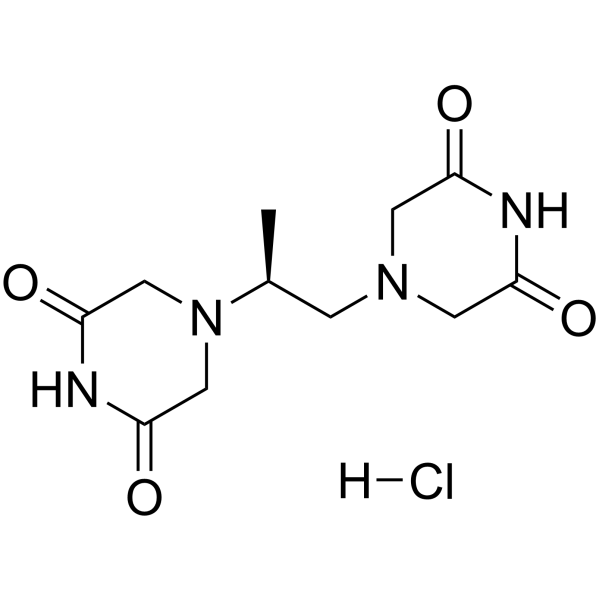
Dexrazoxane monohydrochloride is the hydrochloride of Dexrazoxane (HY-B0581). Dexrazoxane can prevent or reduce cardiac damage and is an iron chelator and apoptosis inducer. Dexrazoxane has cardioprotective, anti-inflammatory, antioxidant, anti-tumor and neuroprotective activities and inhibits ferroptosis.
For research use only. We do not sell to patients.
- CAS No.: 1263283-43-7
- Formula: C11H17ClN4O4
- Molecular Weight:304.73
-
Storage:
Please store the product under the recommended conditions in the Certificate of Analysis.

To place orders, for customer services and technical support, please contact: MedChemExpress USA
Tel: 609-228-6898 E-mail: [email protected] [email protected]


-
 Biological Activity
-
Chemical Information
- Protocols
- Purity & Documentation
- References
-
 Help & FAQs


Publications Citing Use of MedChemExpress (MCE) Dexrazoxane monohydrochloride
More- Adv Sci (Weinh). 2023 May;10(15):e2206007. [Abstract]
- Phytomedicine. 2025 Nov 25:148:157357. [Abstract]
- Phytomedicine. 2023 Aug:117:154922. [Abstract]
- Nano Res. 2023 Apr 18.
- Free Radic Biol Med. 2024 Dec 9:227:296-311. [Abstract]
- Free Radic Biol Med. 2020 Nov 20:160:303-318. [Abstract]
- J Ethnopharmacol. 2025 Oct 14:120751. [Abstract]
- Eur J Med Chem. 2025 Feb 5:283:117108. [Abstract]
- Biochem Pharmacol. 2024 Jun:224:116247. [Abstract]
- Mol Med Rep. 2024 May;29(5):84. [Abstract]
- J Cell Mol Med. 2025 Jun;29(11):e70641. [Abstract]
- Cardiovasc Toxicol. 2024 Aug;24(8):818-835. [Abstract]
- Biochem Biophys Res Commun. 2025 Sep 8:778:152417. [Abstract]
- Biomed Pharmacother. 2022 Sep:153:113280. [Abstract]
 Customer Validation & Images

-
Bio/Physico-chemical Assay
-
Flow Cytometry
-
Cell Imaging/Staining
-
RT-PCR
-
In Vivo Efficacy Study


Biological Activity
Description
Clinical Trial
| NCT Number | Sponsor | Condition | Start Date |
Phase

|
|---|---|---|---|---|
| NCT01329991 | Plexxikon| | 2011-05 | PHASE1 |


Chemical Information
-
CAS No. 1263283-43-7
-
Molecular Weight 304.73
-
Formula C11H17ClN4O4
-
SMILES
C[C@H](N(C1)CC(NC1=O)=O)CN(C2)CC(NC2=O)=O.[H]Cl
-
Shipping
Room temperature in continental US; may vary elsewhere.
-
Storage
Please store the product under the recommended conditions in the Certificate of Analysis.


Publications (14)
-
Journal Impact Factor
-
Most Recent
-
Adv Sci (Weinh)
The Imbalance of p53-Park7 Signaling Axis Induces Iron Homeostasis Dysfunction in Doxorubicin-Challenged Cardiomyocytes. [Abstract]2023 May;10(15):e2206007. PMID: 36967569
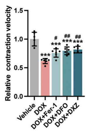
Dexrazoxane monohydrochloride purchased from MedChemExpress. Usage Cited in: Adv Sci (Weinh). 2023 May;10(15):e2206007. [Abstract]
Dexrazoxane (DXZ) (1 µM; 24 h) resulted in a decline in contractile speed in NMCMs.
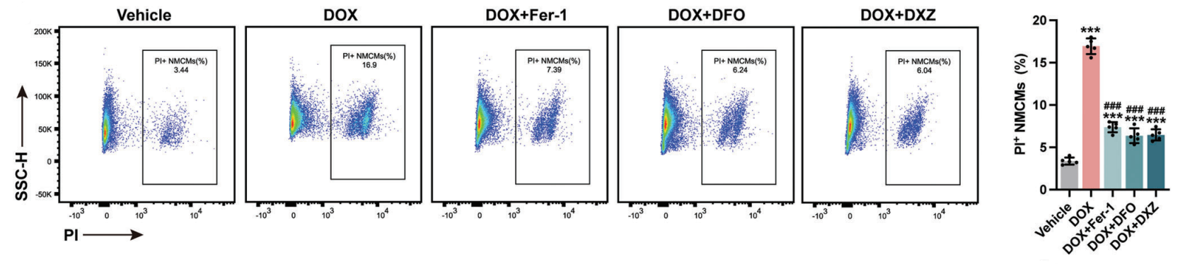
Dexrazoxane monohydrochloride purchased from MedChemExpress. Usage Cited in: Adv Sci (Weinh). 2023 May;10(15):e2206007. [Abstract]
Dexrazoxane (DXZ) (1 µM; 24 h) resulted in a decline in cell viability in NMCMs.
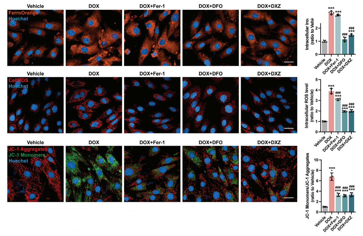
Dexrazoxane monohydrochloride purchased from MedChemExpress. Usage Cited in: Adv Sci (Weinh). 2023 May;10(15):e2206007. [Abstract]
Dexrazoxane (DXZ) (1 µM; 24 h) inhibited DOX-induced iron accumulation and ROS and restored mitochondrial membrane potential in NMCMs.
Dexrazoxane monohydrochloride purchased from MedChemExpress. Usage Cited in: Adv Sci (Weinh). 2023 May;10(15):e2206007. [Abstract]
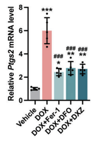
Dexrazoxane (DXZ) (1 µM; 24 h) reversed Ptgs2 mRNA expression in NMCMs.
Dexrazoxane monohydrochloride purchased from MedChemExpress. Usage Cited in: Adv Sci (Weinh). 2023 May;10(15):e2206007. [Abstract]
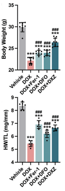
Dexrazoxane (DXZ) (20 mg/kg; i.p.; weekly for 4 weeks) reversed the DOX-induced decrease in body weight and the ratio of heart weight to tibia length (HW/TL) of chronic DoIC (doxorubicin (DOX)-induced cardiotoxicity) mice.
-
Phytomedicine
Quercetin ameliorates doxorubicin-induced atrial fibrillation via EGFR-mediated restoration of autophagic flux. [Abstract]2025 Nov 25:148:157357. PMID: 41056856 -
Phytomedicine
Amentoflavone mitigates doxorubicin-induced cardiotoxicity by suppressing cardiomyocyte pyroptosis and inflammation through inhibition of the STING/NLRP3 signalling pathway. [Abstract]2023 Aug:117:154922. PMID: 37321078 -
-
Free Radic Biol Med
2024 Dec 9:227:296-311. PMID: 39653130 -
Free Radic Biol Med
2020 Nov 20:160:303-318. PMID: 32846217 -
J Ethnopharmacol
Huanglian-ejiao decoction ameliorates doxorubicin-induced cardiomyocyte apoptosis and autophagic flux dysregulation by up-regulating ubiquilin1. [Abstract]2025 Oct 14:120751. PMID: 41101550 -
Eur J Med Chem
Praeruptorin A screened by a ferrous ion probe inhibited DMT1 and ferroptosis to attenuate Doxorubicin-induced cardiomyopathy. [Abstract]2025 Feb 5:283:117108. PMID: 39615370 -
Biochem Pharmacol
Calcium saccharate/DUSP6 suppresses renal cell carcinoma glycolytic metabolism and boosts sunitinib efficacy via the ERK-AKT pathway. [Abstract]2024 Jun:224:116247. PMID: 38697311 -
Mol Med Rep
Food therapy of scutellarein ameliorates pirarubicin‑induced cardiotoxicity in rats by inhibiting apoptosis and ferroptosis through regulation of NOX2‑induced oxidative stress. [Abstract]2024 May;29(5):84. PMID: 38516760 -
J Cell Mol Med
Downregulation of Alox5 Inhibits Ferroptosis to Improve Doxorubicin-Induced Cardiotoxicity via the P53/SLC7A11 Pathway. [Abstract]2025 Jun;29(11):e70641. PMID: 40485049 -
Cardiovasc Toxicol
Marein Alleviates Doxorubicin-Induced Cardiotoxicity through FAK/AKT Pathway Modulation while Potentiating its Anticancer Activity. [Abstract]2024 Aug;24(8):818-835. PMID: 38896162 -
Biochem Biophys Res Commun
The m6A modification reader protein IGF2BP2 regulates ferroptosis in nasopharyngeal carcinoma cells by stabilizing CP expression via an m6A-dependent mechanism. [Abstract]2025 Sep 8:778:152417. PMID: 40743989 -
Biomed Pharmacother
2022 Sep:153:113280. PMID: 35724508

Protocols
-
Apoptosis
Apoptosis, also called programmed cell death, is generally characterized by distinct morphological characteristics.
-
TUNEL staining for apoptotic DNA fragmentation
TUNEL staining detects DNA strand breaks by using terminal deoxynucleotidyl transferase to add labeled nucleotides to exposed 3′-OH DNA termini, generating either microscopic staining in fixed cells or tissue sections, or fluorescence/cytometric signal in cell suspensions. TUNEL positivity reflects DNA fragmentation but should not be interpreted alone as definitive apoptosis, because TUNEL can also label necrotic, autolytic, mechanically damaged, or DNA-repair-associated DNA breaks.
-
Annexin V plus membrane-impermeant dye apoptosis staining
Annexin V-based apoptosis assays rely on the detection of phosphatidylserine (PS) externalization from the inner leaflet of the plasma membrane to the outer leaflet, an early biochemical hallmark of apoptosis. Fluorescently labeled Annexin V binds PS in a calcium-dependent manner, enabling identification of early apoptotic cells by flow cytometry or fluorescence microscopy. When combined with a membrane-impermeant DNA-binding dye (e. g. , propidium iodide), this approach allows discrimination between viable (Annexin V−/dye−), early apoptotic (Annexin V+/dye−), and late apoptotic or necrotic (Annexin V+/dye+) cell populations by assessing membrane integrity and PS exposure.
-
Apoptosis Solutions
Apoptosis is a regulated, generally non-lytic cell-death pathway that removes unwanted, damaged, infected, or abnormal cells through coordinated morphological changes, caspase activation, DNA fragmentation, and membrane remodeling. The intrinsic apoptosis pathway is controlled mainly by mitochondrial outer membrane permeabilization, BCL-2 family proteins, cytochrome c release, apoptosome formation, caspase-9 activation, and downstream executioner caspase-3/7 activation. The extrinsic apoptosis pathway is initiated by death receptors such as Fas, TNFR, and TRAIL receptors, which recruit adaptor proteins and activate caspase-8 before engaging executioner caspases or mitochondrial amplification through BID cleavage. Apoptosis is linked to many phenotypes, including cancer cell killing, tissue homeostasis, immune regulation, neurodegeneration, infection response, and treatment-induced cytotoxicity; unresolved questions include how apoptosis interacts with necroptosis, pyroptosis, ferroptos
-
Research Protocol for Inflammation-related Diseases
The NLRP3 inflammasome is a cytosolic innate immune signaling platform that integrates priming signals and danger-signal activation to promote caspase-1 activation, maturation of IL-1β and IL-18, and gasdermin D-mediated pyroptotic cell death. The core experimental logic is to determine whether inflammatory disease phenotypes are driven by increased NLRP3 expression, ASC-containing inflammasome assembly, caspase-1 cleavage, GSDMD cleavage, and extracellular release of IL-1β/IL-18 rather than by nonspecific cell injury alone. The pathway is strongly linked to inflammation-related disease phenotypes because monosodium urate crystals activate NALP3/NLRP3 inflammasome signaling in gout-like crystal inflammation, cholesterol crystals activate NLRP3 inflammasomes in atherogenesis models, and DSS-induced intestinal inflammation has been reported to involve NLRP3 inflammasome activity. However, experimental colitis studies also show context-dependent protective effects of NLRP3 inflammasome co
-
Ferroptosis Solutions
Ferroptosis is an iron-dependent, non-apoptotic form of regulated cell death characterized by lethal lipid peroxidation and sensitivity to suppression by iron chelators or lipophilic radical-trapping antioxidants. The core pathway links cystine uptake through system Xc−, glutathione availability, GPX4-dependent detoxification of phospholipid hydroperoxides, iron-dependent oxidative reactions, and polyunsaturated-phospholipid metabolism into a cell-death program that is biochemically and morphologically distinct from apoptosis, necrosis, and autophagy. The ferroptosis pathway is experimentally linked to phenotype through chemical and genetic perturbation. Erastin induces ferroptosis by inhibiting cystine uptake through system Xc− and weakening antioxidant defenses, while GPX4 inhibition or depletion causes lipid peroxide accumulation and ferroptotic cancer-cell death. ACSL4 and oxidizable arachidonoyl- or adrenoyl-containing phosphatidylethanolamines shape ferroptosis sensitivity by con


Purity & Documentation
-
Data Sheet (269 KB)
-
SDS (252 KB)
- Français - FR (252 KB)
- Deutsch - DE (252 KB)
- Norwegian - NO (252 KB)
- Español - ES (252 KB)
- Swedish - SV (252 KB)
- Italian - IT (252 KB)
- Korean - KR (252 KB)
- Portuguese - PT (252 KB)
-
Handling Instructions (2659 KB)
References

Calculators
Concentration (start) × Volume (start) = Concentration (final) × Volume (final)


Dexrazoxane monohydrochloride Related Classifications
HY-110075 Related Classifications
 Powered by Bioz