



DHODH-IN-26
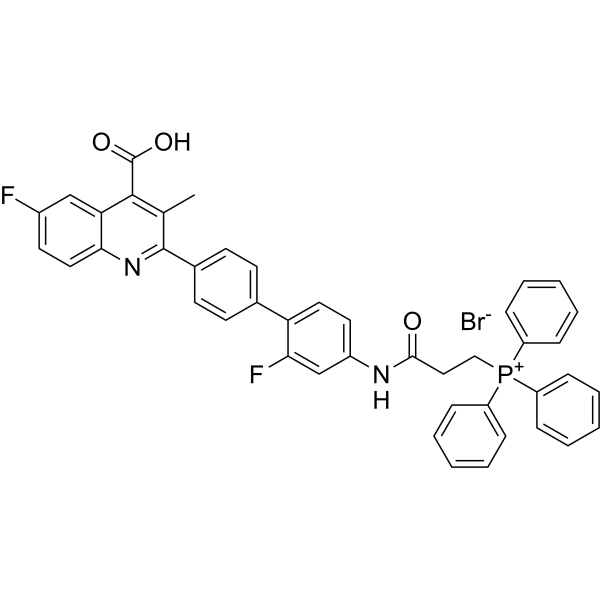
DHODH-IN-26 (compound B2) is a mitochondria-targeting DHODH inhibitor. DHODH-IN-26 shows anticancer activity, triggers the formation of reactive oxygen species (ROS), promots mitochondrial lipid peroxidation, and induces ferroptosis.
For research use only. We do not sell to patients.
- Formula: C44H34BrF2N2O3P
- Molecular Weight:787.63
-
Storage:
Please store the product under the recommended conditions in the Certificate of Analysis.

To place orders, for customer services and technical support, please contact: MedChemExpress USA
Tel: 609-228-6898 E-mail: [email protected] [email protected]


-
 Biological Activity
-
Chemical Information
- Protocols
- Purity & Documentation
- References
-
 Help & FAQs




Biological Activity
Description
In Vitro
DHODH-IN-26 (compound B2) (0.01-100 μM, 72 h) inhibits cell growth of 4T1, B16F10, U251, GL261, SH-SY5Y, U87, and A375 cells[1].
DHODH-IN-26 (50 and 100 nM) inhibits colony formation in B16F10 and A375 cells[1].
DHODH-IN-26 (50 and 100 nM, 48 h) decreases the expression of PD-L1 in B16F10 and A375 cells[1].
MedChemExpress (MCE) has not independently confirmed the accuracy of these methods. They are for reference only. Further protocols information, click here.
-
Cell Line:4T1, B16F10, U251, GL261, SH-SY5Y, U87, and A375 cells
-
Concentration:0.01, 0.1, 1, 10 and100 μM
-
Incubation Time:72 h
-
Result:Inhibited cell growth.
-
Cell Line:B16F10 and A375 cells
-
Concentration:50 and 100 nM
-
Incubation Time:48 h
-
Result:Decreased the expression of PD-L1.
In Vivo
MedChemExpress (MCE) has not independently confirmed the accuracy of these methods. They are for reference only.
-
Animal Model:B16F10 xenograft model in C57BL/6 mice[1]
-
Dosage:50 mg/kg
-
Administration:I.p.; for 14 days
-
Result:Reduced tumor volume.


Chemical Information
-
Molecular Weight 787.63
-
Formula C44H34BrF2N2O3P
-
SMILES
CC1=C(C2=CC(F)=CC=C2N=C1C3=CC=C(C=C3)C4=CC=C(C=C4F)NC(CC[P+](C5=CC=CC=C5)(C6=CC=CC=C6)C7=CC=CC=C7)=O)C(O)=O.[Br-]
-
Shipping
Room temperature in continental US; may vary elsewhere.
-
Storage
Please store the product under the recommended conditions in the Certificate of Analysis.

Protocols
-
ROS/oxidative-stress fluorescent staining
ROS/oxidative-stress fluorescent staining uses cell-permeant fluorogenic probes that become fluorescent after oxidation inside cells or tissues; commonly used examples include DCFH-DA/DCFDA for broad cellular oxidant detection, DHE for superoxide-related signal detection, MitoSOX for mitochondrial superoxide-related signal detection, and CellROX probes for oxidative-stress-associated fluorescence readouts. The assay detects probe oxidation rather than a single ROS species unless the probe and analysis method have been chemically validated for that species. DCFH-DA enters cells, is deacetylated by intracellular esterases to DCFH, and produces fluorescent DCF after oxidation, so the readout is used as an operational measure of total cellular oxidative stress rather than a species-specific ROS measurement. DHE and MitoSOX can report superoxide-related oxidation, but red fluorescence alone can include non-specific ethidium-like oxidation products; HPLC or optimized spectral approaches are
-
Ferroptosis Solutions
Ferroptosis is an iron-dependent, non-apoptotic form of regulated cell death characterized by lethal lipid peroxidation and sensitivity to suppression by iron chelators or lipophilic radical-trapping antioxidants. The core pathway links cystine uptake through system Xc−, glutathione availability, GPX4-dependent detoxification of phospholipid hydroperoxides, iron-dependent oxidative reactions, and polyunsaturated-phospholipid metabolism into a cell-death program that is biochemically and morphologically distinct from apoptosis, necrosis, and autophagy. The ferroptosis pathway is experimentally linked to phenotype through chemical and genetic perturbation. Erastin induces ferroptosis by inhibiting cystine uptake through system Xc− and weakening antioxidant defenses, while GPX4 inhibition or depletion causes lipid peroxide accumulation and ferroptotic cancer-cell death. ACSL4 and oxidizable arachidonoyl- or adrenoyl-containing phosphatidylethanolamines shape ferroptosis sensitivity by con


Purity & Documentation
References

Calculators
Concentration (start) × Volume (start) = Concentration (final) × Volume (final)


DHODH-IN-26 Related Classifications
HY-158023 Related Classifications
 Powered by Bioz