



Disitamab vedotin
Based on 5 publication(s) in Google Scholar
Disitamab vedotin (RC48) is a HER2-targeted antibody-drug conjugate (ADC), formed by conjugation of the humanized anti-HER2 antibody Disitamab (HY-P99854) with drug-linker VcMMAE (HY-15575). VcMMAE consists of a cleavable MC-Val-Cit-PAB linker and the microtubule inhibitor MMAE (HY-15162). After binding to HER2 and undergoing endocytosis, Disitamab vedotin releases MMAE in lysosomes, which further inhibits microtubule assembly, arrests mitosis and induces apoptosis, while also exerting a bystander effect. Disitamab vedotin can be used in studies of HER2-positive breast cancer.
For research use only. We do not sell to patients.
- Purity : 99.06%
- CAS No.: 2136633-23-1
- Molecular Weight:149000 (average)
-
Storage:
Please store the product under the recommended conditions in the Certificate of Analysis.


To place orders, for customer services and technical support, please contact: MedChemExpress USA
Tel: 609-228-6898 E-mail: [email protected] [email protected]


-
 Biological Activity
-
Chemical Information
- Protocols
- Purity & Documentation
- References
-
 Help & FAQs


-

HY-138298A
Trastuzumab deruxtecanHER2 ADC -

HY-P9921
Trastuzumab emtansineAntibody-drug Conjugate -

HY-132254
Sacituzumab govitecanTrop-2 ADC -

HY-138298
Trastuzumab deruxtecan (solution)HER2 ADC -

HY-141598
Datopotamab deruxtecan (solution)TROP2-Targeted ADC -

HY-P99016B
Enfortumab vedotin-ejfv (solution)Nectin-4 ADC -

HY-141601
Telisotuzumab vedotinAnti-c-MET ADC -

HY-P99107A
Brentuximab vedotin (solution)ADC -

HY-132254A
Sacituzumab govitecan (solution)Trop-2 ADC -

HY-152963
Tisotumab vedotinAntibody-Drug Conjugate -

HY-132253
Polatuzumab vedotinMicrotubule Inhibitor -

HY-P3239
Belantamab mafodotinAnti-BCMA ADC
Publications Citing Use of MedChemExpress (MCE) Disitamab vedotin
More Customer Validation & Images

-
Cell Proliferation/Viability Assay
-
Cell Proliferation/Viability Assay
-
Flow Cytometry
-
In Vivo Efficacy Study
-
IHC
All EGFR Isoforms
More

Biological Activity
Description
IC50 & Target
[2]|
HER2 |
Cellular Effect
|
Cell Line

|
Type | Value | Description | References |
|---|---|---|---|---|
| E0771 | IC50 |
90 ng/mL
|
Reduction of murine breast cancer E0771-hHER2 cell viability incubated for 96 hrs assessed via Cell Counting Kit-8 (CCK-8) metabolic activity assay.
Reduction of murine breast cancer E0771-hHER2 cell viability incubated for 96 hrs assessed via Cell Counting Kit-8 (CCK-8) metabolic activity assay.
|
34657203 |
In Vitro
Disitamab vedotin (RC48) exerts a significant bystander killing effect in vitro against heterogeneous HER2-expressing cancer cell populations, enhancing its efficacy by targeting both HER2-positive and adjacent HER2-negative tumor cells[3].
Disitamab vedotin (0.0001-10 µg/mL; 48-96 h) potently inhibits E0771-hHER2 murine breast cancer cell viability with an IC50 of 90 ng/mL, with target-specific activity and no effect on hHER2-negative E0771-WT cells[2].
Disitamab vedotin (10.0 μg/mL) shows specific cross-reactivity to normal human tissues including bladder, skin, breast, pancreas, placenta, kidney, prostate, ureter, fallopian tube, and stomach[5].
MedChemExpress (MCE) has not independently confirmed the accuracy of these methods. They are for reference only. Further protocols information, click here.
-
Cell Line:E0771-hHER2; E0771-WT
-
Concentration:100, 500 ng/mL
-
Incubation Time:48, 72 h
-
Result:Preferentially reduced viability of E0771-hHER2 cells compared with E0771-WT cells.
In Vivo
Combination treatment with Disitamab vedotin (5 mg/kg; i.v.; Days 0, 7) and anti-PD-1 antibody (HY-P9902A) (10 mg/kg; i.v.; Days 0, 3, 7, 10) significantly inhibits the growth of E0771-hHER2 tumors and leads to tumor regression in the combination group within approximately 2-3 weeks[2].
Disitamab vedotin (10 mg/kg; single i.v. administration) increases T cell infiltration in tumor tissues of E0771-hHER2 tumor-bearing mice[2].
Combination therapy with disitamab vedotin and a PD-1/PD-L1 checkpoint inhibitor induces durable immune protection in mice with completely regressed E0771-hHER2 tumors; complete tumor rejection occurs upon rechallenge with E0771-hHER2 cells, and a weaker rejection effect is also observed upon rechallenge with E0771-WT cells[2].
Disitamab vedotin (3-12 mg/kg; intravenous injection; once every 2 weeks; for 12 consecutive weeks) induces dose-dependent, reversible hematological and lymphoid organ toxicity in healthy cynomolgus monkeys, with a maximum non-severe toxic dose of 6 mg/kg, and the positive rate of anti-drug antibodies decreases with increasing dose[5].
Disitamab vedotin (4-16 mg/kg; intravenous injection; single administration) produces no treatment-related effects on the central nervous system of healthy Sprague Dawley rats at doses up to 16 mg/kg[5].
MedChemExpress (MCE) has not independently confirmed the accuracy of these methods. They are for reference only.
-
Animal Model:Human PD-1 transgenic C57BL/6 (5 to 6 weeks old; subcutaneous inoculation of 2 × 10⁶ E0771-hHER2 cells; treatment initiated when average tumor volume reached 100-200 mm3)[2]
-
Dosage:10 mg/kg
-
Administration:i.v.; single dose on day 5 post-tumor inoculation
-
Result:Reduced mean tumor volume to 244.82 mm³ on day 9 after administration.
Increased intratumoral T cell infiltration relative to vehicle and parental anti-hHER2 antibody groups.
-
Animal Model:Human PD-1 transgenic C57BL/6 (5 to 6 weeks old; subcutaneous inoculation of 2 × 10⁶ E0771-hHER2 cells; treatment initiated when average tumor volume reached 150-200 mm3)[2]
-
Dosage:5 mg/kg (monotherapy); 5 mg/kg + 10 mg/kg (anti-PD-1/PD-L1 antibody, combination)
-
Administration:i.v.;Disitamab vedotin on days 0 and 7; anti-PD-1 on days 0, 3, 7, and 10
-
Result:Combination markedly inhibited tumor growth and resulted in tumor disappearance within approximately 2-3 weeks.
-
Animal Model:Cynomolgus monkeys (3.5-5 years old)[5]
-
Dosage:3 mg/kg; 6 mg/kg; 12 mg/kg
-
Administration:i.v.; every 2 weeks; 12 weeks
-
Result:Produced dose-dependent hematological changes; the highest non-severely toxic dose was 6 mg/kg.
-
Animal Model:Sprague Dawley rats (6-9 weeks old, n=5/sex/group)[5]
-
Dosage:4 mg/kg; 8 mg/kg; 16 mg/kg
-
Administration:i.v.; single dose
-
Result:Showed no administration-related abnormalities in home-cage, hand-held, or open-field observations, stimulus responses, rearing counts, defecation counts, forelimb grip strength, or body temperature, with all parameters statistically similar to the negative control group.
Clinical Trial
| NCT Number | Sponsor | Condition | Start Date |
Phase
|
|---|---|---|---|---|
| NCT01329991 | Plexxikon| | 2011-05 | PHASE1 |


Chemical Information
-
CAS No. 2136633-23-1
-
Appearance Solid
-
Molecular Weight 149000 (average)
-
Color White to light yellow
-
SMILES
[Disitamab vedotin]
-
Synonyms
RC48
-
Preparation Instructions
The product can be reconstituted/diluted with sterile PBS or saline.
-
Shipping
Shipping with dry ice.
-
Storage
Please store the product under the recommended conditions in the Certificate of Analysis.


Publications (5)
-
Journal Impact Factor
-
Most Recent
-
Nat Commun
Supramolecular coiled-coil peptide platform for site-specific antibody drug conjugate engineering. [Abstract]2026 Mar 2. PMID: 41771920 -
Cancer Lett
Drug-tolerant persisting polyploid giant cancer cells mediate resistance to HER2-targeting antibody-drug conjugates. [Abstract]2025 Oct 10:630:217900. PMID: 40614854
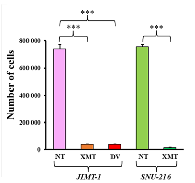
Disitamab vedotin purchased from MedChemExpress. Usage Cited in: Cancer Lett. 2025 Oct 10:630:217900. [Abstract]
Disitamab vedotin (DV, 0.25 μg/mL) reduced the overall cell number of HER2-positive JIMT-1 breast cancer cells.
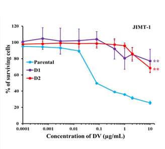
Disitamab vedotin purchased from MedChemExpress. Usage Cited in: Cancer Lett. 2025 Oct 10:630:217900. [Abstract]
Unlike the parental cells, which were sensitive to XMT-1522 and Disitamab vedotin (DV, 0.0001, 0.0006, 0.003, 0.016, 0.08, 0.4, 1, 2, and 10 μg/mL; 5 days), both D1 and D2 daughter cells that emerged from the DTP-PGCC were resistant to these drugs. D1, daughter-1 cells (one prior ADC treatment); D2, daughter-2 cells (two prior ADC treatments). ∗∗, p < 0.01; ∗∗∗, p < 0.001.
-
Mol Cancer Ther
Antibody-drug-conjugates prepared with Peptide Asparaginyl Ligases achieve strong in vitro and in vivo anti-tumor activity. [Abstract]2026 Mar 26. PMID: 41889275 -
Transl Oncol
Disitamab vedotin in preclinical models of HER2-positive breast and gastric cancers resistant to trastuzumab emtansine and trastuzumab deruxtecan. [Abstract]2025 Mar:53:102284. PMID: 39837059
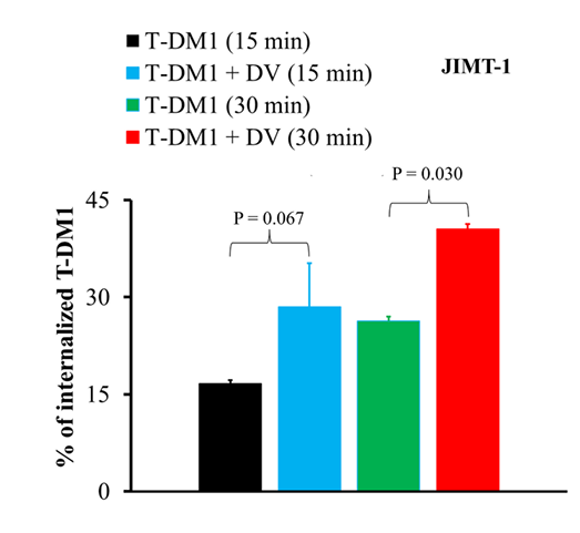
Disitamab vedotin purchased from MedChemExpress. Usage Cited in: Transl Oncol. 2025 Mar:53:102284. [Abstract]
Disitamab vedotin (DV, 50 μg/mL, 15 or 30 min) increased T-DM1 internalization into JIMT-1 breast cancer cells.
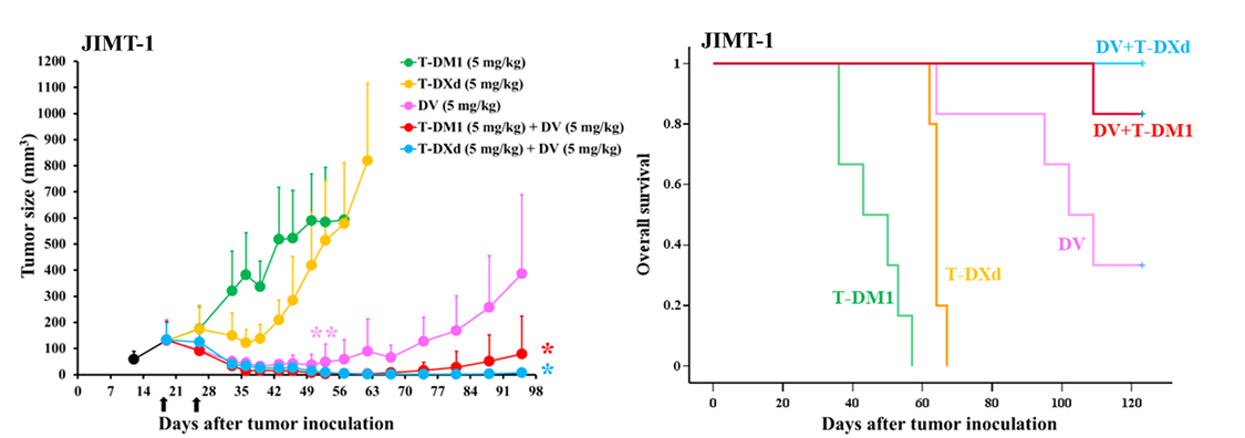
Disitamab vedotin purchased from MedChemExpress. Usage Cited in: Transl Oncol. 2025 Mar:53:102284. [Abstract]
In JIMT-1 xenografts, the combination of Disitamab vedotin (DV, 0.5 mg/kg or 5 mg/kg, administered intravenously twice at 7-day intervals) plus T-DM1, as well as DV plus T-DXd, inhibited tumor growth more effectively than the corresponding single treatments.
Disitamab vedotin purchased from MedChemExpress. Usage Cited in: Transl Oncol. 2025 Mar:53:102284. [Abstract]
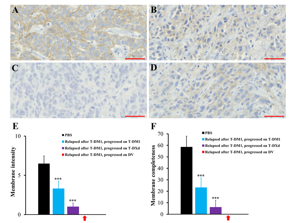
In macroscopic JIMT-1 xenografts, tumors that relapsed after T-DM1 treatment and subsequently responded to Disitamab vedotin (DV, 0.5 mg/kg or 5 mg/kg, administered intravenously twice at 7-day intervals) but later progressed had lost HER2 expression. A: PBS; B: relapsed after T-DM1, progressed on T-DM1; C: relapsed after T-DM1, progressed on DV; D: relapsed after T-DM1, progressed on T-DXd.
-
Clin Exp Metastasis
Comparison of trastuzumab emtansine, trastuzumab deruxtecan, and disitamab vedotin in a multiresistant HER2-positive breast cancer lung metastasis model. [Abstract]2024 Apr;41(2):91-102. PMID: 38367127

Protocols
-
Apoptosis
Apoptosis, also called programmed cell death, is generally characterized by distinct morphological characteristics.
-
TUNEL staining for apoptotic DNA fragmentation
TUNEL staining detects DNA strand breaks by using terminal deoxynucleotidyl transferase to add labeled nucleotides to exposed 3′-OH DNA termini, generating either microscopic staining in fixed cells or tissue sections, or fluorescence/cytometric signal in cell suspensions. TUNEL positivity reflects DNA fragmentation but should not be interpreted alone as definitive apoptosis, because TUNEL can also label necrotic, autolytic, mechanically damaged, or DNA-repair-associated DNA breaks.
-
Annexin V plus membrane-impermeant dye apoptosis staining
Annexin V-based apoptosis assays rely on the detection of phosphatidylserine (PS) externalization from the inner leaflet of the plasma membrane to the outer leaflet, an early biochemical hallmark of apoptosis. Fluorescently labeled Annexin V binds PS in a calcium-dependent manner, enabling identification of early apoptotic cells by flow cytometry or fluorescence microscopy. When combined with a membrane-impermeant DNA-binding dye (e. g. , propidium iodide), this approach allows discrimination between viable (Annexin V−/dye−), early apoptotic (Annexin V+/dye−), and late apoptotic or necrotic (Annexin V+/dye+) cell populations by assessing membrane integrity and PS exposure.
-
Apoptosis Solutions
Apoptosis is a regulated, generally non-lytic cell-death pathway that removes unwanted, damaged, infected, or abnormal cells through coordinated morphological changes, caspase activation, DNA fragmentation, and membrane remodeling. The intrinsic apoptosis pathway is controlled mainly by mitochondrial outer membrane permeabilization, BCL-2 family proteins, cytochrome c release, apoptosome formation, caspase-9 activation, and downstream executioner caspase-3/7 activation. The extrinsic apoptosis pathway is initiated by death receptors such as Fas, TNFR, and TRAIL receptors, which recruit adaptor proteins and activate caspase-8 before engaging executioner caspases or mitochondrial amplification through BID cleavage. Apoptosis is linked to many phenotypes, including cancer cell killing, tissue homeostasis, immune regulation, neurodegeneration, infection response, and treatment-induced cytotoxicity; unresolved questions include how apoptosis interacts with necroptosis, pyroptosis, ferroptos
-
Breast Cancer Modeling
Breast cancer is a heterogeneous cancer, and it has been distinguished into four subtypes: luminal A, luminal B, HER2-positive and basal-like. Molecular mutations, epigenetic alterations, hormone exposure and immune microenvironment are related to the progression of breast cancer.


Purity & Documentation
-
Data Sheet (282 KB)
-
SDS (251 KB)
- English - EN (251 KB)
- Français - FR (251 KB)
- Deutsch - DE (251 KB)
- Norwegian - NO (251 KB)
- Español - ES (251 KB)
- Swedish - SV (251 KB)
- Italian - IT (251 KB)
- Korean - KR (251 KB)
- Portuguese - PT (251 KB)
-
Handling Instructions (2659 KB)
References
[1]. Deeks ED, et al. Disitamab Vedotin: First Approval. Drugs. 2021 Nov;81(16):1929-1935. [Content Brief]
[2]. Huang L, et al. A HER2 target antibody drug conjugate combined with anti-PD-(L)1 treatment eliminates hHER2+ tumors in hPD-1 transgenic mouse model and contributes immune memory formation. Breast cancer research and treatment. 2022 Jan;191(1):51-61. [Content Brief]
[5]. Jiang J, et al. Preclinical safety profile of Disitamab vedotin: a novel anti-HER2 antibody conjugated with MMAE. Toxicology Letters. 2019. [Content Brief]

Calculators
Concentration (start) × Volume (start) = Concentration (final) × Volume (final)


Disitamab vedotin Related Classifications
HY-P9985 Related Classifications
 Powered by Bioz