



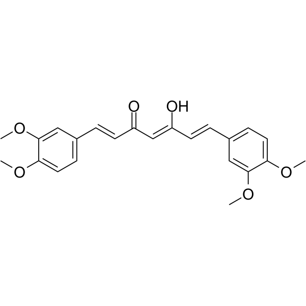
Dimethylcurcumin
Based on 14 publication(s) in Google Scholar
Dimethylcurcumin (ASC-J9) is an androgen receptor degradation enhancer that effectively suppresses castration resistant prostate cancer cell proliferation and invasion.
For research use only. We do not sell to patients.
- Purity : 98.91%
- CAS No.: 52328-98-0
- Formula: C23H24O6
- Molecular Weight:396.43
-
Storage:Powder -20°C, 3 years , 4°C, 2 years ; In solvent -80°C, 2 years , -20°C, 1 year

To place orders, for customer services and technical support, please contact: MedChemExpress USA
Tel: 609-228-6898 E-mail: [email protected] [email protected]


-
 Biological Activity
-
Chemical Information
-
Solvent & Solubility
- Protocols
- Purity & Documentation
- References
-
 Help & FAQs


-
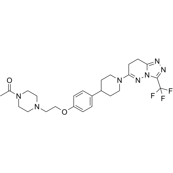
HY-16079
AZD3514AR Inhibitor -
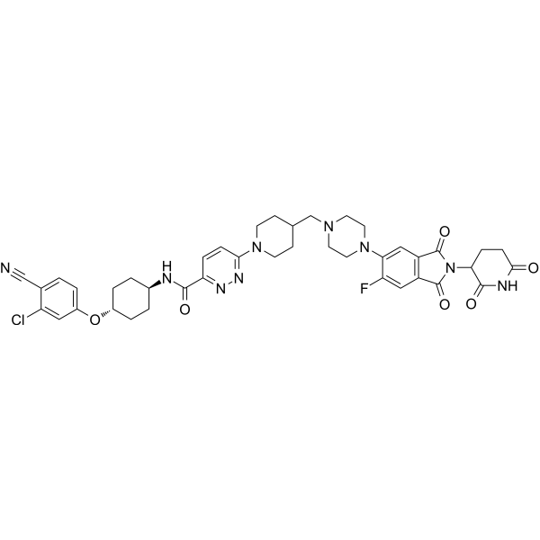
HY-138641
BavdegalutamideAR PROTAC Degrader -
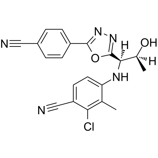
HY-14383
VosilasarmAndrogen Receptor Modulator -
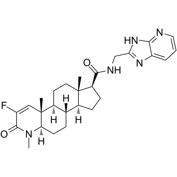
HY-11027
MK-0773Androgen Receptor Inhibitor -
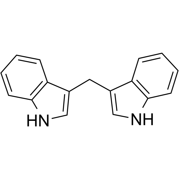
HY-15758
3,3'-DiindolylmethaneAndrogen Receptor Antagonist -
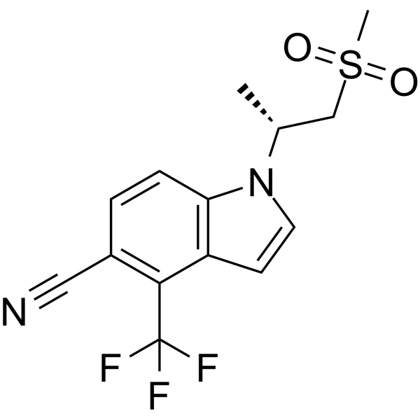
HY-100186
GSK-2881078Androgen Receptor Modulator -

HY-13259
MG-132Proteasome Inhibitor -
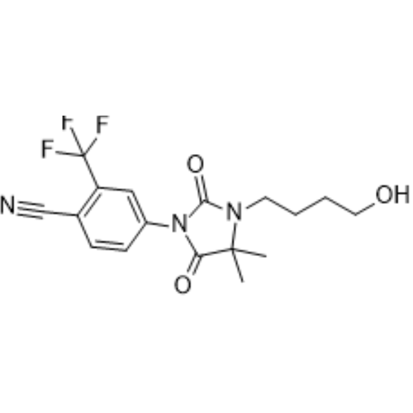
HY-10561
RU 58841Androgen Receptor Antagonist -
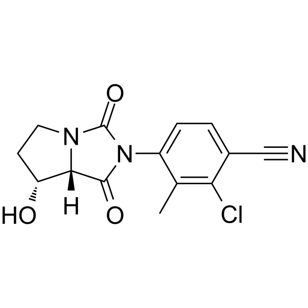
HY-12111
BMS-564929Androgen Receptor Agonist -
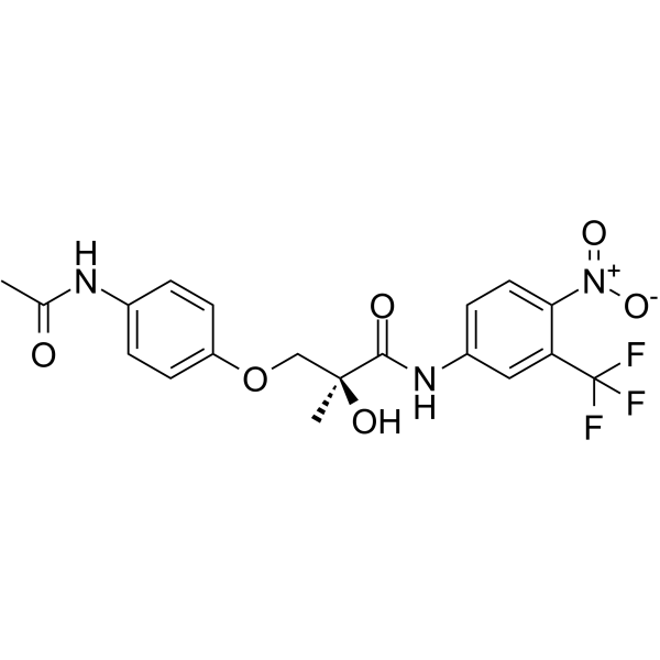
HY-12023
GTx-007Androgen Receptor SARM -
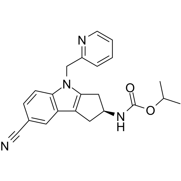
HY-114530
LY2452473Androgen Receptor Modulator -
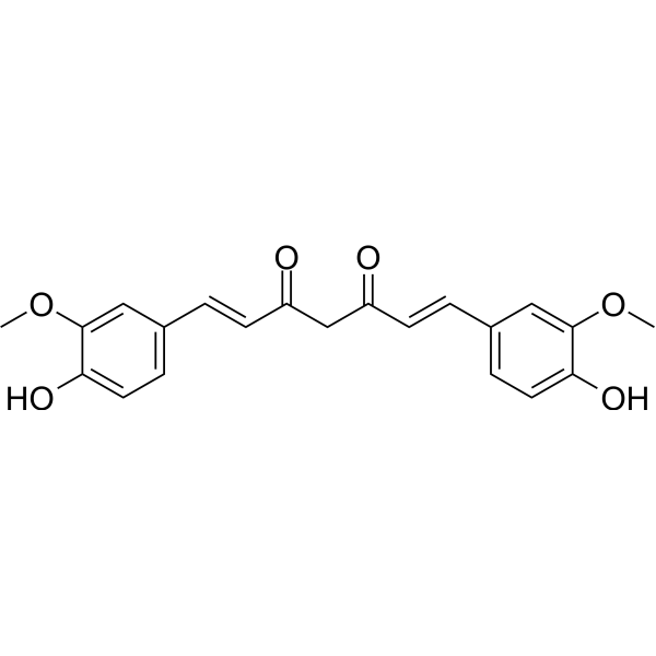
HY-N0005
Curcuminp300 Histone Acetylatransferase Inhibitor
Publications Citing Use of MedChemExpress (MCE) Dimethylcurcumin
More- Clin Transl Med. 2022 Apr;12(4):e797. [Abstract]
- Mol Cancer Ther. 2016 Jul;15(7):1702-12. [Abstract]
- Biochem Pharmacol. 2017 Sep 15:140:73-88. [Abstract]
- EXCLI J. 2019 Jan 24;18:21-29.
- Biol Sex Differ. 2020 Mar 29;11(1):12. [Abstract]
- J Ovarian Res. 2018 May 2;11(1):36. [Abstract]
- Transl Oncol. 2021 Aug;14(8):101115. [Abstract]
- Lab Invest. 2023 Jul;103(7):100148. [Abstract]
- Endocrinology. 2019 Apr 1;160(4):947-963. [Abstract]
- J Steroid Biochem Mol Biol. 2021 Jul:211:105906. [Abstract]
- Biochem Biophys Res Commun. 2018 Nov 17;506(1):130-136. [Abstract]
- Biochem Biophys Res Commun. 2017 Apr 15;485(4):746-752. [Abstract]
- Oncotarget. 2016 Jun 14;7(24):36814-36828. [Abstract]
- Tumour Biol. 2015 Nov;36(11):8727-33. [Abstract]
 Customer Validation & Images

-
WB
-
WB
-
RT-PCR
-
WB
-
WB


Biological Activity
Description
Cellular Effect
|
Cell Line

|
Type | Value | Description | References |
|---|---|---|---|---|
| A549 | GI50 |
5.8 μM
Compound: 3
|
Growth inhibition of human A549 cells after 48 hrs by sulforhodamine B assay
Growth inhibition of human A549 cells after 48 hrs by sulforhodamine B assay
|
[PMID: 21070043] |
| A549 | IC50 |
>40 μM
Compound: 3
|
Inhibition of TNF-alpha-induced NFkappaB nuclear translocation in human A549 cells preincubated for 30 mins before TNFalpha challenge and measured 30 post TNFalpha challenge by fluorescence method
Inhibition of TNF-alpha-induced NFkappaB nuclear translocation in human A549 cells preincubated for 30 mins before TNFalpha challenge and measured 30 post TNFalpha challenge by fluorescence method
|
[PMID: 21070043] |
| HCT-116 | GI50 |
2 μM
Compound: GO-Y025
|
Growth inhibition of human HCT116 cells after 48 hrs by MTS assay
Growth inhibition of human HCT116 cells after 48 hrs by MTS assay
|
[PMID: 20060305] |
| HEK293 | EC50 |
8.3 μM
Compound: 1b
|
Inhibition of Wnt3A/beta-catenin signaling in HEK293 cells after 24 hrs by firefly/renilla dual luciferase reporter gene assay
Inhibition of Wnt3A/beta-catenin signaling in HEK293 cells after 24 hrs by firefly/renilla dual luciferase reporter gene assay
|
[PMID: 24275249] |
| KB | CC50 |
2 μM
Compound: 11a
|
Compound concentration required to reduce the exponential growth of KB cells by 50%
Compound concentration required to reduce the exponential growth of KB cells by 50%
|
[PMID: 9767632] |
| LNCaP | IC50 |
1.3 μM
Compound: 71, JC-9, ASC-J9
|
Cytotoxicity against human LNCAP cells after 2 days
Cytotoxicity against human LNCAP cells after 2 days
|
[PMID: 20187635] |
| LNCaP | IC50 |
1.3 μM
Compound: 2, DMC
|
Cytotoxicity against human LNCaP cell line
Cytotoxicity against human LNCaP cell line
|
[PMID: 16789753] |
| LNCaP | IC50 |
15.3 μM
Compound: 21
|
Cytotoxicity against androgen-dependent human LNCAP cells after 72 hrs by SRB assay
Cytotoxicity against androgen-dependent human LNCAP cells after 72 hrs by SRB assay
|
[PMID: 22672984] |
| LNCaP | IC50 |
3.9 μM
Compound: 6
|
Antiproliferative activity against human LNCAP cells after 72 hrs by MTT assay
Antiproliferative activity against human LNCAP cells after 72 hrs by MTT assay
|
[PMID: 19249204] |
| MCF-10A | IC50 |
31.5 μM
Compound: 6
|
Antiproliferative activity against human MCF10A cells after 72 hrs by MTT assay
Antiproliferative activity against human MCF10A cells after 72 hrs by MTT assay
|
[PMID: 19249204] |
| MCF7 | IC50 |
5.4 μM
Compound: 6
|
Antiproliferative activity against human MCF7 cells after 72 hrs by MTT assay
Antiproliferative activity against human MCF7 cells after 72 hrs by MTT assay
|
[PMID: 19249204] |
| MDA-MB-231 | IC50 |
4.9 μM
Compound: 6
|
Antiproliferative activity against human MDA-MB-231 cells after 72 hrs by MTT assay
Antiproliferative activity against human MDA-MB-231 cells after 72 hrs by MTT assay
|
[PMID: 19249204] |
| MT4 | CC50 |
4.1 μM
Compound: 11a
|
Compound concentration required to reduce the exponential growth of MT-4 cells by 50%
Compound concentration required to reduce the exponential growth of MT-4 cells by 50%
|
[PMID: 9767632] |
| PC-3 | IC50 |
1.1 μM
Compound: 71, JC-9, ASC-J9
|
Cytotoxicity against human PC3 cells after 2 days
Cytotoxicity against human PC3 cells after 2 days
|
[PMID: 20187635] |
| PC-3 | IC50 |
1.1 μM
Compound: 2, DMC
|
Cytotoxicity against human PC3 cell line
Cytotoxicity against human PC3 cell line
|
[PMID: 16789753] |
| PC-3 | IC50 |
23.7 μM
Compound: 21
|
Cytotoxicity against androgen-independent human PC3 cells after 72 hrs by SRB assay
Cytotoxicity against androgen-independent human PC3 cells after 72 hrs by SRB assay
|
[PMID: 22672984] |
| PC-3 | IC50 |
5.9 μM
Compound: 6
|
Antiproliferative activity against human PC3 cells after 72 hrs by MTT assay
Antiproliferative activity against human PC3 cells after 72 hrs by MTT assay
|
[PMID: 19249204] |
| SK-OV-3 | IC50 |
3.51 μM
Compound: 15
|
Cytotoxicity against human SKOV3 Cells after 48 hrs by Cell Titer Blue assay
Cytotoxicity against human SKOV3 Cells after 48 hrs by Cell Titer Blue assay
|
[PMID: 31129455] |
In Vitro
Dimethylcurcumin (ASC-J9) is able to degrade fAR and AR3 in a dose-dependent manner in various human PCa cells. Dimethylcurcumin (ASC-J9) can also effectively suppress AR-targeted genes in CWR22Rv1-fARKD cells. Dimethylcurcumin (ASC-J9) (5 or 10 μM) significantly suppresses the DHT-induced cell growth in all three PCa cell lines. Dimethylcurcumin (ASC-J9) suppresses AR-targeted genes and cell growth by degradation of fAR and ectopic AR3 in C81 and C4-2 cells[1]. Dimethylcurcumin (ASC-J9) selectively promotes AR degradation by disrupting the interaction between AR and AR coregulators. ASC-J9 reduces the AR aggregated AR-112Q in cells. Dimethylcurcumin (ASC-J9) suppresses the aggregation of AR-112Q in SBMA PC12/AR-112Q cells[2].
MedChemExpress (MCE) has not independently confirmed the accuracy of these methods. They are for reference only.
In Vivo
MedChemExpress (MCE) has not independently confirmed the accuracy of these methods. They are for reference only.
- BrdU Incorporation Assay
- Somatic Cell Culture
- CFSE Dye Dilution Proliferation Assay
- Dye-dilution cell tracking and proliferation staining
- CCK-8/WST-8 Cell Proliferation Assay
- Protocol for Cell Cycle
- Cell Counting-Based Growth Curve Assay
- Colony Formation (Clonogenic) Assay
- Cell Viability Determination by MTT Colorimetric Assay
- EdU Incorporation Assay (Click Chemistry-Based DNA Synthesis Measurement)
- Ki-67 Immunostaining Proliferation Assay
- PCNA Immunodetection Proliferation Assay
- Protocol for Cell Counting and Cell Density Analysis
- Research Protocol for Endocrine Diseases
- Cell invasion
- MTT Cell Proliferation Assay
Clinical Trial
| NCT Number | Sponsor | Condition | Start Date |
Phase
|
|---|---|---|---|---|
| NCT01329991 | 2011-05 | PHASE1 |


Chemical Information
-
CAS No. 52328-98-0
-
Appearance Solid
-
Molecular Weight 396.43
-
Formula C23H24O6
-
Color Yellow to orange
-
SMILES
O=C(/C=C(O)/C=C/C1=CC=C(OC)C(OC)=C1)/C=C/C2=CC=C(OC)C(OC)=C2
-
Synonyms
ASC-J9; GO-Y025
-
Structure Classification
-
Initial Source
-
Shipping
Room temperature in continental US; may vary elsewhere.
-
Storage
Powder -20°C 3 years 4°C 2 years In solvent -80°C 2 years -20°C 1 year


Publications (14)
-
Journal Impact Factor
-
Most Recent
-
Clin Transl Med
Both SUMOylation and ubiquitination of TFE3 fusion protein regulated by androgen receptor are the potential target in the therapy of Xp11.2 translocation renal cell carcinoma. [Abstract]2022 Apr;12(4):e797. PMID: 35452181 -
Mol Cancer Ther
2016 Jul;15(7):1702-12. PMID: 27196756 -
Biochem Pharmacol
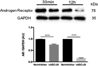
Testosterone regulates 3T3-L1 pre-adipocyte differentiation and epididymal fat accumulation in mice through modulating macrophage polarization. [Abstract]2017 Sep 15:140:73-88. PMID: 28642037
Dimethylcurcumin purchased from MedChemExpress. Usage Cited in: Biochem Pharmacol. 2017 Sep 15:140:73-88. [Abstract]
Androgen receptors levels in RAW264.7 cells treated with ASC-J9. Cells are treated with or without ASC-J9 for 30 min or 12 h and cells lysate is examined by western blotting using antibodies for the Androgen Receptor and GAPDH.
-
-
Biol Sex Differ
Neuroprotective and neurotoxic outcomes of androgens and estrogens in an oxidative stress environment. [Abstract]2020 Mar 29;11(1):12. PMID: 32223745 -
J Ovarian Res
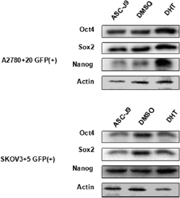
Nanog interaction with the androgen receptor signaling axis induce ovarian cancer stem cell regulation: studies based on the CRISPR/Cas9 system. [Abstract]2018 May 2;11(1):36. PMID: 29716628
Dimethylcurcumin purchased from MedChemExpress. Usage Cited in: J Ovarian Res. 2018 May 2;11(1):36. [Abstract]
Oct4 and Sox2 expression in the GFP (+) cells clearly increased under DHT treatment compared with ASC-J9 treatment based on western blot analysis, which is also consistent with the Nanog expression trend.
-
Transl Oncol
Breaking androgen receptor addiction of prostate cancer by targeting different functional domains in the treatment of advanced disease. [Abstract]2021 Aug;14(8):101115. PMID: 33993099 -
Lab Invest
Androgen receptor transcriptionally inhibits programmed death ligand-1 (PD-L1) expression and influences immune escape in bladder cancer. [Abstract]2023 Jul;103(7):100148. PMID: 37059268 -
Endocrinology
2019 Apr 1;160(4):947-963. PMID: 30811529 -
J Steroid Biochem Mol Biol
(-)-Epicatechin acts as a potent agonist of the membrane androgen receptor, ZIP9 (SLC39A9), to promote apoptosis of breast and prostate cancer cells. [Abstract]2021 Jul:211:105906. PMID: 33989703 -
Biochem Biophys Res Commun
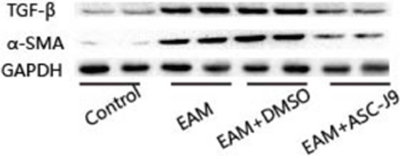
Androgen receptor regulates cardiac fibrosis in mice with experimental autoimmune myocarditis by increasing microRNA-125b expression. [Abstract]2018 Nov 17;506(1):130-136. PMID: 30340830
Dimethylcurcumin purchased from MedChemExpress. Usage Cited in: Biochem Biophys Res Commun. 2018 Nov 17;506(1):130-136. [Abstract]
The protein expression level of AR in EAM mice with/without ASC-J9 treatment is analysed.
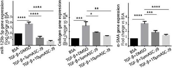
Dimethylcurcumin purchased from MedChemExpress. Usage Cited in: Biochem Biophys Res Commun. 2018 Nov 17;506(1):130-136. [Abstract]
Treatment with ASC-J9 in the presence of TGF-b decreases the mRNA expression of collagen I and a-SMA.
-
Biochem Biophys Res Commun
Targeting androgen receptor with ASC-J9 attenuates cardiac injury and dysfunction in experimental autoimmune myocarditis by reducing M1-like macrophage. [Abstract]2017 Apr 15;485(4):746-752. PMID: 28246012
Dimethylcurcumin purchased from MedChemExpress. Usage Cited in: Biochem Biophys Res Commun. 2017 Apr 15;485(4):746-752. [Abstract]
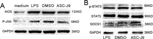
ASC-J9 inhibits M1 polarization of Raw264.7 cells. (A) Western blotting and analysis of iNOS in Raw264.7 cells stimulated with LPS in each group. (B) Western blotting and analysis of SOCS1 and p-STAT5 expression in Raw264.7 cells stimulated with LPS in each group.
-
Oncotarget
Androgen/androgen receptor axis maintains and promotes cancer cell stemness through direct activation of Nanog transcription in hepatocellular carcinoma. [Abstract]2016 Jun 14;7(24):36814-36828. PMID: 27167111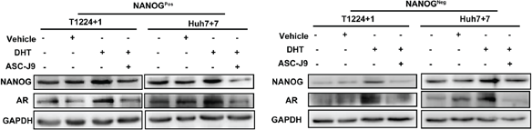
Dimethylcurcumin purchased from MedChemExpress. Usage Cited in: Oncotarget. 2016 Jun 14;7(24):36814-36828. [Abstract]
Protein levels of AR and NANOG expression is measured in NanogPos and NanogNeg cells from T1224+1 and Huh7+7 after treatment with DHT and with or without ASC-J9 for 24h. The addition of ASC-J9 attenuates the DHT effect and reduces the expression of AR and Nanog in both groups.
-
Tumour Biol
2015 Nov;36(11):8727-33. PMID: 26050226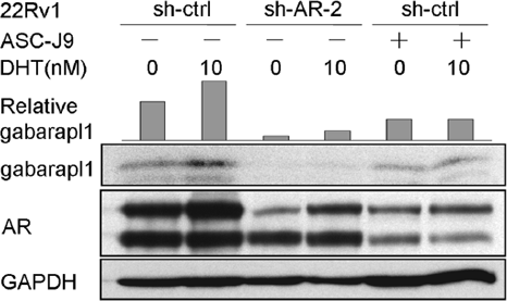
Dimethylcurcumin purchased from MedChemExpress. Usage Cited in: Tumour Biol. 2015 Nov;36(11):8727-33. [Abstract]
Application of ASC-J9 (an AR degradation enhancer) to 22Rv1 cell line also represses androgen-induced Gabarapl1 upregulation.


Solvent & Solubility
In Vitro:
DMSO : ≥ 50 mg/mL (126.13 mM; Hygroscopic DMSO has a significant impact on the solubility of product, please use newly opened DMSO)
* "≥" means soluble, but saturation unknown.
Please refer to the solubility information to select the appropriate solvent. Once prepared, please aliquot and store the solution to prevent product inactivation from repeated freeze-thaw cycles.
Storage method and period of stock solution: -80°C, 2 years; -20°C, 1 year. When stored at -80°C, please use it within 2 years. When stored at -20°C, please use it within 1 year.
Please refer to the solubility information to select the appropriate solvent. Once prepared, please aliquot and store the solution to prevent product inactivation from repeated freeze-thaw cycles.
Storage method and period of stock solution: -80°C, 2 years; -20°C, 1 year. When stored at -80°C, please use it within 2 years. When stored at -20°C, please use it within 1 year.
Concentration (start) × Volume (start) = Concentration (final) × Volume (final)
In Vivo:
Select the appropriate dissolution method based on your experimental animal and administration route.
- For the following dissolution methods, please ensure to first prepare a clear stock solution using an In Vitro approach and then sequentially add co-solvents:
- To ensure reliable experimental results, the clarified stock solution can be appropriately stored based on storage conditions. As for the working solution for In Vivo experiments, it is recommended to prepare freshly and use it on the same day.
- The percentages shown for the solvents indicate their volumetric ratio in the final prepared solution. If precipitation or phase separation occurs during preparation, heat and/or sonication can be used to aid dissolution.
Add each solvent one by one: 10% DMSO 40% PEG300 5% Tween-80 45% Saline
Solubility: ≥ 2.17 mg/mL (5.47 mM); Clear solution
This protocol yields a clear solution of ≥ 2.17 mg/mL (saturation unknown).
Taking 1 mL working solution as an example, add 100 μL DMSO stock solution (21.7 mg/mL) to 400 μL PEG300, and mix evenly; then add 50 μL Tween-80 and mix evenly; then add 450 μL Saline to adjust the volume to 1 mL.
Preparation of Saline: Dissolve 0.9 g sodium chloride in ddH₂O and dilute to 100 mL to obtain a clear Saline solution.
In Vivo Dissolution Calculator
Please enter the basic information of animal experiments:
-
-
-
-
Recommended: Prepare an additional quantity of animals to account for potential losses during experiments.
Please enter your animal formula composition:
-
%DMSO +
Recommended: Keep the proportion of DMSO in working solution below 2% if your animal is weak.
-
%+
-
+%Tween-80 + +
-
%Saline +
The co-solvents required include: DMSO, . All of co-solvents are available by MedChemExpress (MCE). , Tween 80. All of co-solvents are available by MedChemExpress (MCE).
Working solution concentration: 0.22 mg/mL
Method for preparing stock solution: mg drug dissolved in μL DMSO. Stock solution concentration: mg/mL.
1. Take μL DMSO stock solution;
2. Add μL .
μL , mix evenly;
3. Then add μL Tween 80, mix evenly;
4. Then add μL
Please ensure that the stock solution in the first step is dissolved to a clear state, and add co-solvents in sequence. You can use ultrasonic heating (ultrasonic cleaner, recommended frequency 20-40 kHz), vortexing, etc. to assist dissolution.


Purity & Documentation
-
Data Sheet (283 KB)
-
SDS (396 KB)
- English - EN (396 KB)
- Français - FR (396 KB)
- Deutsch - DE (396 KB)
- Norwegian - NO (396 KB)
- Español - ES (396 KB)
- Swedish - SV (396 KB)
- Italian - IT (396 KB)
- Korean - KR (396 KB)
- Portuguese - PT (396 KB)
-
Handling Instructions (2659 KB)
References
[1]. Yamashita S, et al. ASC-J9 suppresses castration-resistant prostate cancer growth through degradation of full-length and splice variant androgen receptors. Neoplasia. 2012 Jan;14(1):74-83. [Content Brief]
[2]. Yang Z, et al. ASC-J9 ameliorates spinal and bulbar muscular atrophy phenotype via degradation of androgen receptor. Nat Med. 2007 Mar;13(3):348-53. [Content Brief]
[3]. Lee SO, et al. New therapy targeting differential androgen receptor signaling in prostate cancer stem/progenitor vs non-stem/progenitor cells. J Mol Cell Biol. 2012 Jul 24. [Content Brief]
[4]. Ma W, et al. Targeting androgen receptor with ASC-J9 attenuates cardiac injury and dysfunction in experimental autoimmune myocarditis by reducing M1-like macrophage. Biochem Biophys Res Commun. 2017 Apr 15;485(4):746-752. doi: 10.1016/j.bbrc.2017.02.123. Epub 2017 Feb 27. [Content Brief]


Complete Stock Solution Preparation Table
Please refer to the solubility information to select the appropriate solvent. Once prepared, please aliquot and store the solution to prevent product inactivation from repeated freeze-thaw cycles.
Storage method and period of stock solution: -80°C, 2 years; -20°C, 1 year. When stored at -80°C, please use it within 2 years. When stored at -20°C, please use it within 1 year.
| Optional Solvent | Concentration Solvent Mass | 1 mg | 5 mg | 10 mg | 25 mg |
|---|---|---|---|---|---|
| DMSO | 1 mM | 2.5225 mL | 12.6126 mL | 25.2251 mL | 63.0628 mL |
| 5 mM | 0.5045 mL | 2.5225 mL | 5.0450 mL | 12.6126 mL | |
| 10 mM | 0.2523 mL | 1.2613 mL | 2.5225 mL | 6.3063 mL | |
| 15 mM | 0.1682 mL | 0.8408 mL | 1.6817 mL | 4.2042 mL | |
| 20 mM | 0.1261 mL | 0.6306 mL | 1.2613 mL | 3.1531 mL | |
| 25 mM | 0.1009 mL | 0.5045 mL | 1.0090 mL | 2.5225 mL | |
| 30 mM | 0.0841 mL | 0.4204 mL | 0.8408 mL | 2.1021 mL | |
| 40 mM | 0.0631 mL | 0.3153 mL | 0.6306 mL | 1.5766 mL | |
| 50 mM | 0.0505 mL | 0.2523 mL | 0.5045 mL | 1.2613 mL | |
| 60 mM | 0.0420 mL | 0.2102 mL | 0.4204 mL | 1.0510 mL | |
| 80 mM | 0.0315 mL | 0.1577 mL | 0.3153 mL | 0.7883 mL | |
| 100 mM | 0.0252 mL | 0.1261 mL | 0.2523 mL | 0.6306 mL |


Dimethylcurcumin Related Classifications
HY-15194 Related Classifications
 Powered by Bioz