



GEM-5
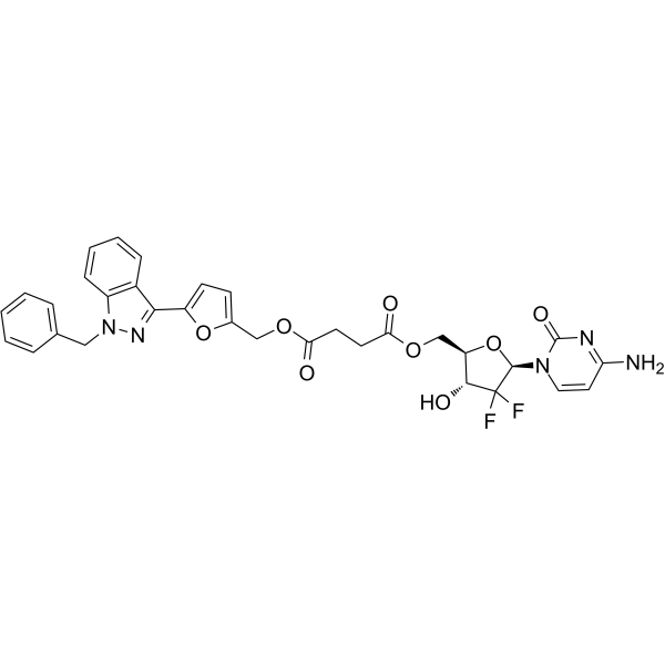
GEM-5 is a gemcitabine-based conjugate containing a HIF-1α inhibitor (YC-1) (IC50=30 nM). GEM-5 can significantly down-regulate the expression of HIF-1α and up-regulate the expression of tumor suppressor p53. GEM-5 induces the apoptosis of A2780 cells and inhibits tumor growth.
For research use only. We do not sell to patients.
- CAS No.: 2233543-49-0
- Formula: C32H29F2N5O8
- Molecular Weight:649.60
-
Storage:
Please store the product under the recommended conditions in the Certificate of Analysis.

To place orders, for customer services and technical support, please contact: MedChemExpress USA
Tel: 609-228-6898 E-mail: [email protected] [email protected]


-
 Biological Activity
-
Chemical Information
- Protocols
- Purity & Documentation
- References
-
 Help & FAQs




Biological Activity
Description
IC50 & Target
IC50: 30 nM (HIF-1α) in A2780[1]
Cellular Effect
|
Cell Line

|
Type | Value | Description | References |
|---|---|---|---|---|
| A2780 | IC50 |
0.03 μM
Compound: GEM-5
|
Cytotoxicity against human A2780 cells assessed as reduction in cell viability incubated for 72 hrs in hypoxic condition by MTT assay
Cytotoxicity against human A2780 cells assessed as reduction in cell viability incubated for 72 hrs in hypoxic condition by MTT assay
|
[PMID: 33992863] |
| A2780 | IC50 |
0.13 μM
Compound: GEM-5
|
Cytotoxicity against human A2780 cells assessed as reduction in cell viability incubated for 72 hrs by MTT assay
Cytotoxicity against human A2780 cells assessed as reduction in cell viability incubated for 72 hrs by MTT assay
|
[PMID: 33992863] |
| A549 | IC50 |
0.37 μM
Compound: GEM-5
|
Cytotoxicity against human A549 cells assessed as reduction in cell viability incubated for 72 hrs by MTT assay
Cytotoxicity against human A549 cells assessed as reduction in cell viability incubated for 72 hrs by MTT assay
|
[PMID: 33992863] |
| A549 | IC50 |
3.03 μM
Compound: GEM-5
|
Cytotoxicity against human A549 cells assessed as reduction in cell viability incubated for 72 hrs in hypoxic condition by MTT assay
Cytotoxicity against human A549 cells assessed as reduction in cell viability incubated for 72 hrs in hypoxic condition by MTT assay
|
[PMID: 33992863] |
| L02 | IC50 |
3.04 μM
Compound: GEM-5
|
Cytotoxicity against human L02 cells assessed as reduction in cell viability incubated for 72 hrs in hypoxic condition by MTT assay
Cytotoxicity against human L02 cells assessed as reduction in cell viability incubated for 72 hrs in hypoxic condition by MTT assay
|
[PMID: 33992863] |
| L02 | IC50 |
4.43 μM
Compound: GEM-5
|
Cytotoxicity against human L02 cells assessed as reduction in cell viability incubated for 72 hrs by MTT assay
Cytotoxicity against human L02 cells assessed as reduction in cell viability incubated for 72 hrs by MTT assay
|
[PMID: 33992863] |
| MCF7 | IC50 |
2.6 μM
Compound: GEM-5
|
Cytotoxicity against human MCF7 cells assessed as reduction in cell viability incubated for 72 hrs by MTT assay
Cytotoxicity against human MCF7 cells assessed as reduction in cell viability incubated for 72 hrs by MTT assay
|
[PMID: 33992863] |
In Vitro
GEM-5 (0-80 μM; 12 hours) exhibits excellent antitumor activity toward A2780 cells under hypoxic condition with IC50 of 0.03 μM[1].
GEM-5 (0.5 μM; 72 hours) rises the apoptotic population to 52.67% under normoxic condition and 80.89% in A2780 cells under hypoxic condition[1].
GEM-5 (0.5 μM; 24 hours) arrests the cell cycle at the S phase (63.02% under normoxia and 72.64% under hypoxia)[1].
GEM-5 (0.1 and 1 μM; 24 hours) decreases the levels of HIF-1α and increases the levels of p53 in a dose dependent manner under hypoxic condition[1].
MedChemExpress (MCE) has not independently confirmed the accuracy of these methods. They are for reference only.
-
Cell Line:A2780 cells[1]
-
Concentration:0.5 μM
-
Incubation Time:72 hours
-
Result:Rose the apoptotic population to 52.67% under normoxic condition and 80.89% under hypoxic condition.
-
Cell Line:A2780 cells[1]
-
Concentration:0.5 μM
-
Incubation Time:24 hours
-
Result:Arrested the cell cycle at the S phase (63.02% under normoxia and 72.64% under hypoxia).
-
Cell Line:A2780 cells[1]
-
Concentration:0.1 and 1 μM
-
Incubation Time:24 hours
-
Result:Decreased the levels of HIF-1α and increased the levels of p53 in a dose dependent manner
In Vivo
MedChemExpress (MCE) has not independently confirmed the accuracy of these methods. They are for reference only.
-
Animal Model:GEM-5 (125 or 271 mg/kg; tail vein injection, once a week for 4 weeks) effectively inhibits tumor growth in the A2780 xenograft mouse model and exhibited low toxicity[1].
-
Dosage:125 or 271 mg/kg
-
Administration:Tail vein injection, once a week for 4 weeks
-
Result:Effectively inhibited tumor growth in the A2780 xenograft mouse model and exhibited low toxicity.
- RNA extraction experimental
- Detection of Apoptosis
- TUNEL staining for apoptotic DNA fragmentation
- Annexin V plus membrane-impermeant dye apoptosis staining
- Patient-Derived Xenograft (PDX)
- Apoptosis Solutions
- Patient-Derived Orthotopic Xenograft (PDOX)
- Subcutaneous Cell-Line-Derived Xenograft
- Orthotopic Cell-Line Xenograft


Chemical Information
-
CAS No. 2233543-49-0
-
Molecular Weight 649.60
-
Formula C32H29F2N5O8
-
SMILES
NC(C=CN1[C@@H]2O[C@@H]([C@H](C2(F)F)O)COC(CCC(OCC3=CC=C(O3)C4=NN(C5=C4C=CC=C5)CC6=CC=CC=C6)=O)=O)=NC1=O
-
Shipping
Room temperature in continental US; may vary elsewhere.
-
Storage
Please store the product under the recommended conditions in the Certificate of Analysis.


Purity & Documentation
References

Calculators
Concentration (start) × Volume (start) = Concentration (final) × Volume (final)


GEM-5 Related Classifications
HY-146540 Related Classifications
 Powered by Bioz