



Orcein

Based on 1 Customer Validation
Orcein is an irreversible stain that specifically targets elastic fibers and can interact hydrophobically with the protein components in elastic fibers. Orcein makes elastic fibers in tissues appear purple or purple-red. Orcein can be used for morphological studies of Drosophila polytene chromosomes and for qualitative and quantitative analysis of elastic fibers, collagen fibers and other components in atherosclerotic plaques.
For research use only. We do not sell to patients.
- CAS No.: 1400-62-0
-
Storage:
4°C, protect from light
* In solvent : -80°C, 6 months; -20°C, 1 month (protect from light)


To place orders, for customer services and technical support, please contact: MedChemExpress USA
Tel: 609-228-6898 E-mail: [email protected] [email protected]


-
 Biological Activity
-
Chemical Information
-
Solvent & Solubility
- Protocols
- Purity & Documentation
- References
-
 Help & FAQs


-

HY-10071
Y-27632ROCK Inhibitor -
HY-101888
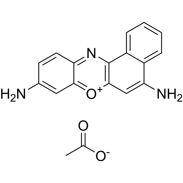
Cresyl Violet acetateRed Fluorescent Stain -
HY-D0714
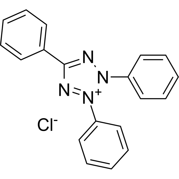
Tetrazolium RedBiochemical Assay Reagent -
HY-B1571
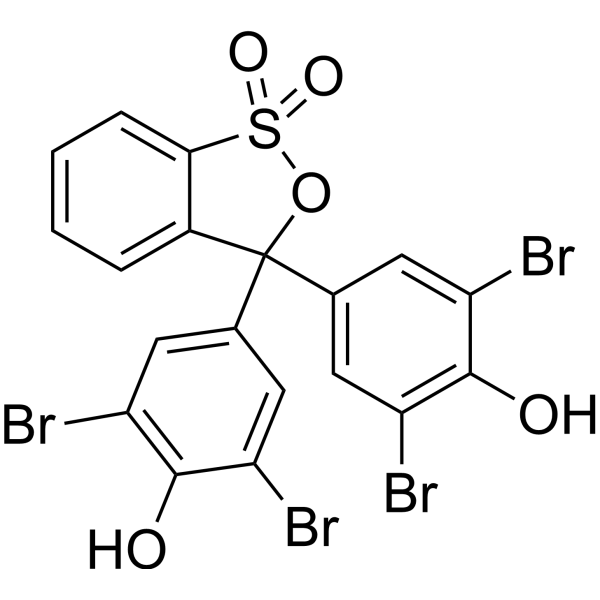
Bromophenol bluepH Indicator -
HY-D0003
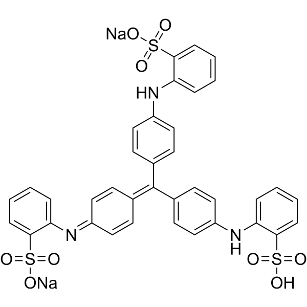
Methyl BlueAntiseptic Dye -
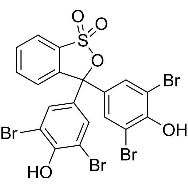
HY-W110798
Bromophenol blue indicator (3.0-4.6)Biochemical Assay Reagent -
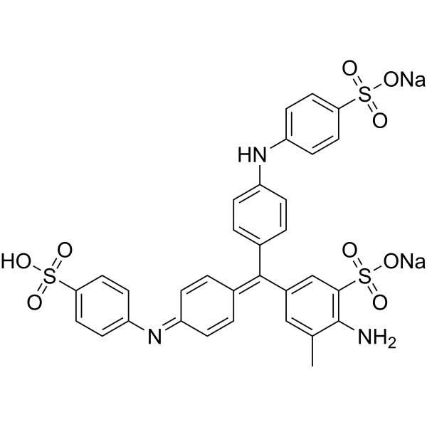
HY-W133919
Aniline Blue sodiumBiochemical Assay Reagent -
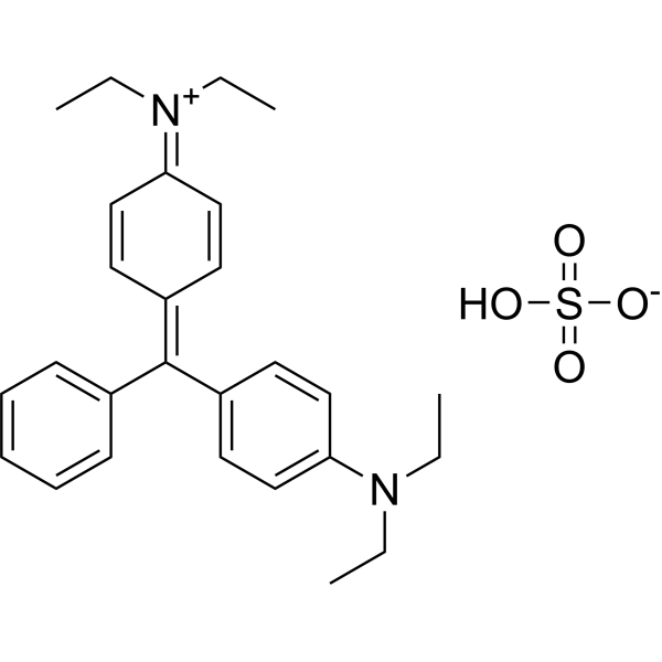
HY-D0008
Brilliant greenCationic Dye -

HY-101888R
Cresyl Violet acetate (Standard)Reference Standard


Biological Activity
Description
In Vitro
Orcein dyes bind to negatively charged groups of chromatin or through hydrophobic interactions under acidic conditions, staining chromosomes. Orcein is mainly used to stain polytene chromosomes in the salivary glands of third-instar larvae of Drosophila. It can also be used to observe chromosomes in tissues such as the midgut, hindgut, and fat body[1].
Orcein can be used to stain tissue samples. In histology and cell biology, it is mainly used to stain elastic fibers, collagen, nucleic acids, and cellular components[2].
Guide (Following is our recommended protocol. This protocol only provides a guideline, and should be modified according to your specific needs)[1].
Aceto-orcein staining:
When staining with Orcein, adding acetic acid to fix the chromosomes can stretch the chromosomes in the interband region when pressing the slides, improving the band resolution; the addition of lactic acid can soften the glands and promote chromosome spreading.
1. Materials
1.1 Culture medium: Texas banana agar, yeast-glucose-agar, Carolina instant Drosophila feed.
1.2 Buffer/fixative: Drosophila Ringers solution, PBS, 0.8% NaCl, 45% acetic acid.
1.3 Staining solution: lactic acid-acetic acid-orcein staining solution (prepared according to Lim's method: 1 g natural orcein dissolved in 50 mL lactic acid, filtered; 1 g natural orcein dissolved in 50 mL glacial acetic acid, heated and filtered; the three were mixed at a ratio of 1:1:1).
1.4 Consumables: No. 5 tweezers, siliconized coverslips, coated slides, Probe-On-Plus slides, Permount mounting medium.
2. Operation steps
2.1 Larva culture and selection:
Use the above culture medium to culture third-instar larvae. Cultivate at 18°C to obtain larger chromosomes. Select larvae that have not pupated and are full-bodied, and rinse the culture medium on the body surface with PBS or Ringers solution.
2.2 Dissection and gland separation:
Place the larvae on a slide containing 45% acetic acid. Under a dissecting microscope, use a pair of forceps to clamp the head and tail respectively, and pull the head off quickly to expose the salivary gland (a transparent long cyst-like structure with attached adipose tissue).
Strip the gland, remove the anterior duct and excess fat, and fix in 45% acetic acid for 2-5 minutes.
2.3 Staining and pressing:
Transfer the gland to lactic acid-acetic acid-orcein staining solution and stain for 5 minutes (avoid evaporation of the staining solution).
Transfer to a slide containing 1:2:3 fixative, cover with a coverslip, tap the coverslip with a dissecting needle to spread the chromosomes, and then press lightly with absorbent paper for blotting (to prevent the coverslip from shifting).
2.4 Seal and observe:
Temporary seal: seal the edges of the coverslip with nail polish; permanent seal: peel off the siliconized coverslip after freezing with dry ice-ethanol, dehydrate with 95% and 100% ethanol in turn, and seal with Permount.
Microscope observation: identify chromosome arms (such as X, 2L, 2R, 3L, 3R) by telomere morphology and banding characteristics (such as puff, constriction area).
3. Precautions
3.1 45% acetic acid fixation can reduce chromosome puffing, which is better than physiological saline dissection.
3.2 Avoid excessive force when pressing the slice to cause chromosome breakage. Light spiral or zigzag pressure can promote uniform spreading.
3.3 Different fruit fly strains and culture conditions may lead to differences in banding patterns, which need to be combined with standard pattern analysis.
MedChemExpress (MCE) has not independently confirmed the accuracy of these methods. They are for reference only. Further protocols information, click here.
In Vivo
MedChemExpress (MCE) has not independently confirmed the accuracy of these methods. They are for reference only.
Clinical Trial
| NCT Number | Sponsor | Condition | Start Date |
Phase

|
|---|---|---|---|---|
| NCT01329991 | Plexxikon| | 2011-05 | PHASE1 |


Chemical Information
-
CAS No. 1400-62-0
-
Appearance Solid
-
Color Reddish brown to black
-
SMILES
[Orcein]
-
Shipping
Room temperature in continental US; may vary elsewhere.
-
Storage
4°C, protect from light
* In solvent : -80°C, 6 months; -20°C, 1 month (protect from light)


Solvent & Solubility
In Vitro:
DMSO : 100 mg/mL (ultrasonic and warming and heat to 60°C; Hygroscopic DMSO has a significant impact on the solubility of product, please use newly opened DMSO)
H2O : < 0.1 mg/mL (insoluble)
Ethanol : < 1 mg/mL (insoluble)

Protocols
-
Fibers: Elastic Fiber Staining
Elastic fiber staining is a histochemical technique used to selectively visualize elastin-rich structures such as elastic fibers in connective tissues (e. g. , blood vessels, lung, dermis) based on the affinity of specific dyes or oxidation products for elastin-associated amino acid residues and cross-linked elastic matrix components. Classical methods such as Verhoeff-Van Gieson (VVG), resorcin-fuchsin (Weigert-type stains), or aldehyde fuchsin rely on differential binding of dye complexes to elastic fibers, allowing them to be distinguished from collagen and other extracellular matrix components by contrast staining (typically black or deep purple elastic fibers against red collagen counterstain). These methods are widely used in histopathology to evaluate elastic fiber integrity, fragmentation, or remodeling in vascular diseases, pulmonary pathology, and connective tissue disorders.


Purity & Documentation
-
Data Sheet (275 KB)
-
SDS (393 KB)
- English - EN (393 KB)
- Français - FR (393 KB)
- Deutsch - DE (393 KB)
- Norwegian - NO (393 KB)
- Español - ES (393 KB)
- Swedish - SV (393 KB)
- Italian - IT (393 KB)
- Korean - KR (393 KB)
- Portuguese - PT (393 KB)
-
Handling Instructions (2659 KB)
References
[1]. Tonzetich J. Orcein staining and the identification of polytene chromosomes. Methods Mol Biol. 2004;247:249-56. [Content Brief]
[2]. Gajda M, et al. Combined orcein and martius scarlet blue (OMSB) staining for qualitative and quantitative analyses of atherosclerotic plaques in brachiocephalic arteries in apoE/LDLR-/- mice. Histochem Cell Biol. 2017 Jun;147(6):671-681. [Content Brief]

Calculators
Concentration (start) × Volume (start) = Concentration (final) × Volume (final)


Orcein Related Classifications
HY-122266 Related Classifications
 Powered by Bioz