



Phenanthrene
Based on 1 publication(s) in Google Scholar
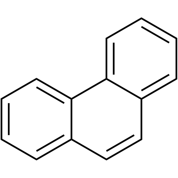
Phenanthrene is an orally active polycyclic aromatic hydrocarbon (PAH) that induces inflammation, oxidative stress, and apoptosis. Additionally, phenanthrene is commonly used to detect or assess PAH pollution in the environment.
For research use only. We do not sell to patients.
- Purity : 99.82%
- CAS No.: 85-01-8
- Formula: C14H10
- Molecular Weight:178.23
-
Storage:
Store at room temperature 3 years.
In solvent -80°C, 2 years , -20°C, 1 year

To place orders, for customer services and technical support, please contact: MedChemExpress USA
Tel: 609-228-6898 E-mail: [email protected] [email protected]


-
 Biological Activity
-
Chemical Information
-
Solvent & Solubility
- Protocols
- Purity & Documentation
- References
-
 Help & FAQs


-
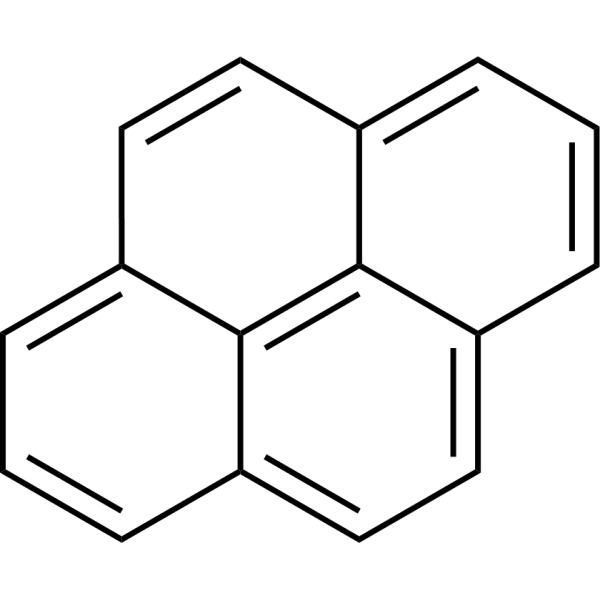
HY-103609
PyreneBiochemical Assay Reagent -
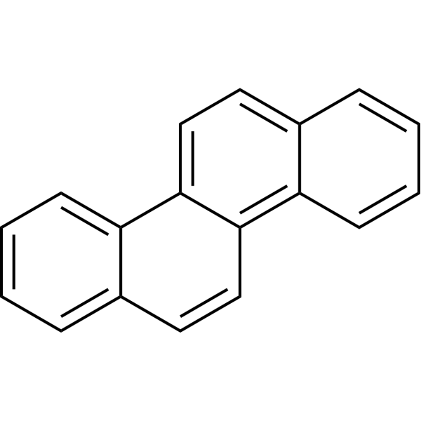
HY-121107
ChryseneCarcinogenic Inducer -
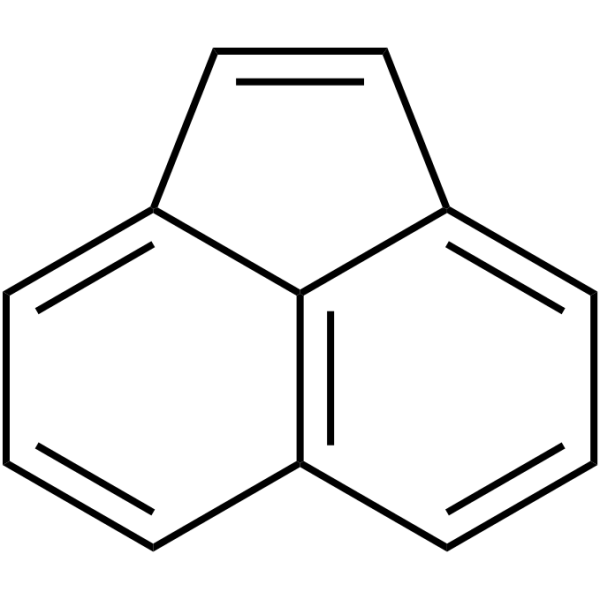
HY-W013570
AcenaphthylenePAH -
HY-W026772
FluorenePolycyclic Aromatic Hydrocarbon -

HY-B1727R
Phenanthrene (Standard)Polycyclic Aromatic Hydrocarbon -

HY-P9963
BlinatumomabAntineoplastic Agent -
![Benzo[a]pyrene](https://file.medchemexpress.com/product_pic/hy-107377.gif)
HY-107377
Benzo[a]pyreneNoxious Pollutant -
![Benzo[k]fluoranthene](https://file.medchemexpress.com/product_pic/hy-23498.gif)
HY-23498
Benzo[k]fluoranthenePAH -

HY-W013973
p-TerphenylBiochemical Assay Reagent -
HY-W010800
Cholesteryl hemisuccinateTopoisomerase Inhibitor
Publications Citing Use of MedChemExpress (MCE) Phenanthrene
More

Biological Activity
Description
In Vitro
In Vivo
MedChemExpress (MCE) has not independently confirmed the accuracy of these methods. They are for reference only.
-
Animal Model:Wistar female rats (180-200 g)[3]
-
Dosage:90, 180, 450, 900 mg/kg
-
Administration:Oral gavage (p.o.), Intraperitoneal injection (i.p.) (p.o. on day 1, once (180 and 900 mg/kg), i.p. for the next 2 days , once a day (90 and 450 mg/kg), dissolved in 0.01% acetone)
-
Result:Significantly increased superoxide dismutase (SOD) and malondialdehyde (MDA) levels in the lungs and liver, serum 8-hydroxy-2'-deoxyguanosine (8-OHdG), and the expression of tumor necrosis factor-α (TNF-α) and interleukin-6 (IL-6) (high dose group: 450 mg/kg + 900 mg/kg). Significantly decreased the expression of transforming growth factor-β (TGF-β) and forkhead box transcription factor 3 (Foxp3) (high dose group: 450 mg/kg + 900 mg/kg).
-
Animal Model:Chinese rare minnows (Gobiocypris rarus)[4]
-
Dosage:10, 100, 1000 μg/L
-
Administration:Exposure for 30 days
-
Result:Increased the frequency of liver damage and hepatocyte apoptosis, as well as the enzyme activity of caspase 9 and caspase 3 in liver tissues.
Clinical Trial
| NCT Number | Sponsor | Condition | Start Date |
Phase

|
|---|---|---|---|---|
| NCT01329991 | 2011-05 | PHASE1 |


Chemical Information
-
CAS No. 85-01-8
-
Appearance Solid
-
Molecular Weight 178.23
-
Formula C14H10
-
Color Off-white to light brown
-
SMILES
C12=CC=CC=C1C=CC3=CC=CC=C23
-
Shipping
Room temperature in continental US; may vary elsewhere.
-
Storage
Store at room temperature 3 years
In solvent -80°C 2 years -20°C 1 year


Publications (1)
-
Journal Impact Factor
-
Most Recent


Solvent & Solubility
In Vitro:
DMSO : 200 mg/mL (1122.15 mM; Need ultrasonic; Hygroscopic DMSO has a significant impact on the solubility of product, please use newly opened DMSO)
Please refer to the solubility information to select the appropriate solvent. Once prepared, please aliquot and store the solution to prevent product inactivation from repeated freeze-thaw cycles.
Storage method and period of stock solution: -80°C, 2 years; -20°C, 1 year. When stored at -80°C, please use it within 2 years. When stored at -20°C, please use it within 1 year.
Please refer to the solubility information to select the appropriate solvent. Once prepared, please aliquot and store the solution to prevent product inactivation from repeated freeze-thaw cycles.
Storage method and period of stock solution: -80°C, 2 years; -20°C, 1 year. When stored at -80°C, please use it within 2 years. When stored at -20°C, please use it within 1 year.
Concentration (start) × Volume (start) = Concentration (final) × Volume (final)
In Vivo:
Select the appropriate dissolution method based on your experimental animal and administration route.
- For the following dissolution methods, please ensure to first prepare a clear stock solution using an In Vitro approach and then sequentially add co-solvents:
- To ensure reliable experimental results, the clarified stock solution can be appropriately stored based on storage conditions. As for the working solution for In Vivo experiments, it is recommended to prepare freshly and use it on the same day.
- The percentages shown for the solvents indicate their volumetric ratio in the final prepared solution. If precipitation or phase separation occurs during preparation, heat and/or sonication can be used to aid dissolution.
Add each solvent one by one: 10% DMSO 40% PEG300 5% Tween-80 45% Saline
Solubility: ≥ 2.08 mg/mL (11.67 mM); Clear solution
This protocol yields a clear solution of ≥ 2.08 mg/mL (saturation unknown).
Taking 1 mL working solution as an example, add 100 μL DMSO stock solution (20.8 mg/mL) to 400 μL PEG300, and mix evenly; then add 50 μL Tween-80 and mix evenly; then add 450 μL Saline to adjust the volume to 1 mL.
Preparation of Saline: Dissolve 0.9 g sodium chloride in ddH₂O and dilute to 100 mL to obtain a clear Saline solution.
In Vivo Dissolution Calculator
Please enter the basic information of animal experiments:
-
-
-
-
Recommended: Prepare an additional quantity of animals to account for potential losses during experiments.
Please enter your animal formula composition:
-
%DMSO +
Recommended: Keep the proportion of DMSO in working solution below 2% if your animal is weak.
-
%+
-
+%Tween-80 + +
-
%Saline +
The co-solvents required include: DMSO, . All of co-solvents are available by MedChemExpress (MCE). , Tween 80. All of co-solvents are available by MedChemExpress (MCE).
Working solution concentration: 0.22 mg/mL
Method for preparing stock solution: mg drug dissolved in μL DMSO. Stock solution concentration: mg/mL.
1. Take μL DMSO stock solution;
2. Add μL .
μL , mix evenly;
3. Then add μL Tween 80, mix evenly;
4. Then add μL
Please ensure that the stock solution in the first step is dissolved to a clear state, and add co-solvents in sequence. You can use ultrasonic heating (ultrasonic cleaner, recommended frequency 20-40 kHz), vortexing, etc. to assist dissolution.


Purity & Documentation
-
Data Sheet (280 KB)
-
SDS (537 KB)
- English - EN (537 KB)
- Français - FR (537 KB)
- Deutsch - DE (537 KB)
- Norwegian - NO (537 KB)
- Español - ES (537 KB)
- Swedish - SV (537 KB)
- Italian - IT (537 KB)
- Korean - KR (537 KB)
- Portuguese - PT (537 KB)
-
Handling Instructions (2659 KB)
References
[1]. Sun K, et al. Subcellular distribution and biotransformation of phenanthrene in pakchoi after inoculation with endophytic Pseudomonas sp. as probed using HRMS coupled with isotope-labeling. Environ Pollut. 2018 Jun;237:858-867. [Content Brief]
[2]. Dupuy J, et al. Effect and localization of phenanthrene in maize roots. Chemosphere. 2016 Apr;149:130-6. [Content Brief]
[3]. Ma H, et al. Effects of phenanthrene on oxidative stress and inflammation in lung and liver of female rats. Environ Toxicol. 2020 Jan;35(1):37-46. [Content Brief]
[4]. Hong X, et al. Phenanthrene-Induced Apoptosis and Its Underlying Mechanism. Environ Sci Technol. 2017 Dec 19;51(24):14397-14405. [Content Brief]


Complete Stock Solution Preparation Table
Please refer to the solubility information to select the appropriate solvent. Once prepared, please aliquot and store the solution to prevent product inactivation from repeated freeze-thaw cycles.
Storage method and period of stock solution: -80°C, 2 years; -20°C, 1 year. When stored at -80°C, please use it within 2 years. When stored at -20°C, please use it within 1 year.
| Optional Solvent | Concentration Solvent Mass | 1 mg | 5 mg | 10 mg | 25 mg |
|---|---|---|---|---|---|
| DMSO | 1 mM | 5.6107 mL | 28.0536 mL | 56.1073 mL | 140.2682 mL |
| 5 mM | 1.1221 mL | 5.6107 mL | 11.2215 mL | 28.0536 mL | |
| 10 mM | 0.5611 mL | 2.8054 mL | 5.6107 mL | 14.0268 mL | |
| 15 mM | 0.3740 mL | 1.8702 mL | 3.7405 mL | 9.3512 mL | |
| 20 mM | 0.2805 mL | 1.4027 mL | 2.8054 mL | 7.0134 mL | |
| 25 mM | 0.2244 mL | 1.1221 mL | 2.2443 mL | 5.6107 mL | |
| 30 mM | 0.1870 mL | 0.9351 mL | 1.8702 mL | 4.6756 mL | |
| 40 mM | 0.1403 mL | 0.7013 mL | 1.4027 mL | 3.5067 mL | |
| 50 mM | 0.1122 mL | 0.5611 mL | 1.1221 mL | 2.8054 mL | |
| 60 mM | 0.0935 mL | 0.4676 mL | 0.9351 mL | 2.3378 mL | |
| 80 mM | 0.0701 mL | 0.3507 mL | 0.7013 mL | 1.7534 mL | |
| 100 mM | 0.0561 mL | 0.2805 mL | 0.5611 mL | 1.4027 mL |


Phenanthrene Related Classifications
HY-B1727 Related Classifications
 Powered by Bioz