



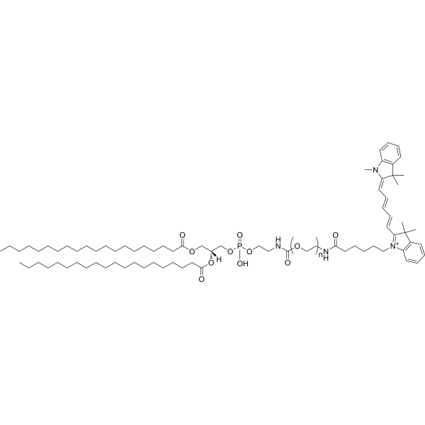
DSPE-PEG5000-Cy5
DSPE-PEG5000-Cy5 is a fluorescently labeled PEGylated phospholipid. After intravenous injection, DSPE-PEG5000-Cy5 shows only extremely low fluorescence accumulation at tumor sites in orthotopic tumor-bearing mice, and can be used as a negative control for evaluating tumor accumulation of nanoparticles. DSPE-PEG5000-Cy5 can serve as a fluorescent marker to prepare Cy5-labeled NPs-DPPA and NPs-DPPA (C3F8) for pharmacokinetic and biodistribution studies in mice. DSPE-PEG5000-Cy5 is widely applicable to research in fields related to triple-negative breast cancer, hepatocellular carcinoma and so on.
Nur für Forschungszwecke. Wir verkaufen nicht an Patienten.
- Molecular Weight:5000 (Average)
-
Speicherung:
Please store the product under the recommended conditions in the Certificate of Analysis.

To place orders, for customer services and technical support, please contact: MedChemExpress USA
Tel: 609-228-6898 E-mail: [email protected] [email protected]


-
 Biologische Aktivität
-
Chemical Information
- Protokoll
- Reinheit & Dokumentation
- Verweise
-
 Help & FAQs




Biologische Aktivität
Beschreibung
In Vitro
DSPE-PEG-Cy5 (2 mg; 30 min) can be used to coat SnSNPs via probe sonication, resulting in fluorescently labeled nanoparticles suitable for biological imaging studies[1].
DSPE-PEG-Cy5 functions as a fluorescent labeling agent for NPs-DPPA and NPs-DPPA(C3F8), enabling visualization and quantification of the nanoparticles in biological systems[2].
MedChemExpress (MCE) has not independently confirmed the accuracy of these methods. They are for reference only. Further protocols information, click here.
In Vivo
Free DSPE-PEG-Cy5 (MW 5000) (50 μg per mouse; i.v.; single dose) exhibits rapid blood clearance in healthy male BALB/c mice[2].
MedChemExpress (MCE) has not independently confirmed the accuracy of these methods. They are for reference only.
-
Animal Model:BALB/c (male, 4-6 weeks old)[2]
-
Dosage:50 μg per mouse
-
Administration:i.v.; single dose
-
Result:Exhibited rapid blood clearance, with no detectable fluorescence remaining in blood samples by 4 hours post-injection.


Chemical Information
-
Molecular Weight 5000 (Average)
-
SMILES
O=C(CCCCCCCCCCCCCCCCCCC)OC[C@](OC(CCCCCCCCCCCCCCCCCCC)=O)([H])COP(O)(OCCNC(OCCNC(CCCCC[N+]1=C(/C=C/C=C/C=C2C(C)(C)C(C=CC=C3)=C3N\2C)C(C)(C)C4=C1C=CC=C4)=O)=O)=O.[n]
-
Versand
Room temperature in continental US; may vary elsewhere.
-
Speicherung
Please store the product under the recommended conditions in the Certificate of Analysis.

Protokoll
-
How to Select the Route of Administration for Mammals
Route-of-administration selection in mammals is a pharmacokinetic, pharmacodynamic, formulation, animal-welfare, and translational decision, not a default technical choice. The selected route should match the study goal: intravenous dosing is most useful when complete systemic exposure and rapid onset are required, oral dosing is most translational for orally intended medicines but is affected by absorption and first-pass metabolism, subcutaneous or intramuscular dosing can provide slower systemic exposure, and intraperitoneal dosing can be useful in rodent proof-of-concept studies but may have limited clinical translation. Published route-comparison studies show that the same compound can produce different exposure, onset, bioavailability, tissue distribution, and tolerability depending on route; therefore, route choice should be supported by pilot pharmacokinetic or pharmacodynamic evidence when the literature is insufficient. Unresolved questions include how to standardize route sel
-
Liver Cancer Modeling
Liver cancer can be classified into primary liver cancer and secondary liver cancer. Secondary liver cancer is the metastatic liver cancer. Primary liver cancer includes hepatocellular carcinoma (HCC), intrahepatic cholangiocarcinoma (ICC) and fibrolamellar HCC, of which HCC is the most common form, accounting for approximately 90% of primary liver cancers[1]. HCC mouse models include chemical agent-induced models, transplanted tumor models, and genetic engineered models.
-
Breast Cancer Modeling
Breast cancer is a heterogeneous cancer, and it has been distinguished into four subtypes: luminal A, luminal B, HER2-positive and basal-like. Molecular mutations, epigenetic alterations, hormone exposure and immune microenvironment are related to the progression of breast cancer.
-
Orthotopic Cell-Line Xenograft
Orthotopic cell-line xenograft models involve implantation of human cancer cell lines into the anatomically corresponding organ of immunodeficient mice to reproduce tumor growth within a native microenvironment, enabling more clinically relevant tumor behavior compared with subcutaneous models. These models are widely used because orthotopic placement better recapitulates tumor progression, including invasion and metastatic spread, which are often underrepresented in heterotopic implantation systems. Compared with conventional xenografts, orthotopic implantation is described as more technically complex but provides improved simulation of tumor-microenvironment interactions and metastatic behavior, making it particularly valuable for translational oncology research. Surgical orthotopic implantation approaches have been emphasized as enabling faithful reproduction of clinical cancer features, including metastasis and disease progression patterns that align with the tumor’s organ of origi


Reinheit & Dokumentation
Verweise
[1]. Li Y, et al. Nanosensitizer-mediated augmentation of sonodynamic therapy efficacy and antitumor immunity. Nat Commun. 2023;14(1):6973. Published 2023 Nov 1. [Content Brief]
[2]. Fang J, et al. Bioactive Nanotherapeutic Ultrasound Contrast Agent for Concurrent Breast Cancer Ultrasound Imaging and Treatment. Adv Healthc Mater. 2024;13(26):e2401436. [Content Brief]

Calculators
Konzentration (Stammlösung) × Volumen (Stammlösung) = Konzentration (Ziellösung) × Volumen (Ziellösung)


DSPE-PEG5000-Cy5 Related Classifications
HY-W440913 Related Classifications
 Powered by Bioz
Keywords
- DSPE-PEG5000-Cy5
- Liposome
- Fluorescent Dye
- orthotopic tumor-bearing mice
- fluorescence imaging
- hepatocellular carcinoma
- nanoparticle tumor accumulation
- in vivo pharmacokinetic studies
- triple-negative breast cancer
- BALB/c mice
- BALB/cJ mice
- biological imaging studies
- biodistribution studies
- Inhibitor
- inhibitor
- inhibit