



DX2-201
Based on 1 publication(s) in Google Scholar
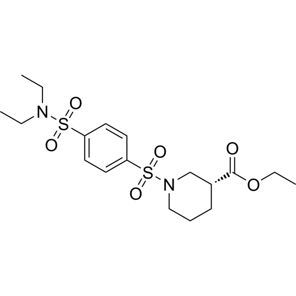
DX2-201 is a potent and selective oxidative phosphorylation (OXPHOS) complex I inhibitor with an IC50 of 312 nM. DX2-201 has anticancer effects.
For research use only. We do not sell to patients.
- CAS No.: 2749554-00-3
- Formula: C18H28N2O6S2
- Molecular Weight:432.55
-
Storage:
Please store the product under the recommended conditions in the Certificate of Analysis.

To place orders, for customer services and technical support, please contact: MedChemExpress USA
Tel: 609-228-6898 E-mail: [email protected] [email protected]


-
 Biological Activity
-
Chemical Information
- Protocols
- Purity & Documentation
- References
-
 Help & FAQs


Publications Citing Use of MedChemExpress (MCE) DX2-201
More

Biological Activity
Description
IC50 & Target
IC50: 312 nM (OXPHOS complex I)[1]
Cellular Effect
|
Cell Line

|
Type | Value | Description | References |
|---|---|---|---|---|
| BXPC-3 | IC50 |
0.6 μM
Compound: 2; DX2-201
|
Cytotoxicity against human BXPC-3 cells assessed as inhibition of cell growth incubated for 7 days in glucose-containing medium by MTT assay
Cytotoxicity against human BXPC-3 cells assessed as inhibition of cell growth incubated for 7 days in glucose-containing medium by MTT assay
|
[PMID: 34982568] |
| MIA PaCa-2 | GI50 |
0.6 μM
Compound: 1
|
Cytotoxicity against human MIA PaCa-2 cells assessed as inhibition of cell growth incubated for overnight in glucose-containing medium followed by compound addition and measured after 7 days by MTT assay
Cytotoxicity against human MIA PaCa-2 cells assessed as inhibition of cell growth incubated for overnight in glucose-containing medium followed by compound addition and measured after 7 days by MTT assay
|
[PMID: 35167303] |
| MIA PaCa-2 | IC50 |
>10 μM
Compound: 2; DX2-201
|
Cytotoxicity against human MIA PaCa-2 cells assessed as inhibition of cell growth incubated for 3 days in galactose-containing medium by MTT assay
Cytotoxicity against human MIA PaCa-2 cells assessed as inhibition of cell growth incubated for 3 days in galactose-containing medium by MTT assay
|
[PMID: 34982568] |
| MIA PaCa-2 | IC50 |
0.09 μM
Compound: 2; DX2-201
|
Cytotoxicity against human MIA PaCa-2 cells assessed as inhibition of cell growth incubated for 3 days in glucose-containing medium by MTT assay
Cytotoxicity against human MIA PaCa-2 cells assessed as inhibition of cell growth incubated for 3 days in glucose-containing medium by MTT assay
|
[PMID: 34982568] |
| MIA PaCa-2 | IC50 |
0.4 μM
Compound: 2; DX2-201
|
Cytotoxicity against human MIA PaCa-2 cells assessed as inhibition of cell growth incubated for 7 days in glucose-containing medium by MTT assay
Cytotoxicity against human MIA PaCa-2 cells assessed as inhibition of cell growth incubated for 7 days in glucose-containing medium by MTT assay
|
[PMID: 34982568] |
| MIA PaCa-2 | IC50 |
118.5 nM
Compound: 2; DX2-201
|
Inhibition of oxidative phosphorylation in human MIA PaCa-2 cells assessed as reduction in ATP production incubated for 24 hrs in galactose-containing medium by CellTiter-glo luminescence assay
Inhibition of oxidative phosphorylation in human MIA PaCa-2 cells assessed as reduction in ATP production incubated for 24 hrs in galactose-containing medium by CellTiter-glo luminescence assay
|
[PMID: 34982568] |
In Vitro
DX2-201 (cmpound 2) potently inhibits the mitochondrial function by depleting ATP production in MIA PaCa-2 cells when glucose is replaced by galactose in the medium, a condition that forces mammalian cells to rely on OXPHOS[1].
DX2-201 depletes ATP production with an IC50 value of 118.5 nM in the galactose-containing medium[1].
DX2-201 significantly inhibits OXPHOS complex I as determined by its inhibition of oxidation of NADH to NAD+ in the NAD/NADH assay (IC50 = 312 nM)[1].
DX2-201 inhibits MIA PaCa-2 and BxPC-3 cells growth with IC50 values of 0.4 μM and 0.6 μM, respectively[1].
MedChemExpress (MCE) has not independently confirmed the accuracy of these methods. They are for reference only.


Chemical Information
-
CAS No. 2749554-00-3
-
Molecular Weight 432.55
-
Formula C18H28N2O6S2
-
SMILES
O=S(N1C[C@@H](CCC1)C(OCC)=O)(C2=CC=C(C=C2)S(N(CC)CC)(=O)=O)=O
-
Shipping
Room temperature in continental US; may vary elsewhere.
-
Storage
Please store the product under the recommended conditions in the Certificate of Analysis.


Publications (1)
-
Journal Impact Factor
-
Most Recent


Purity & Documentation
References

Calculators
Concentration (start) × Volume (start) = Concentration (final) × Volume (final)


DX2-201 Related Classifications
HY-145303 Related Classifications
 Powered by Bioz