



Efercoleukin alfa

Based on 1 Customer Validation
Efercoleukin alfa (XTX-202) is an IL-2 Rβ-biased IL-2 which has a masking domain to prevent IL-2 activity in non-tumor regions along with a half-life extension domain. The masking domain is cleaved by matrix metalloproteases (MMP) in the TME, leading to the release of the IL-2 R agonist. Efercoleukin alfa shows potential immunoregulatory and antineoplastic activities.
Nur für Forschungszwecke. Wir verkaufen nicht an Patienten.
- Reinheit : 99.9%
- CAS. Nr.: 2760549-52-6
- Molecular Weight:93.436 kDa
-
Speicherung:
Please store the product under the recommended conditions in the Certificate of Analysis.


To place orders, for customer services and technical support, please contact: MedChemExpress USA
Tel: 609-228-6898 E-mail: [email protected] [email protected]


-
 Biologische Aktivität
-
Chemical Information
- Protokoll
- Reinheit & Dokumentation
- Verweise
-
 Help & FAQs




Biologische Aktivität
Beschreibung
Isotype
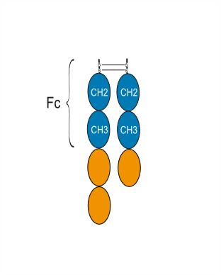
Fusion-IGHG1 Fc-Homo sapiens IL2RB-covalent dimer-IGHG1 Fc-Homo sapiens IL2
Species Reactivity
Human
IC50 & Target
IL2
Conjugated
Unconjugated
Reconsititution
The product can be reconstituted/diluted with sterile PBS or saline.
Format
-
FPIA-FC_[PROTEIN]3
Anwendung
ELISA, FACS, Functional assay
Verified Bioactivity
-
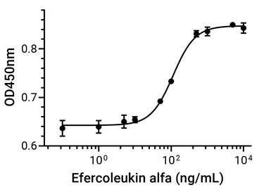 Measured in a cell proliferation assay using CTLL-2 cell. The ED50 for this effect is 113 ng/mL, corresponding to a specific activity is 8849.558 units/mg.


Chemical Information
-
CAS. Nr. 2760549-52-6
-
Appearance Liquid
-
Molecular Weight 93.436 kDa
-
Color Colorless to light yellow
-
Synonyms
XTX-202
-
Versand
Shipping with dry ice.
-
Formulation
Please refer to the lot-specific COA for specific buffer information.
-
Speicherung
Please store the product under the recommended conditions in the Certificate of Analysis.


Reinheit & Dokumentation
-
Data Sheet (255 KB)
-
SDS (251 KB)
- English - EN (251 KB)
- Français - FR (251 KB)
- Deutsch - DE (251 KB)
- Norwegian - NO (251 KB)
- Español - ES (251 KB)
- Swedish - SV (251 KB)
- Italian - IT (251 KB)
- Korean - KR (251 KB)
- Portuguese - PT (251 KB)
-
Inhibitory Antibodies User Guide (603 KB)
Verweise

Calculators
Konzentration (Stammlösung) × Volumen (Stammlösung) = Konzentration (Ziellösung) × Volumen (Ziellösung)


Efercoleukin alfa Related Classifications
HY-P990736 Related Classifications
 Powered by Bioz