



EGFR-IN-49
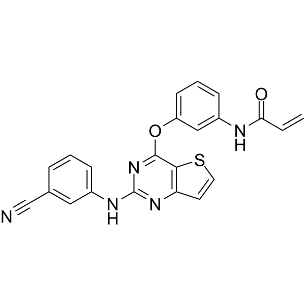
EGFR-IN-49 is a potent and selective EGFR inhibitor with IC50s of 65.0 nM and 13.6 nM for EGFRT790M and EGFRT790M/L858R, respectively. EGFR-IN-49 induces late apoptosis in a dose-dependent manner.
For research use only. We do not sell to patients.
- CAS No.: 2459932-81-9
- Formula: C22H15N5O2S
- Molecular Weight:413.45
-
Storage:
Please store the product under the recommended conditions in the Certificate of Analysis.

To place orders, for customer services and technical support, please contact: MedChemExpress USA
Tel: 609-228-6898 E-mail: [email protected] [email protected]


-
 Biological Activity
-
Chemical Information
- Protocols
- Purity & Documentation
- References
-
 Help & FAQs


All EGFR Isoforms
More

Biological Activity
Description
IC50 & Target
[1]|
EGFRT790M 65.0 nM (IC50) |
EGFRL858R/T790M 13.6 nM (IC50) |
Cellular Effect
|
Cell Line

|
Type | Value | Description | References |
|---|---|---|---|---|
| A-431 | IC50 |
3.79 μM
Compound: 13a
|
Antiproliferative activity against human A-431 cells assessed as inhibition of cell proliferation measured after 72 hrs by MTT assay
Antiproliferative activity against human A-431 cells assessed as inhibition of cell proliferation measured after 72 hrs by MTT assay
|
[PMID: 32679450] |
| A549 | IC50 |
4.34 μM
Compound: 13a
|
Antiproliferative activity against human A549 cells assessed as inhibition of cell proliferation measured after 72 hrs by MTT assay
Antiproliferative activity against human A549 cells assessed as inhibition of cell proliferation measured after 72 hrs by MTT assay
|
[PMID: 32679450] |
| HeLa | IC50 |
6.39 μM
Compound: 13a
|
Antiproliferative activity against human HeLa cells assessed as inhibition of cell proliferation measured after 72 hrs by MTT assay
Antiproliferative activity against human HeLa cells assessed as inhibition of cell proliferation measured after 72 hrs by MTT assay
|
[PMID: 32679450] |
| L02 | IC50 |
>50 μM
Compound: 13a
|
Cytotoxicity against human L02 cells assessed as inhibition of cell proliferation measured after 72 hrs by MTT assay
Cytotoxicity against human L02 cells assessed as inhibition of cell proliferation measured after 72 hrs by MTT assay
|
[PMID: 32679450] |
| MCF7 | IC50 |
18.99 μM
Compound: 13a
|
Antiproliferative activity against human MCF7 cells assessed as inhibition of cell proliferation measured after 72 hrs by MTT assay
Antiproliferative activity against human MCF7 cells assessed as inhibition of cell proliferation measured after 72 hrs by MTT assay
|
[PMID: 32679450] |
| NCI-H1975 | IC50 |
699.2 nM
Compound: 13a
|
Antiproliferative activity against human NCI-H1975 cells harboring EGFR T790M/L858R double mutant assessed as inhibition of cell viability measured after 72 hrs by MTT assay
Antiproliferative activity against human NCI-H1975 cells harboring EGFR T790M/L858R double mutant assessed as inhibition of cell viability measured after 72 hrs by MTT assay
|
[PMID: 32679450] |
In Vitro
EGFR-IN-49 (compound 13a) (1, 10 µM) shows inhibition activity for H1975 cells with an IC50 of 699.2 nM[1].
EGFR-IN-49 (1 µM) shows an strong inhibitory activity to EGFRT790M, EGFRT790M/L858R, EGFRWT with IC50s of 65.0, 13.6, >1000 nM, respectively[1].
EGFR-IN-49 (0.2, 4, 8 µM; 48 h) induces cell apoptosis in a dose-dependent manner in A431 cells[1].
MedChemExpress (MCE) has not independently confirmed the accuracy of these methods. They are for reference only.
-
Cell Line:A549, A431, Hela, MCF7, LO2 cells
-
Concentration:0-50 µM
-
Incubation Time:
-
Result:Showed excellent anti-proliferative activities with IC50s of 4.34, 3.79, 6.39, 18.99, >50 µM for A549, A431, Hela, MCF7, LO2 cells, respectively.
-
Cell Line:A431 cells
-
Concentration:0.2, 4, 8 µM
-
Incubation Time:48 h
-
Result:Induced cell apoptosis in a low concentration (0.33 µM) and exhibited higher percent of 16.41% % in the stage of late apoptotic at concentration of 4 µM.


Chemical Information
-
CAS No. 2459932-81-9
-
Molecular Weight 413.45
-
Formula C22H15N5O2S
-
SMILES
C=CC(NC1=CC(OC2=NC(NC3=CC=CC(C#N)=C3)=NC4=C2SC=C4)=CC=C1)=O
-
Shipping
Room temperature in continental US; may vary elsewhere.
-
Storage
Please store the product under the recommended conditions in the Certificate of Analysis.


Purity & Documentation
References

Calculators
Concentration (start) × Volume (start) = Concentration (final) × Volume (final)


EGFR-IN-49 Related Classifications
HY-146782 Related Classifications
 Powered by Bioz