



FITC-Dextran (MW 150000)
Based on 6 publication(s) in Google Scholar
FITC-Dextran (MW 150000) is a fluorescent probe for fluorescein isothiocyanate (FITC) dextran (Ex=491 nm; Em=518 nm). FITC-Dextran (MW 150000) can be used as a marker to reveal heat shock-induced cell damage and to study the early and late stages of apoptosis. FITC-Dextran (MW 150000) can be used in perfusion studies in animals or in fluorescence microlymphography, to study processes that affect the permeability of the blood brain barrier (BBB). FITC-Dextran (MW 150000) can be used as fluorescent probe to study cell permeability.
For research use only. We do not sell to patients.
- CAS No.: 60842-46-8
- Molecular Weight:150000(Average)
-
Storage:
4°C, protect from light
* In solvent : -80°C, 6 months; -20°C, 1 month (protect from light)

To place orders, for customer services and technical support, please contact: MedChemExpress USA
Tel: 609-228-6898 E-mail: [email protected] [email protected]


-
 Biological Activity
-
Chemical Information
-
Solvent & Solubility
- Protocols
- Purity & Documentation
- References
-
 Help & FAQs


-

HY-128868E
FITC-Dextran (MW 70000)Fluorescent Dye -

HY-128868A
FITC-Dextran (MW 4000)Fluorescent Dye -
HY-128868D
FITC-Dextran (MW 40000)Fluorescent Dyes -

HY-128868
FITC-Dextran (MW 10000)Fluorescent Dyes -

HY-128868H
FITC-Dextran (MW 500000)Fluorescent Dye -

HY-128868C
FITC-Dextran (MW 20000)Fluorescent Dye -
HY-128868I
FITC-Dextran (MW 2000000)Fluorescent Dye -
HY-128868B
FITC-Dextran (MW 3000-5000)Fluorescent Dye -
HY-128868F
FITC-Dextran (MW 110000)Fluorescent Dye -

HY-DY1016
FITC-Dextran (MW 10000) (solution)Fluorescent Dyes -

HY-116282C
Dextran sulfate sodium salt (MW 35000-45000)Colitis Inducer -

HY-116282
Dextran sulfate sodium salt (MW 5000)Complement Inhibitor
Publications Citing Use of MedChemExpress (MCE) FITC-Dextran (MW 150000)
More Customer Validation & Images

-
Cell Imaging/Staining
-
Bio/Physico-chemical Assay
-
Cell Imaging/Staining
-
Bio/Physico-chemical Assay


Biological Activity
Description
In Vitro
Guidelines (Following is our recommended protocol. This protocol only provides a guideline, and should be modified according to your specific needs).
Labeling of cells[1]:
For use with apoptotic HeLa cells and human peripheral blood mononuclear cells (PBMC) (viable HeLa and PBMC can not be stained by FITC-Dextran).
1. Incubate cells at 43.5°C for 1 hour and at 37°C for 8 hours to induce apoptosis.
2. Suspend the cells in 100 μL of medium, and mix in Q-prep tubes with 10 μL of propidium iodide (PI), 10 μL of FITC-Dextran (MW 150000) (the final concentration of PI and FITC-Dextran (MW 150000) is 7.5 μM and 1.13 μM, respectively).
3. Incubate cells for 25 min at room temperature in the dark.
4. Take the labeled cells with 3 mL of medium and centrifuge for 10 min at 500 g.
5. Take centrifuged cells with 1 mL of medium and use flow cytometry or fluorescence microscopy analyze (PI: Ex=500 nm, Em=600 nm; FITC-Dextran (MW 150000): Ex=495 nm, Em=525 nm).
Paracellular permeability measurement[4]
1. Add FITC-dextran (0.1 mg/mL) to the basal media in the transwell chamber.
2. Collect media from the transwell insert after 15 min.
3. Measure the fluorescence signal (Ex=485 nm, Em=538 nm).
4. Calculate FITC-dextran concentration based on fluorescence intensity.
5. Calculate permeability.
MedChemExpress (MCE) has not independently confirmed the accuracy of these methods. They are for reference only. Further protocols information, click here.
In Vivo
For intestinal barrier function assay[5]
1. Fast mice for 4 h.
2. Orally gavage mice with FITC-Dextran MW 150000 (0.6 mg/g).
3. Measure fluorescence intensity of plasma in 4 h (excitation nm/emission 520 nm).
MedChemExpress (MCE) has not independently confirmed the accuracy of these methods. They are for reference only.
Emission (Em)
525
Excitation (Ex)
488


Chemical Information
-
CAS No. 60842-46-8
-
Appearance Solid
-
Molecular Weight 150000(Average)
-
Color Light yellow to yellow
-
SMILES
[FITC-Dextran (MW 150000)]
-
Shipping
Room temperature in continental US; may vary elsewhere.
-
Storage
4°C, protect from light
* In solvent : -80°C, 6 months; -20°C, 1 month (protect from light)


Publications (6)
-
Journal Impact Factor
-
Most Recent
-
-
Biomaterials
Fully synthetic nanofibrillar hydrogel for the growth and enzyme-free release of patient-derived ovarian tumor organoids. [Abstract]2025 Sep 17:326:123723. PMID: 40976136
FITC-Dextran (MW 150000) purchased from MedChemExpress. Usage Cited in: Biomaterials. 2025 Sep 17:326:123723. [Abstract]
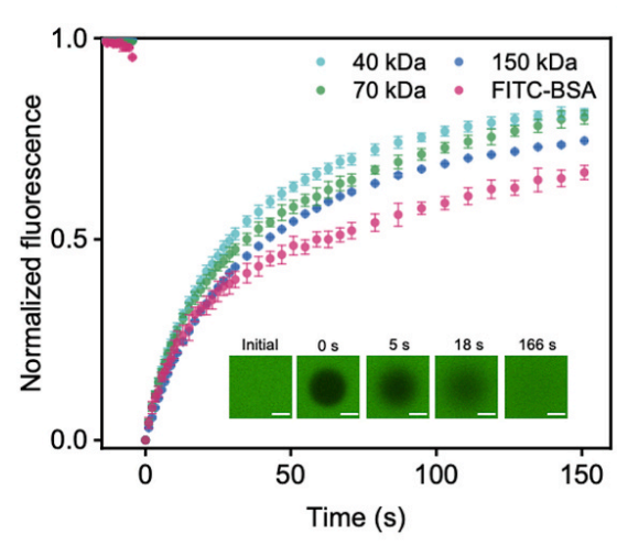
The time-dependent variations of normalized fluorescence of FITC-Dextran (40, 70, 150 kDa) (0.5 mg/mL) and FITC-BSA in TNHs with Cp of 70 mg/mL. The inset shows confocal microscopic images of TNHs containing FITC-BSA at different time points after the photo-bleaching in FRAP experiments. The Cp is 70 mg/mL. Scale bar, 50 μm.
-
FITC-Dextran (MW 150000) purchased from MedChemExpress. Usage Cited in: Engineering. 2026 Jan 21.
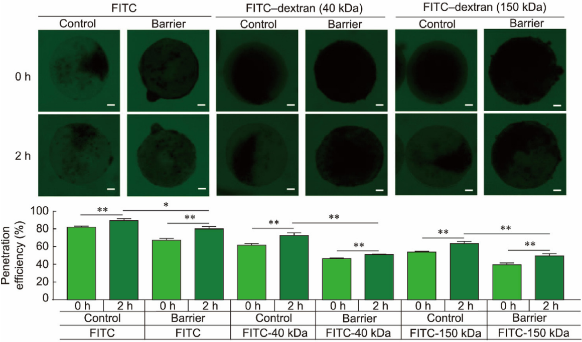
Evaluation of endothelial barrier permeability by monitoring the diffusion of FITC and 40 and 150 kDa FITC-dextran into microspheres in the presence or absence of an HUVEC layer. The results showed that after 2 h of diffusion, the endothelial layer significantly reduced the influx of free FITC compared to the control. Similarly, the penetration of 40 and 150 kDa FITC-dextrans into the hydrogel core was markedly slowed. At the 2-h time point, endothelialized microspheres exhibited significantly lower intramicrosphere fluorescence accumulation than non-endothelialized controls. These findings confirm that the engineered endothelial barrier exerts size-selective regulation over molecular permeation.
-
Phytomedicine
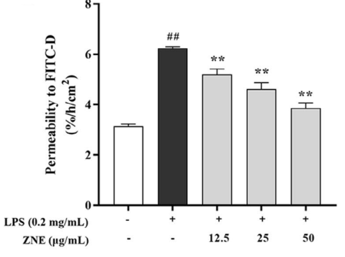
Zanthoxylum nitidum-derived active alkaloid nitidine chloride mitigates ulcerative colitis by remodeling gut microbiota and activating SIRT1 to restore intestinal barrier integrity. [Abstract]2026 Jun 15:159:158435. PMID: 42322763 -
Anal Chem
Biomimetic Microparticles with Myocardial and Endocardial Integration for Drug Toxicity Studies. [Abstract]2025 Apr 8. PMID: 40198907
FITC-Dextran (MW 150000) purchased from MedChemExpress. Usage Cited in: Anal Chem. 2025 Apr 8. [Abstract]
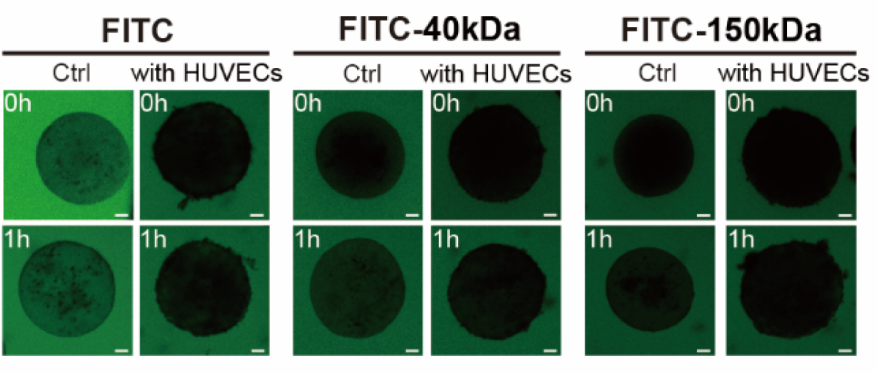
Evaluation of endothelial barrier permeability on the microparticles. Time-lapse fluorescence images of the diffusion of FITC, 40 and 150 kDa FITC-Dextran (25 μg/mL; 1 h) into microparticles with or without HUVECs monolayer.
-
FASEB J
Zanthoxylum Nitidum Ameliorates Intestinal Barrier Dysfunction and Inflammation in TNBS-Induced Colitis Rats and LPS-Stimulated Caco-2 Cells. [Abstract]2025 Oct 15;39(19):e71099. PMID: 41055302
FITC-Dextran (MW 150000) purchased from MedChemExpress. Usage Cited in: FASEB J. 2025 Oct 15;39(19):e71099. [Abstract]
FITC-Dextran (MW 150000) (1 mg/mL; 60 min) flux method was used to determine the permeability of Caco-2 cells.


Solvent & Solubility
In Vitro:
H2O : 66.67 mg/mL (Need ultrasonic)

Protocols
-
Apoptosis
Apoptosis, also called programmed cell death, is generally characterized by distinct morphological characteristics.
-
TUNEL staining for apoptotic DNA fragmentation
TUNEL staining detects DNA strand breaks by using terminal deoxynucleotidyl transferase to add labeled nucleotides to exposed 3′-OH DNA termini, generating either microscopic staining in fixed cells or tissue sections, or fluorescence/cytometric signal in cell suspensions. TUNEL positivity reflects DNA fragmentation but should not be interpreted alone as definitive apoptosis, because TUNEL can also label necrotic, autolytic, mechanically damaged, or DNA-repair-associated DNA breaks.
-
Annexin V plus membrane-impermeant dye apoptosis staining
Annexin V-based apoptosis assays rely on the detection of phosphatidylserine (PS) externalization from the inner leaflet of the plasma membrane to the outer leaflet, an early biochemical hallmark of apoptosis. Fluorescently labeled Annexin V binds PS in a calcium-dependent manner, enabling identification of early apoptotic cells by flow cytometry or fluorescence microscopy. When combined with a membrane-impermeant DNA-binding dye (e. g. , propidium iodide), this approach allows discrimination between viable (Annexin V−/dye−), early apoptotic (Annexin V+/dye−), and late apoptotic or necrotic (Annexin V+/dye+) cell populations by assessing membrane integrity and PS exposure.
-
Apoptosis Solutions
Apoptosis is a regulated, generally non-lytic cell-death pathway that removes unwanted, damaged, infected, or abnormal cells through coordinated morphological changes, caspase activation, DNA fragmentation, and membrane remodeling. The intrinsic apoptosis pathway is controlled mainly by mitochondrial outer membrane permeabilization, BCL-2 family proteins, cytochrome c release, apoptosome formation, caspase-9 activation, and downstream executioner caspase-3/7 activation. The extrinsic apoptosis pathway is initiated by death receptors such as Fas, TNFR, and TRAIL receptors, which recruit adaptor proteins and activate caspase-8 before engaging executioner caspases or mitochondrial amplification through BID cleavage. Apoptosis is linked to many phenotypes, including cancer cell killing, tissue homeostasis, immune regulation, neurodegeneration, infection response, and treatment-induced cytotoxicity; unresolved questions include how apoptosis interacts with necroptosis, pyroptosis, ferroptos
-
Transepithelial/transendothelial electrical resistance assay
TEER measures electrical resistance across epithelial or endothelial monolayers cultured on permeable supports, and the readout reflects ionic conductance through the cell barrier, especially the paracellular pathway regulated by junctional integrity. TEER can be measured without destroying the monolayer and is commonly used before or during transport, permeability, barrier-disruption, and barrier-maturation experiments. TEER values are influenced by biological maturation and technical conditions; reported factors include temperature, medium formulation, passage number, electrode geometry, membrane properties, and junctional length during early monolayer maturation. Therefore, TEER should be interpreted with blank-insert subtraction, area normalization, repeated readings, and, when possible, orthogonal barrier readouts such as FITC-dextran flux or tight-junction staining.


Purity & Documentation
-
Data Sheet (282 KB)
-
SDS (393 KB)
- English - EN (393 KB)
- Français - FR (393 KB)
- Deutsch - DE (393 KB)
- Norwegian - NO (393 KB)
- Español - ES (393 KB)
- Swedish - SV (393 KB)
- Italian - IT (393 KB)
- Korean - KR (393 KB)
- Portuguese - PT (393 KB)
-
Handling Instructions (2659 KB)
References
[2]. Natarajan R, et al. Fluorescein Isothiocyanate (FITC)-Dextran Extravasation as a Measure of Blood-Brain Barrier Permeability. Curr Protoc Neurosci. 2017 Apr 10;79:9.58.1-9.58.15. [Content Brief]
[3]. Eriksson I, et al. Analysis of Lysosomal pH by Flow Cytometry Using FITC-Dextran Loaded Cells. Methods Mol Biol. 2017;1594:179-189. [Content Brief]
[4]. Okabayashi K, et al. Cdc42 activates paracellular transport in polarised submandibular gland cells. Arch Oral Biol. 2021 Dec;132:105276. [Content Brief]
[5]. Yu W, et al. ACE2 contributes to the maintenance of mouse epithelial barrier function. Biochem Biophys Res Commun. 2020 Dec 17;533(4):1276-1282. [Content Brief]
[6]. Bolliner A,et al.,Fluorescence microlymphography: diagnostic potential in lymphedema and basis for the measurement of lymphatic pressure and flow velocity. Lymphology. 2007 Jun;40(2):52-62. [Content Brief]
[7]. Ishii T, et al., Accumulation of macromolecules in brain parenchyma in acute phase of cerebral infarction/reperfusion. Brain Res. 2010 Mar 19;1321:164-8. [Content Brief]

Calculators
Concentration (start) × Volume (start) = Concentration (final) × Volume (final)


FITC-Dextran (MW 150000) Related Classifications
HY-128868G Related Classifications
 Powered by Bioz