



FR-167356
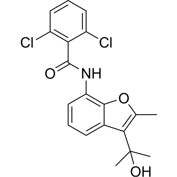
FR-167356 is a potent, orally active and selective vacuolar ATPase inhibitor with IC50 values of 170, 220, 370, and 1200 nM for osteoclast plasma membranes, macrophage microsomes, renal brush border membranes, and liver lysosomal membranes, respectively. FR-167356 inhibits bone resorption and ovariectomy-induced bone loss.
For research use only. We do not sell to patients.
- CAS No.: 174185-16-1
- Formula: C19H17Cl2NO3
- Molecular Weight:378.25
-
Storage:
Please store the product under the recommended conditions in the Certificate of Analysis.

To place orders, for customer services and technical support, please contact: MedChemExpress USA
Tel: 609-228-6898 E-mail: [email protected] [email protected]


-
 Biological Activity
-
Chemical Information
- Protocols
- Purity & Documentation
- References
-
 Help & FAQs


-
HY-103261
SCH28080H+/K+-ATPase Inhibitor -
HY-135474
KM91104v-ATPase a3-B2 Inhibitor -
HY-122632
Ciliobrevin DCytoplasmic Dynein Inhibitor -

HY-103261R
SCH28080 (Standard)H+/K+-ATPase Inhibitor -
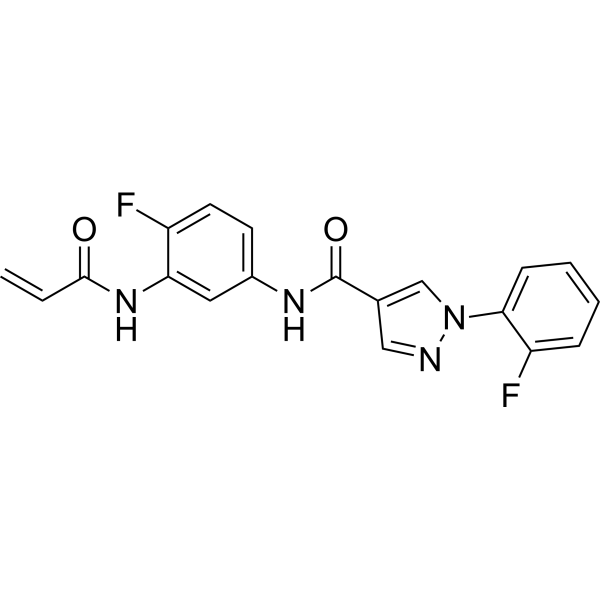
HY-128892
EN6Autophagy Activator -
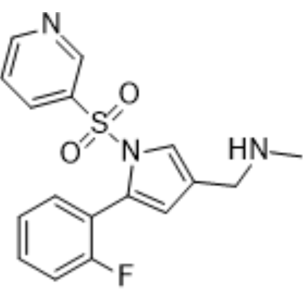
HY-100007
VonoprazanPotassium-Competitive Acid Blocker -
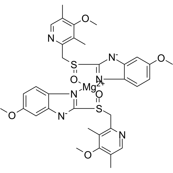
HY-B1446
Esomeprazole magnesiumH+, K+-ATPase Inhibitor -
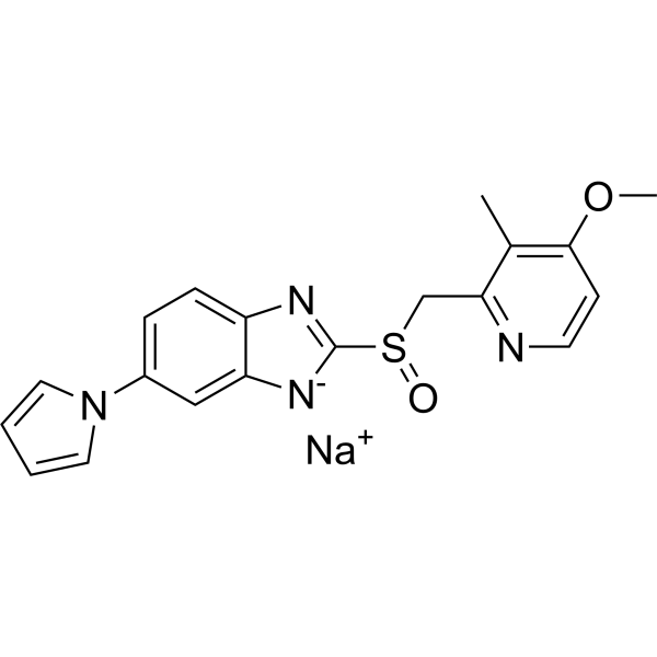
HY-B2145
Ilaprazole sodiumProton Pump Inhibitor -
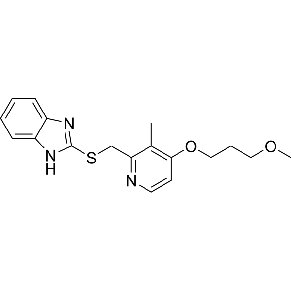
HY-W003467
Rabeprazole SulfideDrug Metabolite -
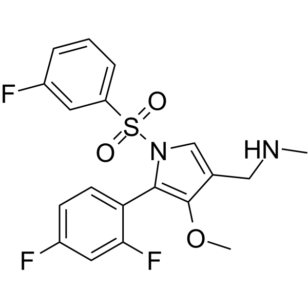
HY-109079
AbeprazanProton Pump/Potassium-competitive Acid Inhibitor -
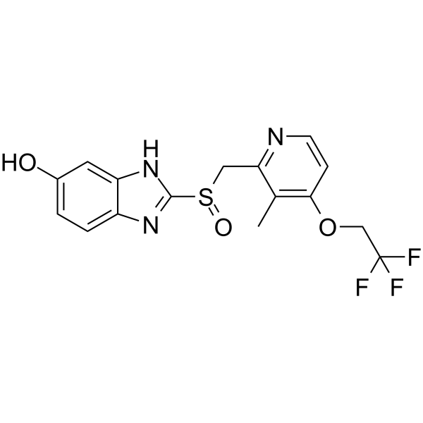
HY-118283
5-HydroxylansoprazoleProton-pump Inhibitor -
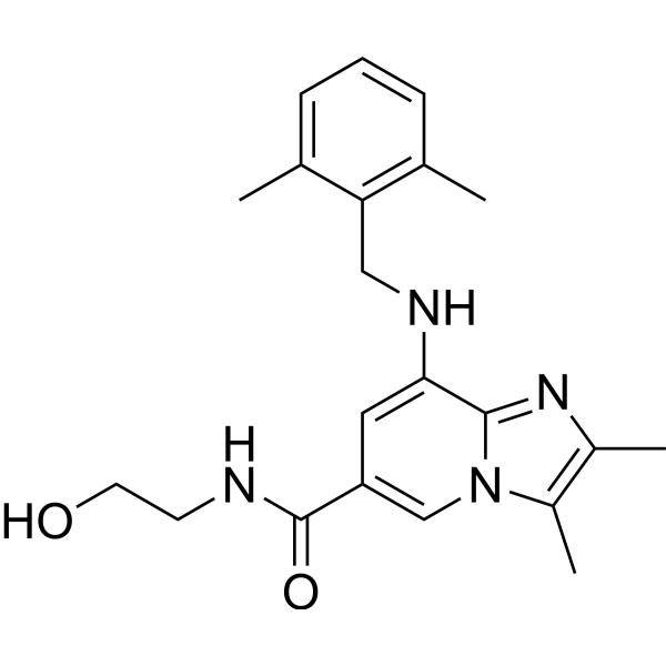
HY-100412
LinaprazanProton Pump Inhibitor


Biological Activity
Description
In Vivo
FR-167356 (200 mg/kg; p.o.; daily, for 10 days; male C57BL/6 mice with B16-F10 xenografts) reduces bone metastasis compared to untreated group[1].
MedChemExpress (MCE) has not independently confirmed the accuracy of these methods. They are for reference only.
-
Animal Model:male C57BL/6 mice with B16-F10 xenografts[1]
-
Dosage:200 mg/kg
-
Administration:Oral administration; daily, for 10 days
-
Result:Reduced the expression of MMP9 and reduced bone metastasis.


Chemical Information
-
CAS No. 174185-16-1
-
Molecular Weight 378.25
-
Formula C19H17Cl2NO3
-
SMILES
O=C(NC1=C(OC(C)=C2C(C)(O)C)C2=CC=C1)C3=C(Cl)C=CC=C3Cl
-
Shipping
Room temperature in continental US; may vary elsewhere.
-
Storage
Please store the product under the recommended conditions in the Certificate of Analysis.

Protocols
-
Cell Cytotoxicity Assay
Cytotoxicity assays are usually based on the assessment of cell membrane damage, which can also be indirectly detected by measuring cell viability. Detection methods include MTT assay, CKK-8 assay, LDH assay and ATP assay, etc.
-
Lysosome and acidic-vesicle live-cell staining
Lysosome and acidic-vesicle live-cell staining detects acidic intracellular compartments by using membrane-permeant acidotropic probes that accumulate in low-pH vesicles, including lysosomes, late endosomes, autolysosomes, and acidic phagosomes. LysoTracker staining is commonly used as an intensity-based readout of acidic lysosomal compartment abundance or enlargement, while acridine orange produces green fluorescence in less concentrated compartments and red fluorescence after concentration-dependent accumulation in acidic vesicular organelles. Loss or reduction of acridine-orange red signal can be used as a readout of lysosomal membrane permeabilization or reduced acidic-vesicle integrity. This protocol is designed for live cultured cells and can be adapted for fluorescence microscopy, high-content imaging, plate-reader readout, or flow cytometry when the selected literature supports the readout. Because these dyes report acidotropic accumulation rather than lysosome identity alone,
-
Osteoclast differentiation from monocyte/macrophage precursors
Osteoclast differentiation is an in vitro induction assay in which monocyte/macrophage-lineage precursors are exposed to macrophage colony-stimulating factor (M-CSF) and receptor activator of NF-κB ligand (RANKL), generating multinucleated osteoclasts that are commonly identified by tartrate-resistant acid phosphatase (TRAP) staining and functionally confirmed by resorption pits on dentin, bone, or mineralized substrates. M-CSF supports survival and expansion of osteoclast precursors, while RANKL binding to RANK drives osteoclast commitment, fusion, maturation, and resorptive function; osteoprotegerin inhibits this pathway by binding RANKL and preventing RANK activation. The main readouts are the number of TRAP-positive multinucleated cells, formation of F-actin rings, and resorbed surface area; TRAP-positive multinucleated cells indicate osteoclast differentiation, whereas pit formation on dentin, bone, or mineralized coating indicates functional bone-resorbing activity.


Purity & Documentation
References
[1]. Nishisho T, et, al. The a3 isoform vacuolar type H⁺-ATPase promotes distant metastasis in the mouse B16 melanoma cells. Mol Cancer Res. 2011 Jul;9(7):845-55. [Content Brief]
[2]. Niikura K, et, al. A vacuolar ATPase inhibitor, FR167356, prevents bone resorption in ovariectomized rats with high potency and specificity: potential for clinical application. J Bone Miner Res. 2005 Sep;20(9):1579-88. [Content Brief]

Calculators
Concentration (start) × Volume (start) = Concentration (final) × Volume (final)


FR-167356 Related Classifications
HY-123370 Related Classifications
 Powered by Bioz