



Cotadutide acetate
Based on 1 publication(s) in Google Scholar
Cotadutide (MEDI0382) acetate is a potent dual agonist of glucagon-like peptide-1 (GLP-1) and GCGR with EC50 values of 6.9 pM and 10.2 pM, respectively. Cotadutide acetate exhibits ability to facilitate both weight loss and glycaemic control, and alleviate fibrosis. Cotadutide acetate can be used in the research of obesity and type 2 diabetes (T2D).
For research use only. We do not sell to patients.
- Purity : 99.30%
- Formula: C167H252N42O55·xC2H4O2
- Molecular Weight:3728.04 (free base)
-
Storage:
Sealed storage, away from moisture.
Powder -80°C, 2 years , -20°C, 1 year* In solvent : -80°C, 6 months; -20°C, 1 month (sealed storage, away from moisture)

To place orders, for customer services and technical support, please contact: MedChemExpress USA
Tel: 609-228-6898 E-mail: [email protected] [email protected]


-
 Biological Activity
-
Chemical Information
-
Solvent & Solubility
- Protocols
- Purity & Documentation
- References
-
 Help & FAQs


-

HY-P3506
RetatrutideGCGR/GIPR/GLP-1R Agonist -
HY-P0276
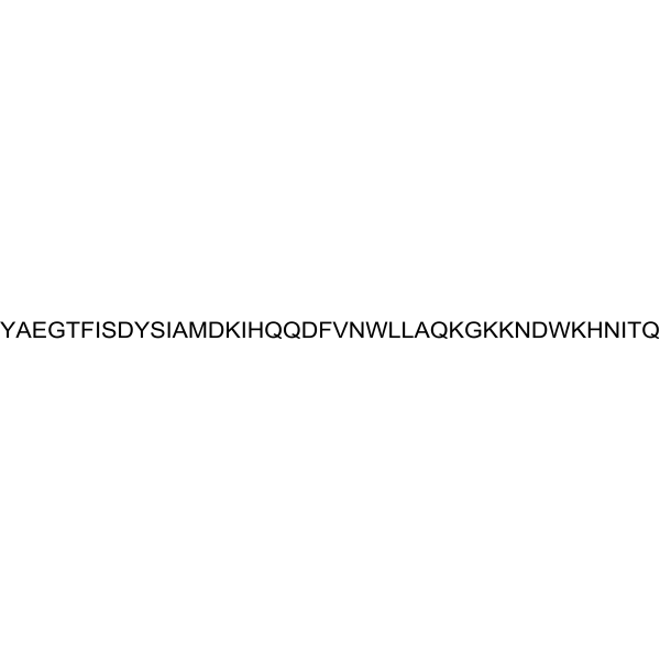
GIP, humanInsulin Secretion Stimulator -

HY-P4146
SurvodutideGCGR/GLP-1R Agonist -

HY-P0059
TeriparatideParathyroid Hormone Agonist -
HY-P0252
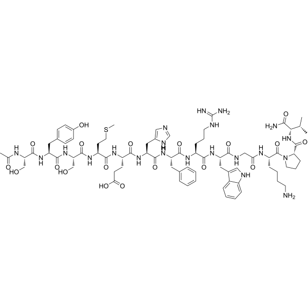
α-MSHMC4R Agonist -
HY-N2466
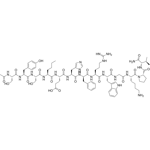
Melanotan IMelanocortin Receptor Activator -
HY-P1071
α-CGRP (human)Vasodilator -
HY-100856
PCO371PTHR1 Agonist -
HY-P1624
TeduglutideGLP-2 Analogue -
HY-P0227
SHU 9119MC3/4R Antagonist -
HY-P0267
Melanotan (MT)-IIMelanotropin Receptor Agonist
Publications Citing Use of MedChemExpress (MCE) Cotadutide acetate
More

Biological Activity
Description
IC50 & Target
EC50: 6.9 pM (GLP-1); 10.2 pM (GCGR)[1]
In Vitro
Cotadutide acetate stimulates a concentration-dependent increase in cAMP accumulation in rat (INS-1 832/3) and human (EndoC-βH1) β-cell lines (EC50: 226 pM and 1051 pM, respectively), as well as rat, mouse and human hepatocytes (EC50: 462 pM, 840 pM, 1447 pM, respectively)[1]. Cotadutide (100 pM-1 μM) acetate potentiates glucose-stimulated insulin secretion in the rat (INS-1 832/3) pancreatic β-cell line and increases glucose output in rat hepatocytes[1]. Cotadutide (100 nM, 2 h) acetate induces mitochondrial turnover and enhances mitochondrial function in mouse primary hepatocytes[2].
MedChemExpress (MCE) has not independently confirmed the accuracy of these methods. They are for reference only.
In Vivo
MedChemExpress (MCE) has not independently confirmed the accuracy of these methods. They are for reference only.
-
Animal Model:DIO mice[1]
-
Dosage:10 nmol/kg
-
Administration:Subcutaneous injection (s.c.)
-
Result:Showed a redction of food intake in mice after an acute administration.
-
Animal Model:Diet-induced obesity (DIO) mice[1]
-
Dosage:10 or 30 nmol/kg
-
Administration:Subcutaneous injection
-
Result:Reduced body weight and food intake, and improved glucose tolerance in DIO mice.
Clinical Trial
| NCT Number | Sponsor | Condition | Start Date |
Phase

|
|---|---|---|---|---|
| NCT01329991 | Plexxikon| | 2011-05 | PHASE1 |


Chemical Information
-
Appearance Solid
-
Molecular Weight 3728.04 (free base)
-
Formula C167H252N42O55·xC2H4O2
-
Color White to light yellow
-
Synonyms
MEDI0382 acetate
-
Sequence
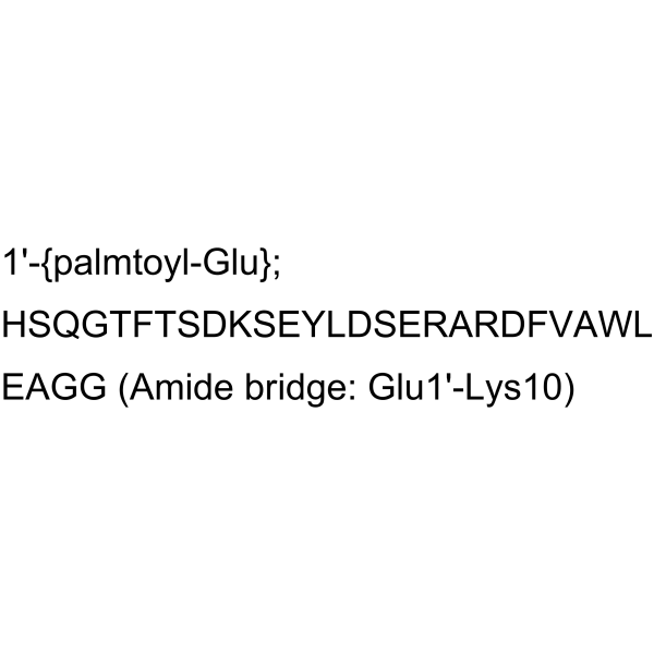
His-Ser-Gln-Gly-Thr-Phe-Thr-Ser-Asp-{Lys(γGlu-C16 acid)}-Ser-Glu-Tyr-Leu-Asp-Ser-Glu-Arg-Ala-Arg-Asp-Phe-Val-Ala-Trp-Leu-Glu-Ala-Gly-Gly
-
Sequence Shortening
HSQGTFTSD-{Lys(γGlu-C16 acid)}-SEYLDSERARDFVAWLEAGG
-
Shipping
Room temperature in continental US; may vary elsewhere.
-
Storage
Sealed storage, away from moisture
Powder -80°C 2 years -20°C 1 year * In solvent : -80°C, 6 months; -20°C, 1 month (sealed storage, away from moisture)


Publications (1)
-
Journal Impact Factor
-
Most Recent
-
Adv Sci (Weinh)
2024 Aug;11(29):e2400819. PMID: 38837628


Solvent & Solubility
In Vitro:
DMSO : 50 mg/mL (Need ultrasonic; Hygroscopic DMSO has a significant impact on the solubility of product, please use newly opened DMSO)
H2O : < 0.1 mg/mL (insoluble)
In Vivo:
Select the appropriate dissolution method based on your experimental animal and administration route.
- For the following dissolution methods, please ensure to first prepare a clear stock solution using an In Vitro approach and then sequentially add co-solvents:
- To ensure reliable experimental results, the clarified stock solution can be appropriately stored based on storage conditions. As for the working solution for In Vivo experiments, it is recommended to prepare freshly and use it on the same day.
- The percentages shown for the solvents indicate their volumetric ratio in the final prepared solution. If precipitation or phase separation occurs during preparation, heat and/or sonication can be used to aid dissolution.
Add each solvent one by one: 10% DMSO 40% PEG300 5% Tween-80 45% Saline
Solubility: 1.25 mg/mL; Suspended solution; Need ultrasonic
This protocol yields a suspended solution of 1.25 mg/mL. Suspended solution can be used for oral and intraperitoneal injection.
Taking 1 mL working solution as an example, add 100 μL DMSO stock solution (12.5 mg/mL) to 400 μL PEG300, and mix evenly; then add 50 μL Tween-80 and mix evenly; then add 450 μL Saline to adjust the volume to 1 mL.
Preparation of Saline: Dissolve 0.9 g sodium chloride in ddH₂O and dilute to 100 mL to obtain a clear Saline solution.
Add each solvent one by one: 10% DMSO 90% (20% SBE-β-CD in Saline)
Solubility: 1.25 mg/mL; Suspended solution; Need ultrasonic
This protocol yields a suspended solution of 1.25 mg/mL. Suspended solution can be used for oral and intraperitoneal injection.
Taking 1 mL working solution as an example, add 100 μL DMSO stock solution (12.5 mg/mL) to 900 μL 20% SBE-β-CD in Saline, and mix evenly.
Preparation of 20% SBE-β-CD in Saline (4°C, storage for one week): 2 g SBE-β-CD powder is dissolved in 10 mL Saline, completely dissolve until clear.
In Vivo Dissolution Calculator
Please enter the basic information of animal experiments:
-
-
-
-
Recommended: Prepare an additional quantity of animals to account for potential losses during experiments.
Please enter your animal formula composition:
-
%DMSO +
Recommended: Keep the proportion of DMSO in working solution below 2% if your animal is weak.
-
%+
-
+%Tween-80 + +
-
%Saline +
The co-solvents required include: DMSO, . All of co-solvents are available by MedChemExpress (MCE). , Tween 80. All of co-solvents are available by MedChemExpress (MCE).
Working solution concentration: 0.22 mg/mL
Method for preparing stock solution: mg drug dissolved in μL DMSO. Stock solution concentration: mg/mL. * In solvent : -80°C, 6 months; -20°C, 1 month (sealed storage, away from moisture)
1. Take μL DMSO stock solution;
2. Add μL .
μL , mix evenly;
3. Then add μL Tween 80, mix evenly;
4. Then add μL
Please ensure that the stock solution in the first step is dissolved to a clear state, and add co-solvents in sequence. You can use ultrasonic heating (ultrasonic cleaner, recommended frequency 20-40 kHz), vortexing, etc. to assist dissolution.


Purity & Documentation
-
Data Sheet (290 KB)
-
SDS (254 KB)
- English - EN (254 KB)
- Français - FR (254 KB)
- Deutsch - DE (254 KB)
- Norwegian - NO (254 KB)
- Español - ES (254 KB)
- Swedish - SV (254 KB)
- Italian - IT (254 KB)
- Korean - KR (254 KB)
- Portuguese - PT (254 KB)
-
Handling Instructions (2659 KB)
References
[1]. Henderson SJ,et al. Robust anti-obesity and metabolic effects of a dual GLP-1/glucagon receptor peptide agonist in rodents and non-human primates.Diabetes Obes Metab. 2016 Dec;18(12):1176-1190. [Content Brief]
[2]. Resolution of NASH and hepatic fibrosis by the GLP-1R/GcgR dual-agonist Cotadutide via modulating mitochondrial function and lipogenesis. Nat Metab. 2020 May;2(5):413-431. [Content Brief]

Calculators
Concentration (start) × Volume (start) = Concentration (final) × Volume (final)


Cotadutide acetate Related Classifications
HY-P2231A Related Classifications
 Powered by Bioz