



HB0030
HB0030 is a TIGIT inhibitor with antibody-dependent cellular cytotoxicity (ADCC) and complement-dependent cytotoxicity (CDC) activities. HB0030 enhances the expression of activation markers in natural killer (NK) cells, promotes the killing of regulatory T cells (Tregs), and reduces the proportion of FoxP3+ Treg in tumor-infiltrating lymphocytes. The combination of HB0030 with the anti-PD-L1/VEGF bispecific antibody HB0025 further enhances tumor suppression efficacy. HB0030 can be used in studies related to colorectal cancer, pancreatic adenocarcinoma, hepatocellular carcinoma, bladder cancer, breast cancer, non-small cell lung cancer, and advanced solid tumors.
For research use only. We do not sell to patients.
-
Storage:
Please store the product under the recommended conditions in the Certificate of Analysis.


To place orders, for customer services and technical support, please contact: MedChemExpress USA
Tel: 609-228-6898 E-mail: [email protected] [email protected]


-
 Biological Activity
-
Chemical Information
- Protocols
- Purity & Documentation
- References
-
 Help & FAQs




Biological Activity
Description
Isotype
Human IgG1 kappa
Recommend Isotype Controls
Species Reactivity
Human
In Vitro
HB0030 (10-5-103 nM; 5 hours) potently mediates NK cell-dependent lysis of human TIGIT-expressing Jurkat cells in vitro, with greater cytotoxic activity than HB0036[1].
MedChemExpress (MCE) has not independently confirmed the accuracy of these methods. They are for reference only.
-
Cell Line:human TIGIT-expressing Jurkat cells, pre-activated human NK cells
-
Concentration:10-5-103 nM
-
Incubation Time:5 hours
-
Result:Mediated dose-dependent NK cell killing of TIGIT-expressing Jurkat cells.
Showed stronger cytotoxic activity compared to HB0036.
In Vivo
HB0030 (10 mg/kg; i.p.; twice weekly; 4 total doses) combined with anti-PD-L1 antibody 900458 significantly reduces tumor volume and weight, and increases intratumoral IFN-γ and TNF-α levels, in CT26-hPD-L1 tumor-bearing humanized BALB/c mice[1].
HB0030 (3 mg/kg; i.p.; twice weekly; 4 total doses) monotherapy significantly inhibits H22-hPD-L1 tumor growth with a 50% partial response rate, and its combination with HB0025 enhances efficacy to an 87.5% partial response rate in humanized BALB/c mice[1].
HB0030 (3 mg/kg; i.p.; twice weekly; 4 total doses) combined with HB0025 significantly enhances tumor inhibition compared to monotherapies in hPD-L1-expressing MC38 tumor-bearing humanized C57BL/6 mice[1].
HB0030 (10 mg/kg; i.p.; twice weekly; 4 total doses) combined with HB0025 significantly enhances tumor inhibition compared to monotherapies in hPD-L1-expressing EMT6 tumor-bearing humanized BALB/c mice[1].
MedChemExpress (MCE) has not independently confirmed the accuracy of these methods. They are for reference only.
-
Animal Model:hTIGIT/hPD-L1/hPD-1 humanized C57BL/6 (subcutaneously inoculated with 1×10⁶ wild-type hPD-L1- MC38 cells on left flank and 1×10⁶ hPD-L1+ MC38 cells on right flank, grown to ~200 mm3 over 13 days)[1]
-
Dosage:500 mg/kg
-
Administration:i.p.; single dose
-
Result:Significantly reduced the proportion of FoxP3+ regulatory T cells (Tregs) in tumor-infiltrating lymphocytes within hPD-L1+ MC38 tumors.
No significant Treg reduction was observed in hPD-L1- MC38 tumors.
-
Animal Model:hPD-1/hPD-L1/hTIGIT humanized BALB/c (subcutaneously inoculated with 1×10⁶ CT26-hPD-L1 cells, grown to average volume of 160 mm3)[1]
-
Dosage:10 mg/kg (in combination with 10 mg/kg anti-PD-L1 antibody 900458)
-
Administration:i.p.; twice weekly; 4 total doses
-
Result:Significantly reduced tumor volume and tumor weight compared to control.
Elevated intratumoral IFN-γ and TNF-α levels compared to control, though lower than levels in the HB0036 monotherapy group.
-
Animal Model:hPD-1/hPD-L1/hTIGIT humanized BALB/c (subcutaneously inoculated with 1×10⁶ H22-hPD-L1 cells, grown to average volume of 100 mm3)[1]
-
Dosage:3 mg/kg (monotherapy; in combination with 2 mg/kg anti-PD-L1/VEGF bispecific antibody HB0025)
-
Administration:i.p.; twice weekly; 4 total doses
-
Result:Showed significant tumor inhibition compared to isotype control, with a partial response rate of 3/6.
Combination with HB0025 showed superior tumor suppression compared to either monotherapy, with a partial response rate of 7/8.
Gene ID
Accession
Target
TIGIT
Conjugated
Unconjugated
Reconsititution
The product can be reconstituted/diluted with sterile PBS or saline.
Format
-
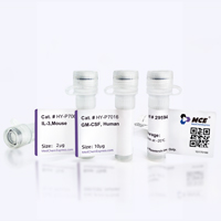
Product Image
Application
ELISA, FACS, Functional assay


Chemical Information
-
Formulation
Please refer to the lot-specific COA for specific buffer information.
-
Storage
Please store the product under the recommended conditions in the Certificate of Analysis.


Purity & Documentation
References

Calculators
Concentration (start) × Volume (start) = Concentration (final) × Volume (final)


HB0030 Related Classifications
HY-P992361 Related Classifications
 Powered by Bioz
Keywords