



CO-1
CO-1 is an IgG4 anti-CD47 antibody. CO-1 not only enhances themacrophage phagocytosis activity but also induces a unique and fast form of programmed celldeath (PCD) in cancer cells directly through ligation of CD47. CO-1 has anticancer activity against hematologic malignancies such as acute lymphoblastic leukemia.
For research use only. We do not sell to patients.
-
Storage:
Please store the product under the recommended conditions in the Certificate of Analysis.


To place orders, for customer services and technical support, please contact: MedChemExpress USA
Tel: 609-228-6898 E-mail: [email protected] [email protected]


-
 Biological Activity
-
Chemical Information
- Protocols
- Purity & Documentation
- References
-
 Help & FAQs




Biological Activity
Description
Isotype
Human IgG4 kappa
Recommend Isotype Controls
Species Reactivity
Human
IC50 & Target
CD47
In Vitro
CO-1 induces rapid (within 3 hours) and potent (≥40%) programmed cell death in several cell lines derived from hematologic malignancies (Jurkat, MOLT-4 and REH)[1].
CO-1 does not cause hemagglutination of red blood cells[1].
MedChemExpress (MCE) has not independently confirmed the accuracy of these methods. They are for reference only.
In Vivo
MedChemExpress (MCE) has not independently confirmed the accuracy of these methods. They are for reference only.
Gene ID
Accession
Conjugated
Unconjugated
Reconsititution
The product can be reconstituted/diluted with sterile PBS or saline.
Format
-
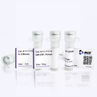
Product Image
Application
ELISA, FACS, Functional assay


Chemical Information
-
Formulation
Please refer to the lot-specific COA for specific buffer information.
-
Storage
Please store the product under the recommended conditions in the Certificate of Analysis.


Purity & Documentation
References

Calculators
Concentration (start) × Volume (start) = Concentration (final) × Volume (final)


CO-1 Related Classifications
HY-P991924 Related Classifications
 Powered by Bioz