



Evernic Acid

Based on 1 Customer Validation
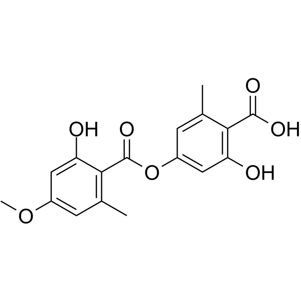
Evernic Acid is an orally active thioredoxin reductase 1 (TrxR1) inhibitor and antiproliferative agent. Evernic Acid inhibits the proliferation and migration of human breast cancer cells. Evernic Acid blocks the NF-κB pathway by inhibiting p65 nuclear translocation and IκBα phosphorylation, thereby suppressing downstream inflammatory mediators. Evernic Acid acts as an antioxidant, anti-inflammatory agent and neuroprotective agent, protects neurons from cell death, mitochondrial dysfunction and oxidative stress damage, reduces astrocyte activation, and ameliorates dopaminergic neuron loss and neuroinflammation. Evernic Acid inhibits enoyl reductases FabI and FabZ of Plasmodium falciparum. Evernic Acid downregulates the expression of lasB and rhlA genes in Pseudomonas aeruginosa, inhibits quorum sensing and biofilm formation, and exerts antibacterial activity against Gram-positive bacteria, Gram-negative bacteria and fungi. Evernic Acid is applicable to research related to breast cancer, Parkinson's disease, bacterial infections and fungal infections.
For research use only. We do not sell to patients.
- Purity : 99.71%
- CAS No.: 537-09-7
- Formula: C17H16O7
- Molecular Weight:332.30
-
Storage:Powder -20°C, 3 years ; In solvent -80°C, 6 months , -20°C, 1 month

To place orders, for customer services and technical support, please contact: MedChemExpress USA
Tel: 609-228-6898 E-mail: [email protected] [email protected]


-
 Biological Activity
-
Chemical Information
-
Solvent & Solubility
- Protocols
- Purity & Documentation
- References
-
 Help & FAQs


-

HY-121362R
Evernic Acid (Standard)Secondary Metabolite -
HY-117769
GSK837149AFASN Inhibitor -
HY-N6707
Triacsin CACSL Inhibitor -
HY-N6742
BorrelidinCDK Inhibitor -
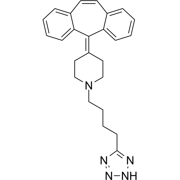
HY-13988
AT-56L-PGDS Inhibitor -
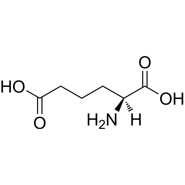
HY-W013665
(S)-2-Aminohexanedioic acidBiochemical Reagent -

HY-W013665R
(S)-2-Aminohexanedioic acid (Standard)Standard of (S)-2-Aminohexanedioic acid -
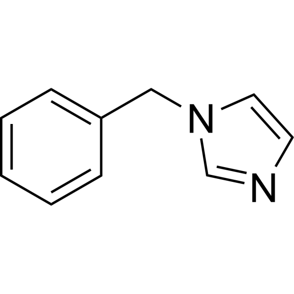
HY-W042985
1-BenzylimidazoleBiochemical Reagent -

HY-B0338A
Rimantadine hydrochlorideAntiviral Agent -

HY-13988R
AT-56 (Standard)L-PGDS Inhibitor -

HY-116535A
DL-threo-PPMP hydrochlorideSphingosine Synthetase Inhibitor -
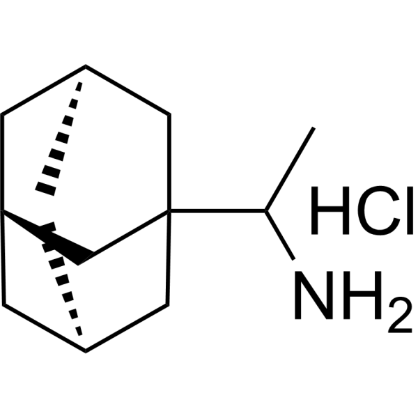
HY-B0338AR
Rimantadine hydrochloride (Standard)Antiviral Agent
All Endogenous Metabolite Isoforms
More

Biological Activity
Description
IC50 & Target
|
Fungal Metabolite |
Cellular Effect
|
Cell Line

|
Type | Value | Description | References |
|---|---|---|---|---|
| Huh-7 | IC50 |
>100 μM
Compound: 1
|
Cytotoxicity against human Huh7.5 cells after 24 hrs by Cell-titerGlo luminescence assay
Cytotoxicity against human Huh7.5 cells after 24 hrs by Cell-titerGlo luminescence assay
|
[PMID: 23806111] |
| Huh-7 | IC50 |
77.3 μM
Compound: 1
|
Antiplasmodial activity against liver stage of Plasmodium berghei ANKA expressing GFP infected in human Huh7.5 cells assessed as reduction in PB18S gene expression after 48 hrs by qRT-PCR analysis
Antiplasmodial activity against liver stage of Plasmodium berghei ANKA expressing GFP infected in human Huh7.5 cells assessed as reduction in PB18S gene expression after 48 hrs by qRT-PCR analysis
|
[PMID: 23806111] |
| KB | IC50 |
190.4 μM
Compound: 1
|
Cytotoxicity against human KB cells after 72 hrs by Alamar Blue assay
Cytotoxicity against human KB cells after 72 hrs by Alamar Blue assay
|
[PMID: 23806111] |
In Vitro
Evernic acid (10-150 μg/mL; 24-52 h) exerts dose-dependent antiproliferative effects on human breast cancer MCF-7 and MDA-MB-453 cell lines, with an IC50 of 33.79 μg/mL at 52 h for MCF-7 cells and 121.40 μg/mL at 48 h for MDA-MB-453 cells[1].
Evernic acid (33.79-121.40 μg/mL; 48-52 h) upregulates TrxR1 gene expression in human breast cancer MCF-7 cells and downregulates TrxR1 gene expression in MDA-MB-453 cells when treated at respective IC50 concentrations for 52 h (MCF-7) and 48 h (MDA-MB-453)[1].
Evernic acid (33.79-121.40 μg/mL; 12-24 h) inhibits migration of human breast cancer MCF-7 and MDA-MB-453 cell lines when treated at respective IC50 concentrations, with significant inhibition observed at 24 h for MCF-7 cells and at 12 and 24 h for MDA-MB-453 cells[1].
Evernic acid (33.79-121.40 μg/mL; 48-52 h) suppresses intracellular TrxR1 enzyme activity by 25% in human breast cancer MCF-7 cells and 30% in MDA-MB-453 cells when treated at respective IC50 concentrations for 52 h (MCF-7) and 48 h (MDA-MB-453)[1].
Evernic acid (1-100 μM; 6 h pre-incubation followed by 24 h co-treatment with MPP+) suppresses MPP+-induced apoptotic morphological changes in primary rat cortical neurons, and 100 μM evernic acid normalizes MPP+-altered Bcl-2/Bax protein expression balance[2].
Evernic acid (100 μM; 6 h pre-incubation followed by 6 h co-treatment with MPP+) protects primary rat cortical neurons from MPP+-induced mitochondrial membrane potential loss[2].
Evernic acid (0.1-100 μM; 6 h pre-incubation prior to MPP+ co-treatment) dose-dependently suppresses MPP+-induced ROS generation in primary rat cortical neurons[2].
Evernic acid (100 μM; 6 h pre-incubation followed by 1 h or 24 h co-treatment with MPP+) inhibits the NF-κB signaling pathway in primary rat astrocytes exposed to MPP+, by blocking p65 nuclear translocation, reducing IκBα phosphorylation, and lowering COX-2 expression[2].
Evernic acid (100 μM; 6 h pre-incubation prior to MPP+ co-treatment) suppresses MPP+-induced increases in pro-inflammatory cytokine (IL-1β, IL-6, TNF-α) and chemokine (CCL2) mRNA expression in primary rat astrocytes[2].
Evernic acid (as the primary component of fraction VI) inhibits the growth of Staphylococcus aureus ATCC 29213 (MIC 0.98 μg/mL), Pseudomonas aeruginosa ATCC 27853 (MIC 31.25 μg/mL), Escherichia coli ATCC 25922 (MIC 125 μg/mL), and Candida albicans ATCC 90028 (MIC 62.5 μg/mL) in vitro[3].
Evernic acid inhibits the expression of quorum-sensing-dependent virulence genes lasB and rhlA in Pseudomonas aeruginosa in vitro[3].
MedChemExpress (MCE) has not independently confirmed the accuracy of these methods. They are for reference only. Further protocols information, click here.
-
Cell Line:Human breast cancer MCF-7 and MDA-MB-453 cell lines
-
Concentration:10-150 μg/mL (24 h); 10-100 μg/mL (52 h, MCF-7); 10-150 μg/mL (48 h, MDA-MB-453)
-
Incubation Time:24 h; 52 h (MCF-7); 48 h (MDA-MB-453)
-
Result:Suppressed the proliferation of MCF-7 and MDA-MB-453 cells in a dose-dependent manner.
Resulted in an IC50 value of 33.79 μg/mL in MCF-7 cells treated for 52 h.
Resulted in an IC50 value of 121.40 μg/mL in MDA-MB-453 cells treated for 48 h.
-
Cell Line:Human breast cancer MCF-7 and MDA-MB-453 cell lines
-
Concentration:33.79 μg/mL (MCF-7, IC50); 121.40 μg/mL (MDA-MB-453, IC50)
-
Incubation Time:52 h (MCF-7); 48 h (MDA-MB-453)
-
Result:Did not change the percentage of apoptotic and necrotic cells in MCF-7 cells compared with the control group.
Caused a very low increase in the percentage of late apoptotic cells but did not affect early apoptotic and necrotic cell percentages in MDA-MB-453 cells.
-
Cell Line:Human breast cancer MCF-7 and MDA-MB-453 cell lines
-
Concentration:33.79 μg/mL (MCF-7, IC50); 121.40 μg/mL (MDA-MB-453, IC50)
-
Incubation Time:52 h (MCF-7); 48 h (MDA-MB-453)
-
Result:Downregulated BAX, BCL2, and BAX/BCL2 ratio gene expression by 4.3-, 2.5-, and 2.4-fold, respectively, but did not change P53 gene expression in MCF-7 cells.
Downregulated BAX, BCL2, P53, and BAX/BCL2 ratio gene expression by 2.1-, 1.2-, 1.9-, and 1.7-fold, respectively, in MDA-MB-453 cells.\nUpregulated TrxR1 mRNA expression by 2-fold compared to the control group in MCF-7 cells.
Downregulated TrxR1 mRNA expression by 1.5-fold compared to the control group in MDA-MB-453 cells.
-
Cell Line:Human breast cancer MCF-7 and MDA-MB-453 cell lines
-
Concentration:33.79 μg/mL (MCF-7, IC50); 121.40 μg/mL (MDA-MB-453, IC50)
-
Incubation Time:52 h (MCF-7); 48 h (MDA-MB-453)
-
Result:Did not change the protein expression of BAX or P53 in MCF-7 cells.
Suppressed P53 protein expression by 5.8-fold but did not change BAX protein expression in MDA-MB-453 cells.\nDid not alter TrxR1 protein expression in either MCF-7 or MDA-MB-453 cells compared to control groups.
-
Cell Line:Human breast cancer MCF-7 and MDA-MB-453 cell lines
-
Concentration:33.79 μg/mL (MCF-7, IC50); 121.40 μg/mL (MDA-MB-453, IC50)
-
Incubation Time:12 h, 24 h
-
Result:Reduced wound closure to 30.0% at 24 h in MCF-7 cells, compared to 38.8% in the control group.
Reduced wound closure to 0.7% at 12 h and resulted in a -4.0% wound closure (negative value indicates wound enlargement) at 24 h in MDA-MB-453 cells, compared to 4.3% and 12.6% in the control group, respectively.
In Vivo
MedChemExpress (MCE) has not independently confirmed the accuracy of these methods. They are for reference only.
-
Animal Model:C57BL/6 (male, 6 weeks old, 20-23 g, MPTP-induced Parkinson's disease)[2]
-
Dosage:5 mg/kg; 80 mg/kg
-
Administration:p.o.; daily; 10 days
-
Result:Significantly accelerated recovery of motor function after MPTP-induced impairment, as measured by rotarod testing (80 mg/kg).
Significantly attenuated MPTP-induced dopaminergic neuronal loss in both the striatum (STR) and substantia nigra (SN), as quantified by tyrosine hydroxylase (TH) densitometry in the STR and TH-positive neuron counts in the SN (80 mg/kg).
Significantly suppressed MPTP-induced astroglial activation in the STR and SN, as measured by GFAP fluorescence intensity; no effect on MPTP-induced microglial activation was observed (80 mg/kg).
Showed partial, less pronounced protective effects on dopaminergic neuronal loss and astroglial activation compared to the 80 mg/kg dose (5 mg/kg).


Chemical Information
-
CAS No. 537-09-7
-
Appearance Solid
-
Molecular Weight 332.30
-
Formula C17H16O7
-
Color White to off-white
-
SMILES
CC1=CC(OC(C2=C(C=C(C=C2O)OC)C)=O)=CC(O)=C1C(O)=O
-
Structure Classification
-
Initial Source
-
Shipping
Room temperature in continental US; may vary elsewhere.
-
Storage
Powder -20°C 3 years In solvent -80°C 6 months -20°C 1 month


Solvent & Solubility
In Vitro:
DMSO : 10 mg/mL (30.09 mM; Need ultrasonic and warming; Hygroscopic DMSO has a significant impact on the solubility of product, please use newly opened DMSO)
Please refer to the solubility information to select the appropriate solvent. Once prepared, please aliquot and store the solution to prevent product inactivation from repeated freeze-thaw cycles.
Storage method and period of stock solution: -80°C, 6 months; -20°C, 1 month. When stored at -80°C, please use it within 6 months. When stored at -20°C, please use it within 1 month.
Please refer to the solubility information to select the appropriate solvent. Once prepared, please aliquot and store the solution to prevent product inactivation from repeated freeze-thaw cycles.
Storage method and period of stock solution: -80°C, 6 months; -20°C, 1 month. When stored at -80°C, please use it within 6 months. When stored at -20°C, please use it within 1 month.
Concentration (start) × Volume (start) = Concentration (final) × Volume (final)

Protocols
-
Western Blot
Western blotting (WB) is a commonly used experimental method in molecular biology, biochemistry, and immunogenetics for identifying and quantifying target proteins. It combines gel electrophoresis with immunoassay, enabling researchers to analyze protein expression, post-translational modifications, and molecular weight.
-
RNA extraction experimental
By lysing cells, releasing RNA, and removing impurities such as proteins and DNA, high-purity RNA products are finally obtained. The commonly used traditional method is the guanidine isothiocyanate/phenol/chloroform method (Trizol), which is suitable for a variety of animal materials including animal tissues, microorganisms, cultured cells, etc., and most plant materials.
-
Research Protocol for Infectious Diseases
Infectious-disease experiments test how pathogens interact with host barriers, innate immune receptors, inflammatory signaling, pathogen replication, and tissue injury; pattern-recognition receptors such as TLRs, RIG-I-like receptors, NOD-like receptors, and inflammasomes detect microbial molecules and activate NF-κB, interferon, and cytokine responses. The central hypothesis is that infection severity reflects the balance between pathogen burden and host response: protective inflammation restricts pathogen growth, whereas excessive or mislocalized inflammation contributes to tissue damage and disease phenotype. Unresolved questions include which host pathways are protective versus pathogenic, why some infection models fail to translate to human disease, and which combined readouts best predict clinically relevant infection outcomes.
-
Mammalian live/dead viability and cytotoxicity staining
Live/dead viability and cytotoxicity staining assays are based on the simultaneous detection of intracellular esterase activity in metabolically active (viable) cells and membrane integrity loss in non-viable cells. In commonly used dual-staining approaches, membrane-permeant fluorogenic substrates are converted by intracellular esterases into fluorescent products in live cells, while impermeant DNA-binding dyes selectively enter cells with compromised plasma membranes and label nucleic acids in dead or dying cells, enabling discrimination between viable and non-viable populations by fluorescence microscopy or flow cytometry.
-
Cell migration
Cell migration is a method that plays an important role in wound healing, cell differentiation, embryonic development, etc.
-
ROS/oxidative-stress fluorescent staining
ROS/oxidative-stress fluorescent staining uses cell-permeant fluorogenic probes that become fluorescent after oxidation inside cells or tissues; commonly used examples include DCFH-DA/DCFDA for broad cellular oxidant detection, DHE for superoxide-related signal detection, MitoSOX for mitochondrial superoxide-related signal detection, and CellROX probes for oxidative-stress-associated fluorescence readouts. The assay detects probe oxidation rather than a single ROS species unless the probe and analysis method have been chemically validated for that species. DCFH-DA enters cells, is deacetylated by intracellular esterases to DCFH, and produces fluorescent DCF after oxidation, so the readout is used as an operational measure of total cellular oxidative stress rather than a species-specific ROS measurement. DHE and MitoSOX can report superoxide-related oxidation, but red fluorescence alone can include non-specific ethidium-like oxidation products; HPLC or optimized spectral approaches are
-
Research Protocol for Inflammation-related Diseases
The NLRP3 inflammasome is a cytosolic innate immune signaling platform that integrates priming signals and danger-signal activation to promote caspase-1 activation, maturation of IL-1β and IL-18, and gasdermin D-mediated pyroptotic cell death. The core experimental logic is to determine whether inflammatory disease phenotypes are driven by increased NLRP3 expression, ASC-containing inflammasome assembly, caspase-1 cleavage, GSDMD cleavage, and extracellular release of IL-1β/IL-18 rather than by nonspecific cell injury alone. The pathway is strongly linked to inflammation-related disease phenotypes because monosodium urate crystals activate NALP3/NLRP3 inflammasome signaling in gout-like crystal inflammation, cholesterol crystals activate NLRP3 inflammasomes in atherogenesis models, and DSS-induced intestinal inflammation has been reported to involve NLRP3 inflammasome activity. However, experimental colitis studies also show context-dependent protective effects of NLRP3 inflammasome co
-
Gram Staining of Tissue Sections
Gram staining of tissue sections is a histochemical technique used to differentiate Gram-positive and Gram-negative bacteria within histological specimens based on differences in bacterial cell wall structure and dye retention, adapted from classical bacteriological Gram staining into tissue-compatible “histological Gram stain” variants. In tissue applications, modifications of the Brown-Hopps and Brown-Brenn methods are commonly used to improve differentiation of microorganisms embedded within host connective tissue and to reduce overstaining or loss of Gram-negative signal, which are known limitations of earlier approaches. The principle relies on crystal violet-iodine complex retention in Gram-positive organisms and subsequent decolorization and counterstaining steps that allow contrast visualization of Gram-negative organisms against tissue background.
-
Research Protocol for Neurological Diseases
PINK1/Parkin-mediated mitophagy pathway is a mitochondrial quality-control signaling axis in which mitochondrial depolarization stabilizes PINK1 on damaged mitochondria, activates Parkin recruitment and E3 ubiquitin ligase activity, promotes ubiquitination of outer mitochondrial membrane proteins, recruits selective autophagy adaptors, and drives lysosomal degradation of damaged mitochondria. In neurological disease research, this pathway is experimentally important because neurons, especially dopaminergic neurons, are highly dependent on mitochondrial integrity, and defective mitochondrial turnover can lead to mitochondrial dysfunction, oxidative stress, impaired neuronal survival, α-synuclein accumulation, and neuroinflammatory damage-associated signals. The genetic disease link is strongest in Parkinson’s disease because mutations in PRKN/parkin cause autosomal recessive juvenile parkinsonism, mutations in PINK1 cause hereditary early-onset Parkinson’s disease, and Drosophila studie
-
Breast Cancer Modeling
Breast cancer is a heterogeneous cancer, and it has been distinguished into four subtypes: luminal A, luminal B, HER2-positive and basal-like. Molecular mutations, epigenetic alterations, hormone exposure and immune microenvironment are related to the progression of breast cancer.
-
Bacterial live/dead nucleic-acid viability staining
The LIVE/DEAD bacterial viability staining method is based on differential permeability of nucleic-acid-binding fluorescent dyes, most commonly SYTO 9 and propidium iodide (PI), which enables discrimination of bacterial populations with intact versus compromised cytoplasmic membranes. SYTO 9 penetrates both intact and damaged bacterial membranes and binds nucleic acids to produce green fluorescence, whereas propidium iodide penetrates only cells with compromised membranes and fluoresces red while also reducing SYTO 9 signal through competitive binding and fluorescence interactions. The resulting fluorescence pattern is interpreted as a proxy for membrane integrity, which is widely used as an indicator of bacterial viability in microscopy, flow cytometry, and spectroscopic platforms. However, mechanistic studies show that SYTO 9 and PI interactions involve displacement and fluorescence resonance energy transfer effects, which can influence signal interpretation depending on dye ratios a
-
Cell Viability Determination by MTT Colorimetric Assay
The following protocol uses the MTT colorimetric assay as a classic literature-established method for assessing cell viability/metabolic activity in cultured mammalian cells. MTT[3-(4,5-dimethylthiazol-2-yl)-2,5-diphenyltetrazolium bromide] is reduced by metabolically active cells to a colored formazan product; the amount of formazan is quantified spectrophotometrically and provides an indirect measure of metabolically active viable cells. Importantly, MTT reduction reflects cellular oxidoreductase/metabolic activity rather than an absolute direct count of living cells, so changes in cellular metabolism can alter the signal independently of cell number.
-
Kinase activity and phosphorylation assays
Kinase activity assays measure the ability of kinases to transfer phosphate groups from ATP to specific substrates, while phosphorylation assays detect the presence and levels of phosphorylated proteins. Common methods include radiolabeled ATP incorporation (e. g. ,), ADP release detection via bioluminescence (e. g. ,[3]), enzyme-linked immunosorbent assays (ELISA) for phospho-specific epitopes (e. g. ,[6]), and microtiter-based formats for high-throughput screening (e. g. ,[8]). The ADP-Glo assay quantifies kinase activity by measuring ADP produced during phosphorylation using a luciferase-based system. Radiometric assays involve autoradiography or scintillation counting after incorporation of 32P-labeled ATP into substrate proteins. ELISA-based approaches rely on phospho-specific antibodies to detect activated kinases in cell lysates or purified samples.
-
Cytoplasmic-Nuclear Fractionated Protein Extraction
Cytoplasmic-nuclear fractionated protein extraction separates soluble cytoplasmic proteins from nuclear-enriched proteins by mild plasma-membrane permeabilization, differential centrifugation, washing of nuclei, and extraction of nuclear proteins for downstream immunoblotting or related molecular analysis. The readout is the relative abundance of a protein in cytoplasmic and nuclear fractions, commonly assessed by western blotting together with compartment markers such as tubulin or pyruvate kinase for cytoplasm and lamin, nucleoporin, hnRNP, H2AX, or Lamin B for nuclear fractions.
-
Cell Cytotoxicity Assay
Cytotoxicity assays are usually based on the assessment of cell membrane damage, which can also be indirectly detected by measuring cell viability. Detection methods include MTT assay, CKK-8 assay, LDH assay and ATP assay, etc.
-
Human pluripotent stem cell midbrain dopaminergic neuron differentiation
Human pluripotent stem cells are directed toward midbrain dopaminergic neurons by first inducing a neural floor-plate-like progenitor state, then patterning cells with ventralizing SHH signaling and midbrain/WNT-FGF cues, and finally maturing progenitors into neurons expressing dopaminergic markers such as TH, NURR1/NR4A2, PITX3, DAT/SLC6A3, VMAT2/SLC18A2, GIRK2/KCNJ6, FOXA2, LMX1A, and EN1. The main readouts are loss of pluripotency, acquisition of FOXA2+/LMX1A+ midbrain floor-plate progenitors, emergence of βIII-tubulin+/MAP2+ neurons, and production of TH+ dopaminergic neurons with molecular, dopamine-release, and electrophysiological features of midbrain dopaminergic identity.
-
Mitochondrial membrane-potential and mitochondrial mass staining
Mitochondrial membrane potential staining measures the electrochemical polarization across the mitochondrial inner membrane in live cells using lipophilic cationic fluorescent probes; early rhodamine-based work showed that selective mitochondrial dye accumulation is lost when the mitochondrial transmembrane potential is dissipated. JC-1 reports mitochondrial polarization by shifting from green monomer fluorescence to red J-aggregate fluorescence as dye concentration increases within energized mitochondria; therefore, the red/green fluorescence ratio is used as a relative readout of mitochondrial membrane potential. TMRE or TMRM staining provides a single-channel relative readout because these cationic rhodamine esters accumulate in polarized mitochondria, and lower fluorescence indicates reduced mitochondrial polarization when acquisition and dye-loading conditions are controlled. Mitochondrial mass staining is commonly performed with MitoTracker Green FM or related MitoTracker dyes as
-
Apoptosis Solutions
Apoptosis is a regulated, generally non-lytic cell-death pathway that removes unwanted, damaged, infected, or abnormal cells through coordinated morphological changes, caspase activation, DNA fragmentation, and membrane remodeling. The intrinsic apoptosis pathway is controlled mainly by mitochondrial outer membrane permeabilization, BCL-2 family proteins, cytochrome c release, apoptosome formation, caspase-9 activation, and downstream executioner caspase-3/7 activation. The extrinsic apoptosis pathway is initiated by death receptors such as Fas, TNFR, and TRAIL receptors, which recruit adaptor proteins and activate caspase-8 before engaging executioner caspases or mitochondrial amplification through BID cleavage. Apoptosis is linked to many phenotypes, including cancer cell killing, tissue homeostasis, immune regulation, neurodegeneration, infection response, and treatment-induced cytotoxicity; unresolved questions include how apoptosis interacts with necroptosis, pyroptosis, ferroptos
-
Protocol for Kinase activity and phosphorylation assays
Kinase activity assays measure transfer of phosphate from ATP to a protein or peptide substrate, generating phosphorylated substrate, ADP, or incorporated radiolabeled phosphate as the readout; phosphorylation assays measure site-specific phosphorylation in cells or tissues as a proxy for kinase-pathway activation, inhibition, or substrate regulation. Phosphorylation can be detected by phospho-specific Western blot, immunoprecipitation kinase assay, phospho-immunofluorescence, phospho-flow cytometry, luminescent ADP detection, radiolabeled ATP incorporation, or reporter-based pathway assays, and these readouts can be applied to cancer cells, primary neurons, mouse tumors, organoids, inflammatory macrophages, ferroptosis studies, and mitophagy studies when the kinase target is biologically relevant.
-
Fungal Biofilm Culture
Fungal biofilm culture is an in vitro method for growing surface-attached fungal communities, most commonly Candida albicans, on abiotic substrates such as polystyrene wells, silicone elastomer, or polymethylmethacrylate; the assay models adhesion, proliferation, filamentation, extracellular-matrix-associated maturation, and dispersion. Biofilm output can be read by optical density at 600 nm for adherent biomass, XTT reduction for metabolic activity, CFU recovery for viable attached or dispersed cells, and microscopy for architecture.


Purity & Documentation
-
Data Sheet (285 KB)
-
SDS (252 KB)
- English - EN (252 KB)
- Français - FR (252 KB)
- Deutsch - DE (252 KB)
- Norwegian - NO (252 KB)
- Español - ES (252 KB)
- Swedish - SV (252 KB)
- Italian - IT (252 KB)
- Korean - KR (252 KB)
- Portuguese - PT (252 KB)
-
Handling Instructions (2659 KB)
References
[1]. Kalın ŞN, et al. Effect of evernic acid on human breast cancer MCF-7 and MDA-MB-453 cell lines via thioredoxin reductase 1: A molecular approach. J Appl Toxicol. 2023;43(8):1148-1158. [Content Brief]
[2]. Lee S, et al. Neuroprotective and Anti-Inflammatory Effects of Evernic Acid in an MPTP-Induced Parkinson's Disease Model. Int J Mol Sci. 2021;22(4):2098. Published 2021 Feb 20. [Content Brief]
[3]. Shcherbakova A, et al. Antimicrobial and antioxidant activity of Evernia prunastri extracts and their isolates. World J Microbiol Biotechnol. 2021;37(8):129. Published 2021 Jul 7. [Content Brief]


Complete Stock Solution Preparation Table
Please refer to the solubility information to select the appropriate solvent. Once prepared, please aliquot and store the solution to prevent product inactivation from repeated freeze-thaw cycles.
Storage method and period of stock solution: -80°C, 6 months; -20°C, 1 month. When stored at -80°C, please use it within 6 months. When stored at -20°C, please use it within 1 month.
| Optional Solvent | Concentration Solvent Mass | 1 mg | 5 mg | 10 mg | 25 mg |
|---|---|---|---|---|---|
| DMSO | 1 mM | 3.0093 mL | 15.0466 mL | 30.0933 mL | 75.2332 mL |
| 5 mM | 0.6019 mL | 3.0093 mL | 6.0187 mL | 15.0466 mL | |
| 10 mM | 0.3009 mL | 1.5047 mL | 3.0093 mL | 7.5233 mL | |
| 15 mM | 0.2006 mL | 1.0031 mL | 2.0062 mL | 5.0155 mL | |
| 20 mM | 0.1505 mL | 0.7523 mL | 1.5047 mL | 3.7617 mL | |
| 25 mM | 0.1204 mL | 0.6019 mL | 1.2037 mL | 3.0093 mL | |
| 30 mM | 0.1003 mL | 0.5016 mL | 1.0031 mL | 2.5078 mL |


Evernic Acid Related Classifications
HY-121362 Related Classifications
 Powered by Bioz