



Glucosylceramide, plant

Based on 1 Customer Validation
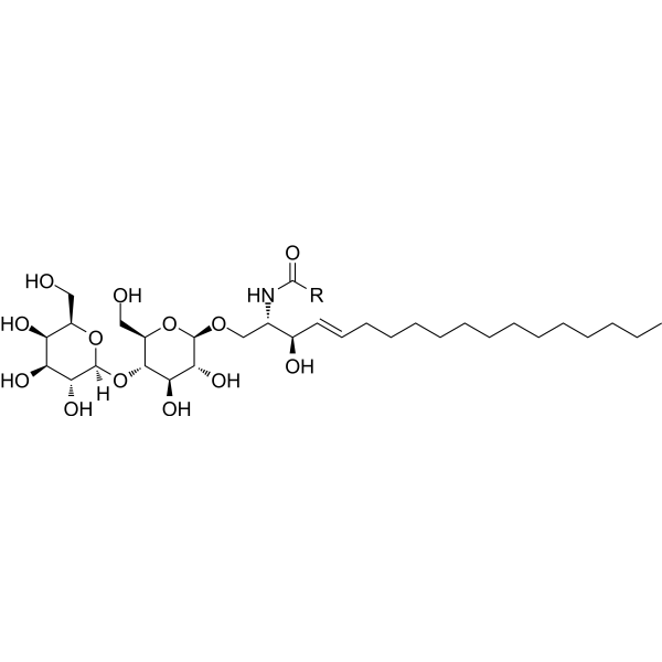
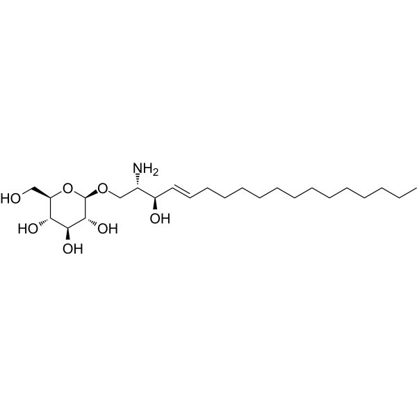
Glucosylceramide, plant (GluCer (Soy)) is an orally active, plant-derived glucosylceramide. Glucosylceramide, plant inhibits lipopolysaccharide-induced cytokine production and Apoptosis. Glucosylceramide, plant suppresses DSS-induced body weight loss, colonic crypt damage and intestinal inflammation in mice. Glucosylceramide, plant can be used in research related to inflammatory bowel disease and colon cancer.
For research use only. We do not sell to patients.
- Purity : 99.9%
- CAS No.: 497155-61-0
- Formula: C40H75NO9
- Molecular Weight:714.02
-
Storage:Powder -20°C, 3 years ; In solvent -80°C, 6 months , -20°C, 1 month


To place orders, for customer services and technical support, please contact: MedChemExpress USA
Tel: 609-228-6898 E-mail: [email protected] [email protected]


-
 Biological Activity
-
Chemical Information
- Protocols
- Purity & Documentation
- References
-
 Help & FAQs


-
HY-154921
Lactosylceramide (bovine buttermilk)Glycosphingolipid -

HY-165035
Glucosylceramide (gaucher's spleen)Sphingolipid/Gaucher Disease Biomarker -

HY-P2149
Concanavalin APlant Lectin -

HY-P2149A
Concanavalin A (agarose)Protein Purification -
HY-133593
Palustric acidNatural Product -
HY-120657
9-PAHSAGPR120 Agonist -

HY-133593R
Palustric acid (Standard)Reference Standard -
HY-P10018
BamadutideGLP-1R/GCGR Agonist -

HY-151240
GalactosylceramideGlucosylceramide -
HY-N7745
GlucosylsphingosineGlucocerebrosidase Inhibitor -

HY-164236
C22 Glucosylceramide (d18:1/22:0)Bioactive Sphingolipid -

HY-153830
LacCer (d18:1/16:0)Sphingolipid


Biological Activity
Description
In Vitro
Glucosylceramide, plant suppresses lipopolysaccharide-induced cytokine production and apoptosis in differentiated Caco-2 cells, exerting potent anti-inflammatory and cytoprotective effects regardless of sphingoid base composition[2].
MedChemExpress (MCE) has not independently confirmed the accuracy of these methods. They are for reference only. Further protocols information, click here.
In Vivo
MedChemExpress (MCE) has not independently confirmed the accuracy of these methods. They are for reference only.
-
Animal Model:BALB/c (female, 4 weeks old, DSS-induced colitis)[1]
-
Dosage:0.1% (1 g/kg diet)
-
Administration:p.o.; daily; 5 or 15 days
-
Result:Suppressed DSS-induced body weight loss in the early phase of treatment.
Reduced DSS-induced chorionic crypt injury observed via toluidine blue-stained colon histology.
Significantly suppressed DSS-elevated myeloperoxidase levels, which were approximately 2-fold higher in DSS-only controls compared to untreated mice.
Normalized DSS-induced increases in inflammatory cytokines (IL-1α, IL-1β, IL-16), chemokines (MIG, IP-10), and adhesion molecules (C5/C5a, sICAM-1) to levels similar to untreated mice.


Chemical Information
-
CAS No. 497155-61-0
-
Appearance Solid
-
Molecular Weight 714.02
-
Formula C40H75NO9
-
Color White to off-white
-
SMILES
[H][C@](/C=C/CC/C=C/CCCCCCCCC)(O)[C@@]([H])(NC(C(O)CCCCCCCCCCCCCC)=O)CO[C@H](O1)[C@H](O)[C@@H](O)[C@@H]([C@H]1CO)O
-
Synonyms
GluCer (Soy)
-
Shipping
Room temperature in continental US; may vary elsewhere.
-
Storage
Powder -20°C 3 years In solvent -80°C 6 months -20°C 1 month

Protocols
-
Apoptosis
Apoptosis, also called programmed cell death, is generally characterized by distinct morphological characteristics.
-
TUNEL staining for apoptotic DNA fragmentation
TUNEL staining detects DNA strand breaks by using terminal deoxynucleotidyl transferase to add labeled nucleotides to exposed 3′-OH DNA termini, generating either microscopic staining in fixed cells or tissue sections, or fluorescence/cytometric signal in cell suspensions. TUNEL positivity reflects DNA fragmentation but should not be interpreted alone as definitive apoptosis, because TUNEL can also label necrotic, autolytic, mechanically damaged, or DNA-repair-associated DNA breaks.
-
Annexin V plus membrane-impermeant dye apoptosis staining
Annexin V-based apoptosis assays rely on the detection of phosphatidylserine (PS) externalization from the inner leaflet of the plasma membrane to the outer leaflet, an early biochemical hallmark of apoptosis. Fluorescently labeled Annexin V binds PS in a calcium-dependent manner, enabling identification of early apoptotic cells by flow cytometry or fluorescence microscopy. When combined with a membrane-impermeant DNA-binding dye (e. g. , propidium iodide), this approach allows discrimination between viable (Annexin V−/dye−), early apoptotic (Annexin V+/dye−), and late apoptotic or necrotic (Annexin V+/dye+) cell populations by assessing membrane integrity and PS exposure.
-
DSS-Induced Colitis
Dextran sulfate sodium (DSS)-induced colitis is generated by administering DSS in mouse drinking water, producing epithelial injury, barrier disruption, weight loss, diarrhea, fecal blood, colon shortening, histologic mucosal damage, and inflammatory mediator changes; the model is mainly used to study acute or chronic intestinal inflammation resembling selected features of ulcerative colitis. DSS injury is interpreted through clinical and tissue readouts rather than a single molecular endpoint: daily body weight, stool consistency, and bleeding are combined into a disease activity index, while colon length, histology, cytokines, myeloperoxidase activity, intestinal permeability, and tight-junction markers provide complementary measures of inflammation and barrier damage.
-
TNBS-Induced Colitis
TNBS-induced colitis is produced by intrarectal delivery of 2,4,6-trinitrobenzene sulfonic acid in ethanol, where ethanol disrupts the mucosal barrier and TNBS haptenates colonic proteins, generating immune-mediated colonic inflammation with weight loss, diarrhea, ulceration, transmural injury, inflammatory-cell infiltration, and cytokine responses. The model is used as an experimental intestinal inflammation model with Crohn’s disease–like features, especially when Th1-type responses, IL-12–dependent inflammation, chronic relapsing inflammation, or fibrosis-related endpoints are studied.
-
Apoptosis Solutions
Apoptosis is a regulated, generally non-lytic cell-death pathway that removes unwanted, damaged, infected, or abnormal cells through coordinated morphological changes, caspase activation, DNA fragmentation, and membrane remodeling. The intrinsic apoptosis pathway is controlled mainly by mitochondrial outer membrane permeabilization, BCL-2 family proteins, cytochrome c release, apoptosome formation, caspase-9 activation, and downstream executioner caspase-3/7 activation. The extrinsic apoptosis pathway is initiated by death receptors such as Fas, TNFR, and TRAIL receptors, which recruit adaptor proteins and activate caspase-8 before engaging executioner caspases or mitochondrial amplification through BID cleavage. Apoptosis is linked to many phenotypes, including cancer cell killing, tissue homeostasis, immune regulation, neurodegeneration, infection response, and treatment-induced cytotoxicity; unresolved questions include how apoptosis interacts with necroptosis, pyroptosis, ferroptos
-
Research Protocol for Inflammation-related Diseases
The NLRP3 inflammasome is a cytosolic innate immune signaling platform that integrates priming signals and danger-signal activation to promote caspase-1 activation, maturation of IL-1β and IL-18, and gasdermin D-mediated pyroptotic cell death. The core experimental logic is to determine whether inflammatory disease phenotypes are driven by increased NLRP3 expression, ASC-containing inflammasome assembly, caspase-1 cleavage, GSDMD cleavage, and extracellular release of IL-1β/IL-18 rather than by nonspecific cell injury alone. The pathway is strongly linked to inflammation-related disease phenotypes because monosodium urate crystals activate NALP3/NLRP3 inflammasome signaling in gout-like crystal inflammation, cholesterol crystals activate NLRP3 inflammasomes in atherogenesis models, and DSS-induced intestinal inflammation has been reported to involve NLRP3 inflammasome activity. However, experimental colitis studies also show context-dependent protective effects of NLRP3 inflammasome co


Purity & Documentation
-
Data Sheet (271 KB)
-
SDS (251 KB)
- English - EN (251 KB)
- Français - FR (251 KB)
- Deutsch - DE (251 KB)
- Norwegian - NO (251 KB)
- Español - ES (251 KB)
- Swedish - SV (251 KB)
- Italian - IT (251 KB)
- Korean - KR (251 KB)
- Portuguese - PT (251 KB)
-
Handling Instructions (2659 KB)
References
[1]. Arai K, et al. Effects of Dietary Plant-Origin Glucosylceramide on Bowel Inflammation in DSS-Treated Mice. Journal of oleo science. 2015;64(7):737-42. [Content Brief]
[2]. Yamashita S, et al. Chemical Properties and Nutritional Value of Plant-Origin Glucosylceramide. Journal of nutritional science and vitaminology. 2019;65(Supplement):S153-S157. [Content Brief]

Calculators
Concentration (start) × Volume (start) = Concentration (final) × Volume (final)


Glucosylceramide, plant Related Classifications
HY-W717504 Related Classifications
 Powered by Bioz