



hMAO-B/MB-COMT-IN-2
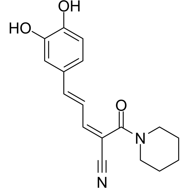
hMAO-B/MB-COMT-IN-2 is a dual MAO-B/MB-COMT inhibitor (IC50s: 4.27 μΜ for hMAO-B, 2.69 μΜ for MB-COMT). hMAO-B/MB-COMT-IN-2 protects cells against oxidative damage. hMAO-B/MB-COMT-IN-2 can be used in the research of neurodegeneration disease, such as Parkinson’s Disease (PD).
For research use only. We do not sell to patients.
- CAS No.: 3033106-46-3
- Formula: C17H18N2O3
- Molecular Weight:298.34
-
Storage:
Please store the product under the recommended conditions in the Certificate of Analysis.

To place orders, for customer services and technical support, please contact: MedChemExpress USA
Tel: 609-228-6898 E-mail: [email protected] [email protected]


-
 Biological Activity
-
Chemical Information
- Protocols
- Purity & Documentation
- References
-
 Help & FAQs




Biological Activity
Description
In Vitro
hMAO-B/MB-COMT-IN-2 (compound 8, 50 μM, 24 h) decreases in resazurin reduction in differentiated SH-SY5Y cells[1].
hMAO-B/MB-COMT-IN-2 (50 μM, 24 h) displays lysosomal toxicity by producing ROS in differentiated SH-SY5Y cells[1].
hMAO-B/MB-COMT-IN-2 (10 μM, 30 min) displays remarkable cytoprotective effects against t-BHP in differentiated SH-SY5Y cells[1].
hMAO-B/MB-COMT-IN-2 is predicted to cross the blood-brain barrier (BBB) by passive diffusion, determined by the parallel artificial membrane permeability assay (PAMPA)-BBB kit[1].
hMAO-B/MB-COMT-IN-2 (30-50 μM, 24 h) has good safety profile in SK-N-SH cells[1].
MedChemExpress (MCE) has not independently confirmed the accuracy of these methods. They are for reference only. Further protocols information, click here.


Chemical Information
-
CAS No. 3033106-46-3
-
Molecular Weight 298.34
-
Formula C17H18N2O3
-
SMILES
OC1=CC(/C=C/C=C(C#N)\C(N2CCCCC2)=O)=CC=C1O
-
Shipping
Room temperature in continental US; may vary elsewhere.
-
Storage
Please store the product under the recommended conditions in the Certificate of Analysis.

Protocols
-
Neurotoxicity Study
This protocol assesses in vitro neurotoxicity by combining neuronal viability, mitochondrial/metabolic activity, neurite outgrowth, and optional neuronal network function readouts. Calcein-AM or resazurin/PrestoBlue readouts estimate viable or metabolically active cells; βIII-tubulin immunofluorescence detects neuronal morphology and neurite networks; TMRE detects mitochondrial membrane potential; and MEA recordings detect functional changes in neuronal network activity.
-
ROS/oxidative-stress fluorescent staining
ROS/oxidative-stress fluorescent staining uses cell-permeant fluorogenic probes that become fluorescent after oxidation inside cells or tissues; commonly used examples include DCFH-DA/DCFDA for broad cellular oxidant detection, DHE for superoxide-related signal detection, MitoSOX for mitochondrial superoxide-related signal detection, and CellROX probes for oxidative-stress-associated fluorescence readouts. The assay detects probe oxidation rather than a single ROS species unless the probe and analysis method have been chemically validated for that species. DCFH-DA enters cells, is deacetylated by intracellular esterases to DCFH, and produces fluorescent DCF after oxidation, so the readout is used as an operational measure of total cellular oxidative stress rather than a species-specific ROS measurement. DHE and MitoSOX can report superoxide-related oxidation, but red fluorescence alone can include non-specific ethidium-like oxidation products; HPLC or optimized spectral approaches are


Purity & Documentation
References

Calculators
Concentration (start) × Volume (start) = Concentration (final) × Volume (final)


hMAO-B/MB-COMT-IN-2 Related Classifications
HY-151390 Related Classifications
 Powered by Bioz