



ICA-105665

Based on 1 Customer Validation
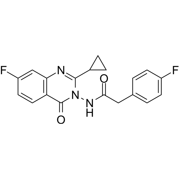
ICA-105665 (PF-04895162) is a potent and orally active neuronal Kv7.2/7.3 and Kv7.3/7.5 potassium channels opener. ICA-105665 inhibits liver mitochondrial function and bile salt export protein (BSEP) transport (IC50 of 311 μM). ICA-105665 can penetrate the blood-brain barrier and has antiseizure effects.
For research use only. We do not sell to patients.
- Purity : 99.85%
- CAS No.: 2694728-63-5
- Formula: C19H15F2N3O2
- Molecular Weight:355.34
-
Storage:Powder -20°C, 3 years , 4°C, 2 years ; In solvent -80°C, 6 months , -20°C, 1 month

To place orders, for customer services and technical support, please contact: MedChemExpress USA
Tel: 609-228-6898 E-mail: [email protected] [email protected]


-
 Biological Activity
-
Chemical Information
-
Solvent & Solubility
- Protocols
- Purity & Documentation
- References
-
 Help & FAQs




Biological Activity
Description
IC50 & Target
In Vitro
ICA-105665 (PF-04895162) does not display potent cytotoxic properties in THLE and HepG2 cell lines (IC50 ~192 μM and 130 μM after 72 hours, respectively) or in human hepatocytes (AC50 for cell loss at 48 hours was >125 μM based on results in three assessments in two different human hepatocyte lots (LBN and HU4165)[1].
Mitochondrial respiratory reserve is compromised in human hepatocytes treated with ICA-105665 (PF-04895162) at concentrations >11 μM for 25 minutes[1].
MedChemExpress (MCE) has not independently confirmed the accuracy of these methods. They are for reference only. Further protocols information, click here.
In Vivo
ICA-105665 (PF-04895162) has demonstrated broad spectrum antiseizure activity in multiple animal models including maximal electroshock, 6 Hz seizures, pentylenetetrazole, and electrical kindling at doses from <1 to 5 mg/kg[3].
MedChemExpress (MCE) has not independently confirmed the accuracy of these methods. They are for reference only.
Clinical Trial
| NCT Number | Sponsor | Condition | Start Date |
Phase

|
|---|---|---|---|---|
| NCT01329991 | Plexxikon| | 2011-05 | PHASE1 |


Chemical Information
-
CAS No. 2694728-63-5
-
Appearance Solid
-
Molecular Weight 355.34
-
Formula C19H15F2N3O2
-
Color White to off-white
-
SMILES
O=C(NN1C(C2CC2)=NC3=C(C=CC(F)=C3)C1=O)CC4=CC=C(F)C=C4
-
Synonyms
PF-04895162
-
Shipping
Room temperature in continental US; may vary elsewhere.
-
Storage
Powder -20°C 3 years 4°C 2 years In solvent -80°C 6 months -20°C 1 month


Solvent & Solubility
In Vitro:
DMSO : 250 mg/mL (703.55 mM; Need ultrasonic; Hygroscopic DMSO has a significant impact on the solubility of product, please use newly opened DMSO)
Please refer to the solubility information to select the appropriate solvent. Once prepared, please aliquot and store the solution to prevent product inactivation from repeated freeze-thaw cycles.
Storage method and period of stock solution: -80°C, 6 months; -20°C, 1 month. When stored at -80°C, please use it within 6 months. When stored at -20°C, please use it within 1 month.
Please refer to the solubility information to select the appropriate solvent. Once prepared, please aliquot and store the solution to prevent product inactivation from repeated freeze-thaw cycles.
Storage method and period of stock solution: -80°C, 6 months; -20°C, 1 month. When stored at -80°C, please use it within 6 months. When stored at -20°C, please use it within 1 month.
Concentration (start) × Volume (start) = Concentration (final) × Volume (final)

Protocols
-
Mitochondrial membrane-potential fluorescent assay
Mitochondrial membrane potential fluorescent assays estimate ΔΨm in living cells using lipophilic cationic dyes such as TMRM, TMRE, rhodamine 123, and JC-1, which accumulate in mitochondria according to membrane polarization; loss of signal after FCCP or CCCP treatment is interpreted as mitochondrial depolarization. TMRM/TMRE and rhodamine 123 are commonly used for semi-quantitative live-cell microscopy or flow cytometry, while JC-1 can report a shift from red aggregate fluorescence to green monomer fluorescence during depolarization; interpretation requires controls because dye concentration, quenching mode, cell type, dye efflux, and mitochondrial mass can affect fluorescence independently of ΔΨm.
-
Cell Cytotoxicity Assay
Cytotoxicity assays are usually based on the assessment of cell membrane damage, which can also be indirectly detected by measuring cell viability. Detection methods include MTT assay, CKK-8 assay, LDH assay and ATP assay, etc.
-
Mitochondrial membrane-potential and mitochondrial mass staining
Mitochondrial membrane potential staining measures the electrochemical polarization across the mitochondrial inner membrane in live cells using lipophilic cationic fluorescent probes; early rhodamine-based work showed that selective mitochondrial dye accumulation is lost when the mitochondrial transmembrane potential is dissipated. JC-1 reports mitochondrial polarization by shifting from green monomer fluorescence to red J-aggregate fluorescence as dye concentration increases within energized mitochondria; therefore, the red/green fluorescence ratio is used as a relative readout of mitochondrial membrane potential. TMRE or TMRM staining provides a single-channel relative readout because these cationic rhodamine esters accumulate in polarized mitochondria, and lower fluorescence indicates reduced mitochondrial polarization when acquisition and dye-loading conditions are controlled. Mitochondrial mass staining is commonly performed with MitoTracker Green FM or related MitoTracker dyes as


Purity & Documentation
-
Data Sheet (273 KB)
-
SDS (393 KB)
- English - EN (393 KB)
- Français - FR (393 KB)
- Deutsch - DE (393 KB)
- Norwegian - NO (393 KB)
- Español - ES (393 KB)
- Swedish - SV (393 KB)
- Italian - IT (393 KB)
- Korean - KR (393 KB)
- Portuguese - PT (393 KB)
-
Handling Instructions (2659 KB)
References
[1]. Aleo MD, et al. Phase I study of PF‐04895162, a Kv7 channel opener, reveals unexpected hepatotoxicity in healthy subjects, but not rats or monkeys: clinical evidence of disrupted bile acid homeostasis. Pharmacol Res Perspect. 2019 Feb;7(1):e00467. [Content Brief]
[2]. Generaux G, et al. Quantitative systems toxicology (QST) reproduces species differences in PF-04895162 liver safety due to combined mitochondrial and bile acid toxicity. Pharmacol Res Perspect. 2019 Oct 9;7(6):e00523. [Content Brief]
[3]. Kasteleijn-Nolst Trenité DG, et al. Kv7 potassium channel activation with ICA-105665 reduces photoparoxysmal EEG responses in patients with epilepsy. Epilepsia. 2013 Aug;54(8):1437-43. [Content Brief]
[4]. Bialer M, et al. Progress report on new antiepileptic drugs: a summary of the Eleventh Eilat Conference (EILAT XI). Epilepsy Res. 2013 Jan;103(1):2-30. [Content Brief]


Complete Stock Solution Preparation Table
Please refer to the solubility information to select the appropriate solvent. Once prepared, please aliquot and store the solution to prevent product inactivation from repeated freeze-thaw cycles.
Storage method and period of stock solution: -80°C, 6 months; -20°C, 1 month. When stored at -80°C, please use it within 6 months. When stored at -20°C, please use it within 1 month.
| Optional Solvent | Concentration Solvent Mass | 1 mg | 5 mg | 10 mg | 25 mg |
|---|---|---|---|---|---|
| DMSO | 1 mM | 2.8142 mL | 14.0710 mL | 28.1421 mL | 70.3552 mL |
| 5 mM | 0.5628 mL | 2.8142 mL | 5.6284 mL | 14.0710 mL | |
| 10 mM | 0.2814 mL | 1.4071 mL | 2.8142 mL | 7.0355 mL | |
| 15 mM | 0.1876 mL | 0.9381 mL | 1.8761 mL | 4.6903 mL | |
| 20 mM | 0.1407 mL | 0.7036 mL | 1.4071 mL | 3.5178 mL | |
| 25 mM | 0.1126 mL | 0.5628 mL | 1.1257 mL | 2.8142 mL | |
| 30 mM | 0.0938 mL | 0.4690 mL | 0.9381 mL | 2.3452 mL | |
| 40 mM | 0.0704 mL | 0.3518 mL | 0.7036 mL | 1.7589 mL | |
| 50 mM | 0.0563 mL | 0.2814 mL | 0.5628 mL | 1.4071 mL | |
| 60 mM | 0.0469 mL | 0.2345 mL | 0.4690 mL | 1.1726 mL | |
| 80 mM | 0.0352 mL | 0.1759 mL | 0.3518 mL | 0.8794 mL | |
| 100 mM | 0.0281 mL | 0.1407 mL | 0.2814 mL | 0.7036 mL |


ICA-105665 Related Classifications
HY-125469 Related Classifications
 Powered by Bioz