



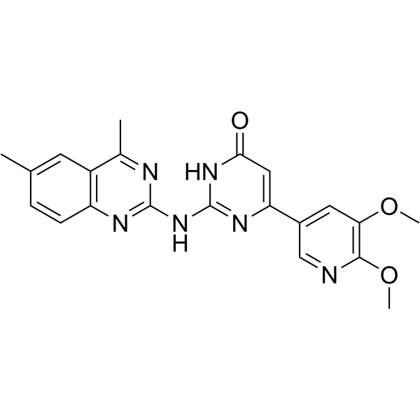
IDD-8E
IDD-8E is an effective anti-pseudomonal agent (MIC =4.4 µM ) with no cytotoxicity. IDD-8E shows significant pseudomonal killing and disruption of pseudomonal biofilm. IDD-8E binds to the ATP-binding pocket of WaaP and also inhibits other ESKAPE pathogens.
For research use only. We do not sell to patients.
- Formula: C21H20N6O3
- Molecular Weight:404.42
-
Storage:
Please store the product under the recommended conditions in the Certificate of Analysis.

To place orders, for customer services and technical support, please contact: MedChemExpress USA
Tel: 609-228-6898 E-mail: [email protected] [email protected]


-
 Biological Activity
-
Chemical Information
- Protocols
- Purity & Documentation
- References
-
 Help & FAQs




Biological Activity
Description
In Vitro
IDD-8E (10-100 µM; 24 h) remains unchanged at higher concentrations compared to untreated controls using RAW cells, showing no cytotoxicity[1].
IDD-8E (2, 4.4 µM; 0-5 h) is observed to significantly reduce the number of Pseudomonas aeruginosaand and is as effective as Rifampicin (HY-B0272). It is also found to have the ability to disrupt biofilm[1].
IDD-8E is synergistic with each of the three antibiotics (Rifampicin (HY-B0272), Lincomycin (HY-117660), Carbenicillin (HY-B0525)) in several combinations. Synergistic combinations effectively reduces the minimum inhibitory concentrations (MICs) of the antibiotics[1].
IDD-8E can inhibit the growth of other ESKAPE pathogens, with MIC values of 6.25 µM, 50 µM, 12.5 µM,12.5 µM and 50 µM against A. baumannii, K. pneumoniae, Methicillin-resistant Staphylococcus aureus (MRSA), Vancomycin-resistant Enterococcus faecalis (VRE) and E. coli, respectively [1].
MedChemExpress (MCE) has not independently confirmed the accuracy of these methods. They are for reference only.
-
Cell Line:RAW cells
-
Concentration:100 µM, 70 µM, 40 µM, and 10 µM
-
Incubation Time:24 h
-
Result:IDD-8E showed no significant cytotoxic effects at the concentrations tested, similar to the negative control, Rifampicin. This indicates that IDD-8E is potentially safe for further development as it does not harm the macrophage cells at effective antibacterial concentrations.


Chemical Information
-
Molecular Weight 404.42
-
Formula C21H20N6O3
-
SMILES
CC(C=C1)=CC2=C1N=C(NC3=NC(C4=CC(OC)=C(OC)N=C4)=CC(N3)=O)N=C2C
-
Shipping
Room temperature in continental US; may vary elsewhere.
-
Storage
Please store the product under the recommended conditions in the Certificate of Analysis.


Purity & Documentation
References

Calculators
Concentration (start) × Volume (start) = Concentration (final) × Volume (final)


IDD-8E Related Classifications
HY-161395 Related Classifications
 Powered by Bioz