



iMAC2
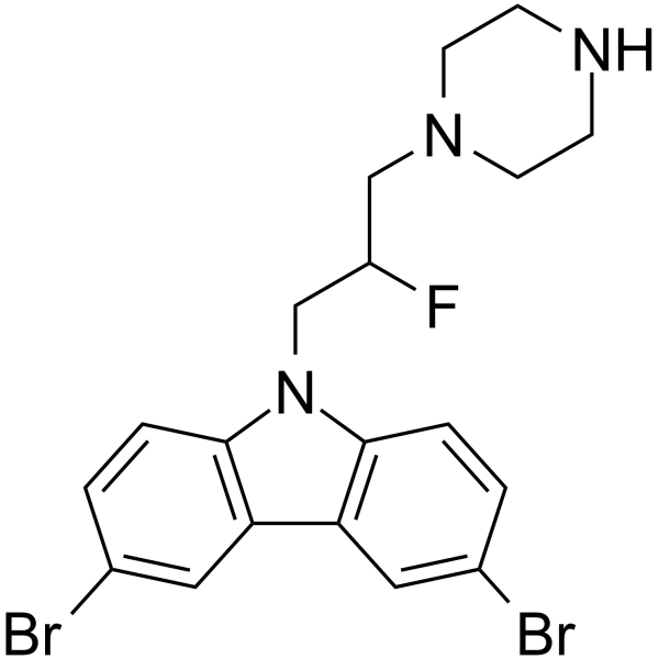
iMAC2 is a potent MAC inhibitor with an IC50 of 28 nM and an LD50 of 15000 nM. iMAC2 shows anti-apoptotic effect. iMAC2 blocks cytochrome c release.
For research use only. We do not sell to patients.
- CAS No.: 335166-36-4
- Formula: C19H20Br2FN3
- Molecular Weight:469.19
-
Storage:
Please store the product under the recommended conditions in the Certificate of Analysis.

To place orders, for customer services and technical support, please contact: MedChemExpress USA
Tel: 609-228-6898 E-mail: [email protected] [email protected]


-
 Biological Activity
-
Chemical Information
- Protocols
- Purity & Documentation
- References
-
 Help & FAQs




Biological Activity
Description
IC50 & Target
IC50: 28 nM (MAC)[1]
Cellular Effect
|
Cell Line

|
Type | Value | Description | References |
|---|---|---|---|---|
| HeLa | IC50 |
0.68 μM
Compound: 13
|
In vitro inhibition of Bax regulated release of cytochrome C was determined using liposome channel assay
In vitro inhibition of Bax regulated release of cytochrome C was determined using liposome channel assay
|
[PMID: 14521400] |
In Vitro
iMAC2 (5 μM; pre-treatment for 1 hour and then treatment for 12 hours) reduces apoptosis in FL5.12 cells[1].
MedChemExpress (MCE) has not independently confirmed the accuracy of these methods. They are for reference only. Further protocols information, click here.
-
Cell Line:FL5.12 cells
-
Concentration:5 μM
-
Incubation Time:Pretreatment 1 h and then treatment with Staurosporine (HY-15141) for 12 hours
-
Result:Reduced apoptosis by more than 50%.


Chemical Information
-
CAS No. 335166-36-4
-
Molecular Weight 469.19
-
Formula C19H20Br2FN3
-
SMILES
FC(CN1CCNCC1)CN2C3=C(C4=C2C=CC(Br)=C4)C=C(Br)C=C3
-
Shipping
Room temperature in continental US; may vary elsewhere.
-
Storage
Please store the product under the recommended conditions in the Certificate of Analysis.

Protocols
-
Apoptosis
Apoptosis, also called programmed cell death, is generally characterized by distinct morphological characteristics.
-
Cell Cytotoxicity Assay
Cytotoxicity assays are usually based on the assessment of cell membrane damage, which can also be indirectly detected by measuring cell viability. Detection methods include MTT assay, CKK-8 assay, LDH assay and ATP assay, etc.
-
Apoptosis Solutions
Apoptosis is a regulated, generally non-lytic cell-death pathway that removes unwanted, damaged, infected, or abnormal cells through coordinated morphological changes, caspase activation, DNA fragmentation, and membrane remodeling. The intrinsic apoptosis pathway is controlled mainly by mitochondrial outer membrane permeabilization, BCL-2 family proteins, cytochrome c release, apoptosome formation, caspase-9 activation, and downstream executioner caspase-3/7 activation. The extrinsic apoptosis pathway is initiated by death receptors such as Fas, TNFR, and TRAIL receptors, which recruit adaptor proteins and activate caspase-8 before engaging executioner caspases or mitochondrial amplification through BID cleavage. Apoptosis is linked to many phenotypes, including cancer cell killing, tissue homeostasis, immune regulation, neurodegeneration, infection response, and treatment-induced cytotoxicity; unresolved questions include how apoptosis interacts with necroptosis, pyroptosis, ferroptos
-
Acute Systemic Toxicity Study
Acute systemic toxicity studies evaluate adverse effects occurring after a single exposure, or repeated exposure within a short acute window, and the main in vivo readouts are mortality, moribund condition, clinical signs, body-weight change, and gross pathological findings; acute oral toxicity methods were developed to replace classical LD50 testing with reduced-animal designs such as fixed-dose procedure, acute toxic class method, and up-and-down procedure. The fixed-dose procedure classifies acute toxicity by administering predefined dose levels and observing evident toxicity rather than using death as the primary endpoint, whereas the acute toxic class method uses sequential groups of three animals per step and the up-and-down procedure doses animals sequentially to estimate an LD50 with fewer animals than conventional LD50 testing.


Purity & Documentation
References

Calculators
Concentration (start) × Volume (start) = Concentration (final) × Volume (final)


iMAC2 Related Classifications
HY-103272 Related Classifications
 Powered by Bioz