



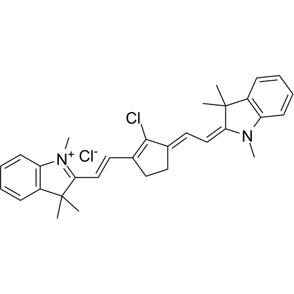
IR-775 chloride

Based on 1 Customer Validation
IR-775 chloride is a cyanine dye with near-infrared absorption (Ex/Em = 775/795 nm). IR-775 chloride acts as a photosensitizer or photothermal agent, and induces apoptosis via generating ROS or exerting photothermal effects upon irradiation; its combination with 2-methoxyestradiol (HY-12033) inhibits SOD2 to enhance photodynamic therapy (PDT) efficacy, and its liposomal encapsulation enables phytotherapy-photothermal therapy (PTT) synergy. IR-775 chloride is applicable for investigating PDT/PTT and near-infrared fluorescence imaging in ovarian cancer and breast cancer.
For research use only. We do not sell to patients.
- CAS No.: 199444-11-6
- Formula: C32H36Cl2N2
- Molecular Weight:519.55
-
Storage:
Store at room temperature, keep dry and cool.
In solvent -80°C, 1 year , -20°C, 6 months


To place orders, for customer services and technical support, please contact: MedChemExpress USA
Tel: 609-228-6898 E-mail: [email protected] [email protected]


-
 Biological Activity
-
Chemical Information
- Protocols
- Purity & Documentation
- References
-
 Help & FAQs


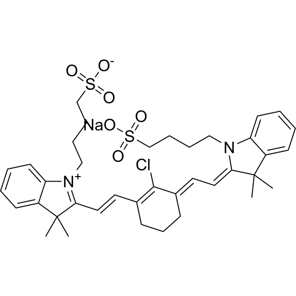
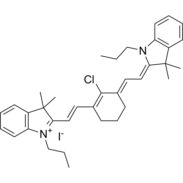
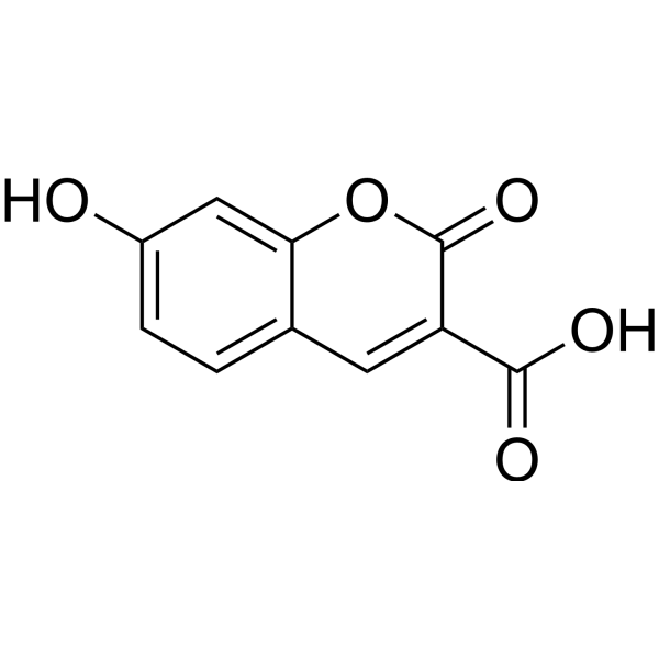
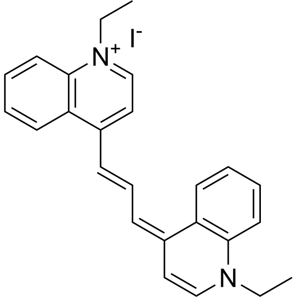


Biological Activity
Description
In Vitro
IR-775 (2-10 μM; 24-72 h) chloride is a potent photosensitizer in proliferative diabetic retinopathy (PDR). It reduces the viability of human ovarian adenocarcinoma cell line (SKOV-3) and human breast adenocarcinoma cell line (MDA-MB-231) in a time-dependent manner, and exhibits stronger cytotoxicity when combined with 2-methoxyestradiol plus irradiation[1].
IR-775 (6 μM; 2-24 h) chloride accumulates in non-nuclear organelles of human ovarian adenocarcinoma cell line (SKOV-3) and human breast adenocarcinoma cell line (MDA MB-231), and an increase in uptake is observed during the 2 h to 24 h incubation period[1].
IR-775 (6 μM; 24-72 h) chloride, when combined with 2-methoxyestradiol in a photodynamic reaction, induces oxidative stress and endoplasmic reticulum-mediated apoptosis in human ovarian adenocarcinoma cell line (SKOV-3) and human breast adenocarcinoma cell line (MDA MB-231), which is evidenced by altered SOD2 expression and increased caspase-12 expression after irradiation[1].
IR-775 (1 mg/mL) chloride can be efficiently encapsulated into spherical Hyptis suaveolens-IR-775-Liposome (HIL) NPs (141 nm), with an encapsulation efficiency of 69.13% and a drug loading rate of 17.29%. These nanoparticles exhibit characteristic near-infrared absorption/emission properties and degrade upon near-infrared laser irradiation[2].
The system of HIL nanoparticles loaded with IR-775 chloride exhibits high intracellular uptake efficiency in 4T1 cells. After irradiation with an 808 nm NIR laser, it induces significant reactive oxygen species (ROS) production, and causes over 85% of 4T1 cell death through a synergistic photothermal-photodynamic effect (CI = 0.6)[2].
HIL-loaded nanoparticles with IR-775 chloride induce > 85% cell death in 4T1 three-dimensional spheroids under 808 nm NIR laser irradiation, and can deeply penetrate into the tumor core[2].
HIL NPs loaded with IR-775 chloride disrupt the mitochondrial membrane potential of 4T1 cells, accumulate in lysosomes, and upregulate γ-H2AX, Cathepsin B, and p53 under 808 nm NIR laser irradiation, thereby inducing DNA damage and apoptosis[2].
MedChemExpress (MCE) has not independently confirmed the accuracy of these methods. They are for reference only. Further protocols information, click here.
-
Cell Line:human ovarian adenocarcinoma (SKOV-3), human breast adenocarcinoma (MDA MB-231)
-
Concentration:2 μM; 4 μM; 6 μM; 8 μM; 10 μM
-
Incubation Time:24 h (pre-incubation); 24 h (post-irradiation incubation); 72 h (post-irradiation incubation)
-
Result:Reduced viability in both cell lines when treated with 2-10 μM for 24 h.
Identified 6 μM as the non-toxic dose.
Reduced MDA MB-231 viability to ~80% and SKOV-3 viability to ~88% after 24 h post-irradiation incubation when used alone in photodynamic reaction (PDR).
Reduced MDA MB-231 viability to 53% after 24 h and 33% after 72 h post-irradiation incubation, and SKOV-3 viability to 60% after 24 h and 45% after 72 h post-irradiation incubation when combined with 2-methoxyestradiol and irradiation.
Showed minimal cytotoxicity alone without irradiation.
-
Cell Line:human ovarian adenocarcinoma (SKOV-3), human breast adenocarcinoma (MDA MB-231)
-
Concentration:6 μM
-
Incubation Time:24 h (pre-incubation); 24 h (post-irradiation incubation); 72 h (post-irradiation incubation)
-
Result:Reduced SOD2 positive staining to 30% (weak intensity) at 24 h post-irradiation and 50% (weak intensity) at 72 h post-irradiation, while increasing caspase-12 positive staining to 80% (strong intensity) at both 24 h and 72 h post-irradiation in SKOV-3 cells.
Reduced SOD2 positive staining to 25% (moderate intensity) at 24 h post-irradiation, which returned to 100% (weak intensity) at 72 h post-irradiation, while increasing caspase-12 positive staining to 25% (weak-moderate intensity) at 24 h post-irradiation and 100% (strong intensity) at 72 h post-irradiation in MDA MB-231 cells.
Showed minimal caspase-12 staining and full SOD2 staining in unirradiated controls of both cell lines.
In Vivo
MedChemExpress (MCE) has not independently confirmed the accuracy of these methods. They are for reference only.


Chemical Information
-
CAS No. 199444-11-6
-
Appearance Solid
-
Molecular Weight 519.55
-
Formula C32H36Cl2N2
-
SMILES
CC1(C)C(/C=C/C2=C(Cl)/C(CCC2)=C/C=C3N(C)C4=C(C=CC=C4)C/3(C)C)=[N+](C)C5=C1C=CC=C5.[Cl-]
-
Synonyms
IR-775 chloride
-
Shipping
Room temperature in continental US; may vary elsewhere.
-
Storage
Store at room temperature, keep dry and cool
In solvent -80°C 1 year -20°C 6 months

Protocols
-
Apoptosis
Apoptosis, also called programmed cell death, is generally characterized by distinct morphological characteristics.
-
TUNEL staining for apoptotic DNA fragmentation
TUNEL staining detects DNA strand breaks by using terminal deoxynucleotidyl transferase to add labeled nucleotides to exposed 3′-OH DNA termini, generating either microscopic staining in fixed cells or tissue sections, or fluorescence/cytometric signal in cell suspensions. TUNEL positivity reflects DNA fragmentation but should not be interpreted alone as definitive apoptosis, because TUNEL can also label necrotic, autolytic, mechanically damaged, or DNA-repair-associated DNA breaks.
-
Annexin V plus membrane-impermeant dye apoptosis staining
Annexin V-based apoptosis assays rely on the detection of phosphatidylserine (PS) externalization from the inner leaflet of the plasma membrane to the outer leaflet, an early biochemical hallmark of apoptosis. Fluorescently labeled Annexin V binds PS in a calcium-dependent manner, enabling identification of early apoptotic cells by flow cytometry or fluorescence microscopy. When combined with a membrane-impermeant DNA-binding dye (e. g. , propidium iodide), this approach allows discrimination between viable (Annexin V−/dye−), early apoptotic (Annexin V+/dye−), and late apoptotic or necrotic (Annexin V+/dye+) cell populations by assessing membrane integrity and PS exposure.
-
ROS/oxidative-stress fluorescent staining
ROS/oxidative-stress fluorescent staining uses cell-permeant fluorogenic probes that become fluorescent after oxidation inside cells or tissues; commonly used examples include DCFH-DA/DCFDA for broad cellular oxidant detection, DHE for superoxide-related signal detection, MitoSOX for mitochondrial superoxide-related signal detection, and CellROX probes for oxidative-stress-associated fluorescence readouts. The assay detects probe oxidation rather than a single ROS species unless the probe and analysis method have been chemically validated for that species. DCFH-DA enters cells, is deacetylated by intracellular esterases to DCFH, and produces fluorescent DCF after oxidation, so the readout is used as an operational measure of total cellular oxidative stress rather than a species-specific ROS measurement. DHE and MitoSOX can report superoxide-related oxidation, but red fluorescence alone can include non-specific ethidium-like oxidation products; HPLC or optimized spectral approaches are
-
Apoptosis Solutions
Apoptosis is a regulated, generally non-lytic cell-death pathway that removes unwanted, damaged, infected, or abnormal cells through coordinated morphological changes, caspase activation, DNA fragmentation, and membrane remodeling. The intrinsic apoptosis pathway is controlled mainly by mitochondrial outer membrane permeabilization, BCL-2 family proteins, cytochrome c release, apoptosome formation, caspase-9 activation, and downstream executioner caspase-3/7 activation. The extrinsic apoptosis pathway is initiated by death receptors such as Fas, TNFR, and TRAIL receptors, which recruit adaptor proteins and activate caspase-8 before engaging executioner caspases or mitochondrial amplification through BID cleavage. Apoptosis is linked to many phenotypes, including cancer cell killing, tissue homeostasis, immune regulation, neurodegeneration, infection response, and treatment-induced cytotoxicity; unresolved questions include how apoptosis interacts with necroptosis, pyroptosis, ferroptos
-
Bioluminescent/Fluorescent Imaging Xenograft
Bioluminescent and fluorescent imaging xenograft models use tumor cells engineered to express optical reporters so tumor engraftment, growth, dissemination, and treatment response can be monitored longitudinally in living animals and validated ex vivo. Bioluminescence imaging usually measures luciferase activity after substrate administration and is commonly used as a surrogate for viable reporter-expressing tumor burden, while fluorescence imaging measures reporter or probe emission and can support tumor localization, ex vivo confirmation, or complementary multimodal analysis.
-
Breast Cancer Modeling
Breast cancer is a heterogeneous cancer, and it has been distinguished into four subtypes: luminal A, luminal B, HER2-positive and basal-like. Molecular mutations, epigenetic alterations, hormone exposure and immune microenvironment are related to the progression of breast cancer.


Purity & Documentation
-
Data Sheet (288 KB)
-
SDS (393 KB)
- English - EN (393 KB)
- Français - FR (393 KB)
- Deutsch - DE (393 KB)
- Norwegian - NO (393 KB)
- Español - ES (393 KB)
- Swedish - SV (393 KB)
- Italian - IT (393 KB)
- Korean - KR (393 KB)
- Portuguese - PT (393 KB)
-
Handling Instructions (2659 KB)
References
[1]. Waszkiewicz M, et al. The photodynamic reaction with IR-775 cyanine combined with 2-methoxyestradiol in ovarian (SKOV-3) and human breast adenocarcinoma (MDA MB-231) cell lines. Photodiagnosis and photodynamic therapy. 2022 Jun;38:102766. [Content Brief]
[2]. Pebam M, et al. IR-775 - Hyptis loaded bioactive nanoparticles for enhanced phyto-photothermal therapy of breast cancer cells. Photodiagnosis and photodynamic therapy. 2023 Dec;44:103872. [Content Brief]

Calculators
Concentration (start) × Volume (start) = Concentration (final) × Volume (final)


IR-775 chloride Related Classifications
HY-W248590 Related Classifications
 Powered by Bioz
Keywords
- IR-775
- 199444-11-6
- IR775
- IR 775
- Fluorescent Dye
- Photosensitizer
- Apoptosis
- Reactive Oxygen Species (ROS)
- SOD
- oxidative stress
- human breast adenocarcinoma (MDA MB-231) cell lines
- human breast adenocarcinoma
- apoptosis
- ovarian carcinoma
- triple-negative breast cancer
- 4T1 cells
- 4T1 3D spheroids
- hyperthermia
- human ovarian adenocarcinoma (SKOV-3) cell lines
- Inhibitor
- inhibitor
- inhibit