



113-N16B

Based on 1 Customer Validation
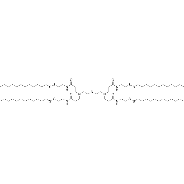
113-N16B is an ionizable cationic lipid that can be used for the synthesis of lipid nanoparticles. 113-N16B delivers mRNA to pulmonary endothelial cells. 113-N16B can be formulated as a component into hybrid lipid nanoparticles for lung-specific delivery of mRNA (including MFSD7C mRNA and luciferase mRNA) in mice. 113-N16B is applicable to research related to lymphangioleiomyomatosis, sickle cell disease, and hemolysis-induced lung injury.
For research use only. We do not sell to patients.
- Purity : 98.0%
- Formula: C73H147N7O4S8
- Molecular Weight:1443.51
-
Storage:Pure form -20°C, 3 years ; In solvent -80°C, 6 months , -20°C, 1 month

To place orders, for customer services and technical support, please contact: MedChemExpress USA
Tel: 609-228-6898 E-mail: [email protected] [email protected]


-
 Biological Activity
-
Chemical Information
- Protocols
- Purity & Documentation
- References
-
 Help & FAQs




Biological Activity
Description
In Vitro
113-N16B (50 molar ratio; overnight) enables the formulation of hybrid LNPs when combined with cholesterol, phospholipids, and DMG-PEG2000 at a molar ratio of 50:38.5:10:1.5[2].
MedChemExpress (MCE) has not independently confirmed the accuracy of these methods. They are for reference only.
In Vivo
113-N16B (LNP formulation; 0.5 mg/kg mRNA; intravenous injection; single administration) specifically delivers fLuc mRNA to the lungs of healthy Balb/c mice[1].
113-N16B formulated lipid nanoparticles (0.5 mg/kg; i.v.) enable lung-specific mRNA delivery, while 113-N16B lipid nanoparticles loaded with MFSD7C mRNA (0.5 mg/kg; i.v.; once weekly for 4 weeks) upregulate MFSD7C expression in mouse lung tissues[2].
MedChemExpress (MCE) has not independently confirmed the accuracy of these methods. They are for reference only.
-
Animal Model:Ai14 Cre reporter mice (transgenic)[1]
-
Dosage:0.75 mg mRNA equiv./kg
-
Administration:i.v.; single dose
-
Result:Achieved tdTomato expression specifically localized to the lungs.
Transfected 69.6% of pulmonary endothelial cells, 7.3% of pulmonary epithelial cells, and 18.9% of pulmonary macrophages.
-
Animal Model:Balb/c mice (female, 6 to 8 weeks old)[1]
-
Dosage:0.5 mg/kg mRNA
-
Administration:i.v.; single dose
-
Result:Detected bioluminescence signals (luciferase expression) primarily in the lungs, confirming lung-targeted mRNA delivery.
-
Animal Model:HbSS-Townes, HbAA-Townes, 7Cflox/flox, 7C-/- (12-month-old HbSS-Townes/HbAA-Townes; 8-week-old 7Cflox/flox/7C-/-; PHZ-challenged 7C-/- for hemolytic lung injury model)[2]
-
Dosage:0.5 mg/kg (luciferase mRNA-loaded LNPs; single dose); 0.5 mg/kg MFSD7C mRNA per dose (MFSD7C mRNA-loaded LNPs; weekly for 4 weeks)
-
Administration:i.v. (single dose for luciferase mRNA-loaded LNPs); i.v.; weekly; 4 weeks (MFSD7C mRNA-loaded LNPs)
-
Result:Confirmed lung-specific delivery of mRNA via increased bioluminescence intensity in the lungs of mice given luciferase mRNA-loaded 113-N16B lipid nanoparticles.
Elevated MFSD7C protein expression in mouse lung tissue following administration of MFSD7C mRNA-loaded 113-N16B lipid nanoparticles.


Chemical Information
-
Appearance Liquid
-
Molecular Weight 1443.51
-
Formula C73H147N7O4S8
-
Color Colorless to light yellow
-
SMILES
O=C(CCN(CCC(NCCSSCCCCCCCCCCCC)=O)CCN(C)CCN(CCC(NCCSSCCCCCCCCCCCC)=O)CCC(NCCSSCCCCCCCCCCCC)=O)NCCSSCCCCCCCCCCCC
-
Shipping
Room temperature in continental US; may vary elsewhere.
-
Storage
Pure form -20°C 3 years In solvent -80°C 6 months -20°C 1 month


Purity & Documentation
-
Data Sheet (272 KB)
-
SDS (251 KB)
- English - EN (251 KB)
- Français - FR (251 KB)
- Deutsch - DE (251 KB)
- Norwegian - NO (251 KB)
- Español - ES (251 KB)
- Swedish - SV (251 KB)
- Italian - IT (251 KB)
- Korean - KR (251 KB)
- Portuguese - PT (251 KB)
-
Handling Instructions (2659 KB)
References
[1]. Qiu M, et al. Lung-selective mRNA delivery of synthetic lipid nanoparticles for the treatment of pulmonary lymphangioleiomyomatosis. Proceedings of the National Academy of Sciences of the United States of America. 2022 Feb 22;119(8):e2116271119. [Content Brief]
[2]. Wang H, et al. MFSD7C protects hemolysis-induced lung impairments by inhibiting ferroptosis. Nature communications. 2024 Sep 19;15(1):8226. [Content Brief]

Calculators
Concentration (start) × Volume (start) = Concentration (final) × Volume (final)


113-N16B Related Classifications
HY-153737 Related Classifications
 Powered by Bioz