



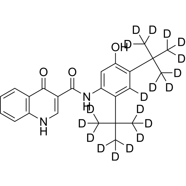
Ivacaftor-d19
Ivacaftor-d9 is a potent CFTR modulator and exhibits an EC50 value of 255 nM for CFTR potentiation in G551D/F508del HBE Cells. Ivacaftor-D9 acts as an orally active and improved deuterated Ivacaftor analog for cystic fibrosis research.
For research use only. We do not sell to patients.
- CAS No.: 1413431-22-7
- Formula: C24H9D19N2O3
- Molecular Weight:411.61
-
Storage:Powder -20°C, 3 years , 4°C, 2 years ; In solvent -80°C, 6 months , -20°C, 1 month

To place orders, for customer services and technical support, please contact: MedChemExpress USA
Tel: 609-228-6898 E-mail: [email protected] [email protected]


-
 Biological Activity
-
Chemical Information
- Protocols
- Purity & Documentation
- References
-
 Help & FAQs


-
HY-13017S
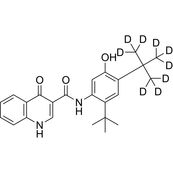
Ivacaftor-d9CFTR Modulator -
HY-17443B
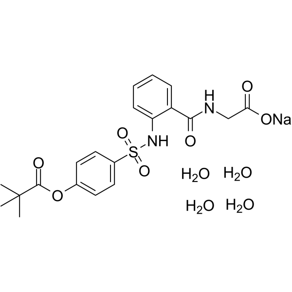
Sivelestat sodium tetrahydrateNeutrophil Elastase Inhibitor -
HY-13017S3
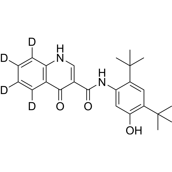
Ivacaftor-d4CFTR Potentiator -
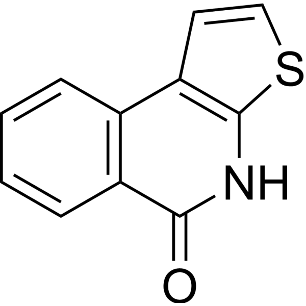
HY-121719
TIQ-ATNKS2 Inhibitor -

HY-17443BR
Sivelestat sodium tetrahydrate (Standard)Neutrophil Elastase Inhibitor -
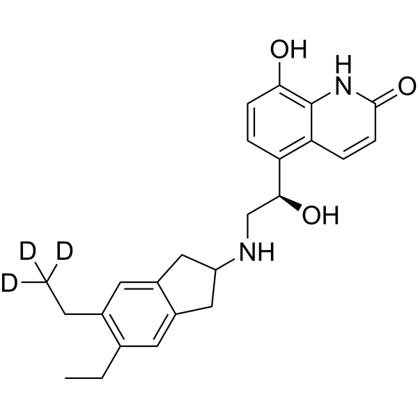
HY-14299S
Indacaterol-d3Stable Isotope -
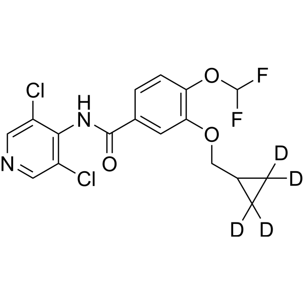
HY-15455S
Roflumilast-d4Stable Isotope -
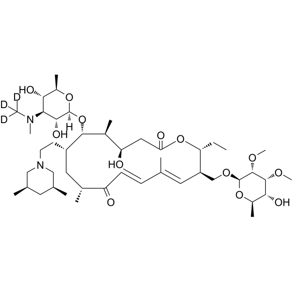
HY-B0905S
Tilmicosin-d3Stable Isotope


Biological Activity
Description
In Vitro
"Stable heavy isotopes of hydrogen, carbon, and other elements have been incorporated into drug molecules, largely as tracers for quantitation during the drug development process. Deuteration has gained attention because of its potential to affect the pharmacokinetic and metabolic profiles of drugs[1].
Potential advantages of deuterated compounds:
(1) Extend the half-life in vivo. Deuterated compounds may be able to prolong the pharmacokinetic characteristics of the compound, that is, prolong the half-life in vivo. This can improve compound safety, efficacy and tolerability, and increase ease of administration.
(2) Improve oral bioavailability. Deuterated compounds may reduce the degree of unwanted metabolism (first-pass metabolism) in the gut wall and liver, allowing a greater proportion of the unmetabolized drug to reach its target site of action. High bioavailability determines its activity at low doses and better tolerance.
(3) Improve metabolic characteristics. Deuterated compounds may reduce the formation of toxic or reactive metabolites and improve drug metabolism.
(4) Improve drug safety. Deuterated compounds may reduce or eliminate adverse side effects of pharmaceutical compounds and are safe.
(5) Preserve the therapeutic properties. Deuterated compounds are expected to retain similar biochemical potency and selectivity to hydrogen analogs in previous studies.
"
MedChemExpress (MCE) has not independently confirmed the accuracy of these methods. They are for reference only. Further protocols information, click here.
Application
1. This compound can be used as a tracer
2. This compound can be used as an internal standard for quantitative analysis by NMR, GC-MS, or LC-MS.


Chemical Information
-
CAS No. 1413431-22-7
-
Unlabeled CAS 873054-44-5
-
Appearance Solid
-
Molecular Weight 411.61
-
Formula C24H9D19N2O3
-
Color White to off-white
-
SMILES
O=C(C1=CNC2=C(C=CC=C2)C1=O)NC3=CC(O)=C(C(C([2H])([2H])[2H])(C([2H])([2H])[2H])C([2H])([2H])[2H])C([2H])=C3C(C([2H])([2H])[2H])(C([2H])([2H])[2H])C([2H])([2H])[2H]
-
Synonyms
VX-770-d19
-
Shipping
Room temperature in continental US; may vary elsewhere.
-
Storage
Powder -20°C 3 years 4°C 2 years In solvent -80°C 6 months -20°C 1 month

Protocols
-
Fibrosis/Collagen Morphometry
Fibrosis and collagen morphometry is based on the quantitative visualization of fibrillar collagen deposition in tissue sections using histochemical stains such as Sirius Red (Picrosirius Red) or Masson's trichrome, followed by image-based or polarization-enhanced analysis to estimate collagen proportional area as a surrogate of extracellular matrix accumulation during fibrotic remodeling. Sirius Red combined with polarized light microscopy enhances detection of collagen fibers due to birefringence properties, enabling more specific visualization of collagen type I and III fibrils compared to conventional bright-field histology, while whole-section or region-restricted digital morphometry reduces field-selection bias in fibrosis assessment. Alternative quantitative approaches include second harmonic generation (SHG) and two-photon excited fluorescence microscopy, which enable label-free detection of fibrillar collagen and have been validated against histological staining and biochemica
-
Collagen: Sirius Red Staining
Sirius Red or picrosirius red staining is a histochemical method for visualizing collagen-rich extracellular matrix in tissue sections, and collagen fibers are detected as red-stained structures under bright-field microscopy with enhanced birefringence under polarized light. Picrosirius red is useful for assessing total collagen organization, distribution, and fibrosis burden, but polarized color should not be interpreted as a definitive collagen type I versus type III readout because color is affected by fiber orientation, thickness, and packing.
-
Connective Tissue: Masson's Trichrome/Collagen Trichrome Staining
Masson’s Trichrome (collagen/trichrome staining) is a histological technique that differentially stains tissue compartments using sequential acidic dyes to distinguish collagen from muscle and cytoplasmic components based on dye affinity and tissue permeability differences, enabling visualization of fibrosis and connective tissue architecture in histological sections. The classical formulation typically uses Weigert's iron hematoxylin for nuclear staining, Biebrich scarlet-acid fuchsin for cytoplasm and muscle, and aniline blue (or light green variants) for collagen, producing a characteristic blue/green collagen signal contrasted against red cytoplasm and dark nuclei. The staining principle relies on selective displacement of smaller dye molecules by larger anionic dyes in collagen-rich regions under controlled acidified conditions, which enhances collagen-specific dye retention. This property makes the method widely used for fibrosis assessment in organs such as heart, liver, lung, a


Purity & Documentation
-
Data Sheet (278 KB)
-
SDS (393 KB)
- English - EN (393 KB)
- Français - FR (393 KB)
- Deutsch - DE (393 KB)
- Norwegian - NO (393 KB)
- Español - ES (393 KB)
- Swedish - SV (393 KB)
- Italian - IT (393 KB)
- Korean - KR (393 KB)
- Portuguese - PT (393 KB)
-
Handling Instructions (2659 KB)
References
[1]. Russak EM, et al. Impact of Deuterium Substitution on the Pharmacokinetics of Pharmaceuticals. Ann Pharmacother. 2019;53(2):211-216. [Content Brief]
[2]. Delaunay JL, et al. Functional defect of variants in the adenosine triphosphate-binding sites of ABCB4 and their rescue by the cystic fibrosis transmembrane conductance regulator potentiator, ivacaftor (VX-770). Hepatology. 2017 Feb;65(2):560-570 [Content Brief]
[3]. Mutyam V, et al. Therapeutic benefit observed with the CFTR potentiator, ivacaftor, in a CF patient homozygous for the W1282X CFTR nonsense mutation. J Cyst Fibros. 2017 Jan;16(1):24-29 [Content Brief]
[4]. Hadida S, et al. Discovery of N-(2,4-di-tert-butyl-5-hydroxyphenyl)-4-oxo-1,4-dihydroquinoline-3-carboxamide (VX-770, ivacaftor), a potent and orally bioavailable CFTR potentiator. J Med Chem. 2014 Dec 11;57(23):9776-9 [Content Brief]
[5]. Van Goor F, et al. Rescue of CF airway epithelial cell function in vitro by a CFTR potentiator, VX-770. Proc Natl Acad Sci U S A. 2009 Nov 3;106(44):18825-30. [Content Brief]

Calculators
Concentration (start) × Volume (start) = Concentration (final) × Volume (final)


Ivacaftor-d19 Related Classifications
HY-13017S1 Related Classifications
 Powered by Bioz