



ボスカリド
Based on 1 publication(s) in Google Scholar
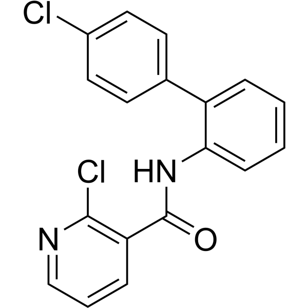
Boscalid is a succinate dehydrogenase (SDHI) inhibitor with antifungal activity. Boscalid binds to the ubiquinone-binding site of fungal mitochondrial complex II, blocks ATP production and aerobic respiration, exhibits good control efficacy against a variety of plant fungal diseases including gray mold, sclerotinia rot and powdery mildew, and is widely used for disease control in agriculture. Boscalid induces apoptosis, altered lipid metabolism, mitochondrial dysfunction, respiratory impairment, oxidative stress, ROS accumulation and neurodevelopmental disorders in zebrafish. Boscalid reduces foraging ability, shortens median death time and causes chronic toxicity in exposed honeybees. Boscalid also possesses genotoxicity, cytotoxicity, elevated mitochondrial superoxide levels and early-stage apoptosis.
商品は「研究用試薬」です。人や動物の医療用・臨床診断用・食品用の製品ではありません。
研究用途以外に使用した場合、当社は一切の責任を負いかねます。
- 純度 : 99.80%
- CAS 番号: 188425-85-6
- 分子式: C18H12Cl2N2O
- 分子量:343.21
-
保管条件:Powder -20°C, 3 years , 4°C, 2 years ; In solvent -80°C, 6 months , -20°C, 1 month

To place orders, for customer services and technical support, please contact: MedChemExpress USA
Tel: 609-228-6898 E-mail: [email protected] [email protected]


-
 生物活性
生物活性
-
化学情報
-
溶剤 & 溶解度
- プロトコル
- 純度とドキュメンテーション
- 参考文献
-
 Help & FAQs


-
HY-B0849
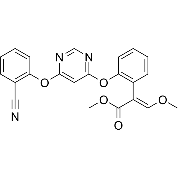
AzoxystrobinFungicide -
HY-100410
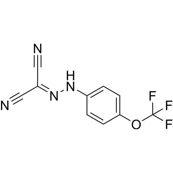
FCCPOXPHOS Uncoupler -
HY-135549
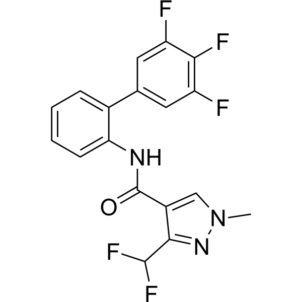
FluxapyroxadAntibiotic -
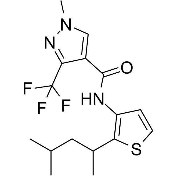
HY-17520
PenthiopyradFungicide -

HY-119976R
Boscalid (Standard)Anti-Fungal Agent -
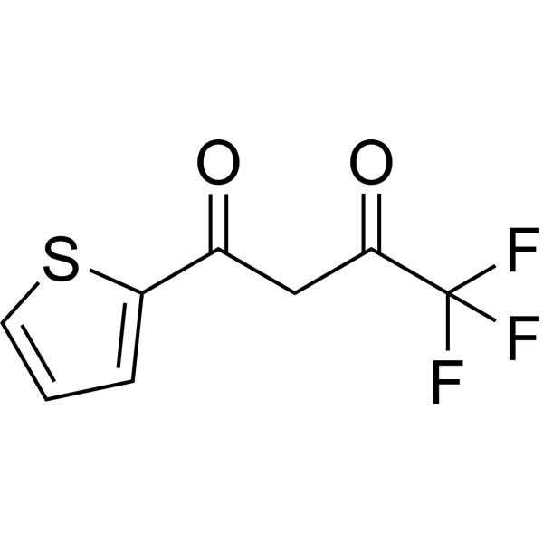
HY-D0190
2-ThenoyltrifluoroacetoneChelating Agent, Mitochondrial Electron Transport Chain Inhibitor, Carboxylesterase Inhibitor -
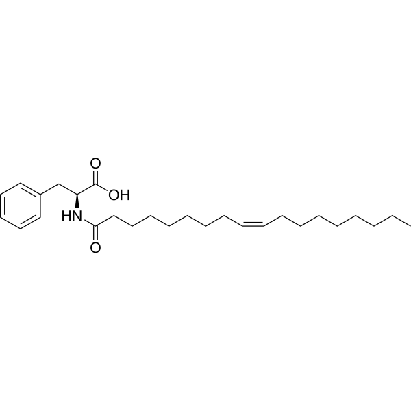
HY-138207
N-Oleoyl-L-phenylalanineMitochondrial Uncoupler -
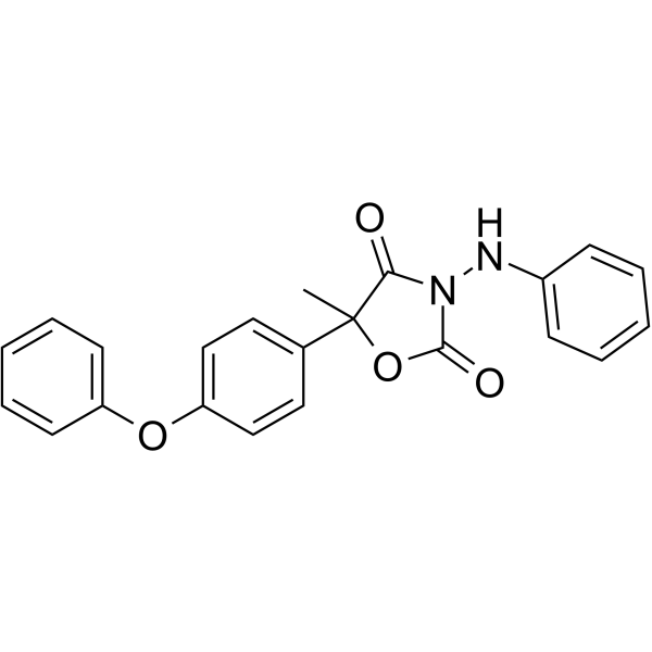
HY-B2008
FamoxadoneFungicide Active -

HY-100410R
FCCP (Standard)OXPHOS Uncoupler -

HY-N12223
Piericidin BAntibiotic -

HY-B2008R
Famoxadone (Standard)Anti-Fungal Agent -

HY-D0190R
2-Thenoyltrifluoroacetone (Standard)Chelating Agent, Mitochondrial Electron Transport Chain Inhibitor, Carboxylesterase Inhibitor
MedChemExpress(MCE)の使用を引用している文献 Boscalid
More

生物活性
製品説明
IC50 & Target
succinate dehydrogenase (SDH)[1]
体内実験
MedChemExpress (MCE) has not independently confirmed the accuracy of these methods. They are for reference only.
-
Animal Model:Tg(elavl3: EGFP) transgenic line, AB strain[2]
-
Dosage:5 mg/L; 15 mg/L; 25 mg/L
-
Administration:waterborne; 6 to 48 hpf (embryos); 24 h (6 dpf larvae)
-
Result:Observed obscure brain regional division, blurred ventricles, malformed eyes, and decreased spontaneous tail coiling frequency at 24 hpf in embryos. Detected enlarged cerebral ventricles, reduced pigmentation, decreased neonatal neurons, defective development around ventricles, increased brain intercellular space, and concentrated nuclei at 48 hpf in embryos. Measured increased Na⁺K⁺-ATPase, Ca²⁺Mg²⁺-ATPase, and total ATPase activities; increased MDA content; accumulated ROS in head and heart; increased CAT activity; decreased SOD activity; decreased tyrosinase activity in embryos. Recorded decreased expression of tfap2a, tfap2e, sox10, gfap, gap43; increased then decreased expression of wnt1, mitfa, pax3a; increased expression of sliva, oca2, nestin, neurogenin1 in embryos. Found increased central area motion proportion, increased angular velocity and sinuosity; decreased total distance, average speed, movement duration, mobile/high mobile frequency and duration in larvae.
- Detection of Apoptosis
- TUNEL staining for apoptotic DNA fragmentation
- Cell Cytotoxicity Assay
- Mammalian live/dead viability and cytotoxicity staining
- Annexin V plus membrane-impermeant dye apoptosis staining
- ROS/oxidative-stress fluorescent staining
- Mitochondrial membrane-potential and mitochondrial mass staining
- Subchronic/Chronic Toxicity Study
- Apoptosis Solutions
- Genotoxicity/Mutagenicity Study
- Cell Viability Determination by MTT Colorimetric Assay


化学情報
-
CAS 番号 188425-85-6
-
性状 Solid
-
分子量 343.21
-
分子式 C18H12Cl2N2O
-
Color White to off-white
-
SMILES
ClC1=CC=C(C=C1)C2=CC=CC=C2NC(C3=C(N=CC=C3)Cl)=O
-
別名
Boscalid
-
輸送条件
Room temperature in continental US; may vary elsewhere.
-
保管条件
Powder -20°C 3 years 4°C 2 years In solvent -80°C 6 months -20°C 1 month


Publications (1)
-
Journal Impact Factor
-
Most Recent
-
J Agric Food Chem
Transcription Repressor SsGATA2 Regulates Broad-Spectrum Resistance to Fungicides and Pathogenicity in Sclerotinia sclerotiorum. [Abstract]2025 Jul 9;73(27):16787-16803. PMID: 40558024


溶剤 & 溶解度
体外:
DMSO : 100 mg/mL (291.37 mM; Need ultrasonic; Hygroscopic DMSO has a significant impact on the solubility of product, please use newly opened DMSO)
Please refer to the solubility information to select the appropriate solvent. Once prepared, please aliquot and store the solution to prevent product inactivation from repeated freeze-thaw cycles.
Storage method and period of stock solution: -80°C, 6 months; -20°C, 1 month. When stored at -80°C, please use it within 6 months. When stored at -20°C, please use it within 1 month.
Please refer to the solubility information to select the appropriate solvent. Once prepared, please aliquot and store the solution to prevent product inactivation from repeated freeze-thaw cycles.
Storage method and period of stock solution: -80°C, 6 months; -20°C, 1 month. When stored at -80°C, please use it within 6 months. When stored at -20°C, please use it within 1 month.
濃度 (開始) × 体積 (開始) = 濃度 (終了) × 体積 (終了)
体内:
Select the appropriate dissolution method based on your experimental animal and administration route.
- For the following dissolution methods, please ensure to first prepare a clear stock solution using an In Vitro approach and then sequentially add co-solvents:
- To ensure reliable experimental results, the clarified stock solution can be appropriately stored based on storage conditions. As for the working solution for In Vivo experiments, it is recommended to prepare freshly and use it on the same day.
- The percentages shown for the solvents indicate their volumetric ratio in the final prepared solution. If precipitation or phase separation occurs during preparation, heat and/or sonication can be used to aid dissolution.
Add each solvent one by one: 10% DMSO 40% PEG300 5% Tween-80 45% Saline
Solubility: ≥ 2.5 mg/mL (7.28 mM); Clear solution
This protocol yields a clear solution of ≥ 2.5 mg/mL (saturation unknown).
Taking 1 mL working solution as an example, add 100 μL DMSO stock solution (25.0 mg/mL) to 400 μL PEG300, and mix evenly; then add 50 μL Tween-80 and mix evenly; then add 450 μL Saline to adjust the volume to 1 mL.
Preparation of Saline: Dissolve 0.9 g sodium chloride in ddH₂O and dilute to 100 mL to obtain a clear Saline solution.
In Vivo Dissolution Calculator
Please enter the basic information of animal experiments:
-
-
-
-
Recommended: Prepare an additional quantity of animals to account for potential losses during experiments.
Please enter your animal formula composition:
-
%DMSO +
Recommended: Keep the proportion of DMSO in working solution below 2% if your animal is weak.
-
%+
-
+%Tween-80 + +
-
%Saline +
The co-solvents required include: DMSO, . All of co-solvents are available by MedChemExpress (MCE). , Tween 80. All of co-solvents are available by MedChemExpress (MCE).
Working solution concentration: 0.22 mg/mL
Method for preparing stock solution: mg drug dissolved in μL DMSO. Stock solution concentration: mg/mL.
1. Take μL DMSO stock solution;
2. Add μL .
μL , mix evenly;
3. Then add μL Tween 80, mix evenly;
4. Then add μL
Please ensure that the stock solution in the first step is dissolved to a clear state, and add co-solvents in sequence. You can use ultrasonic heating (ultrasonic cleaner, recommended frequency 20-40 kHz), vortexing, etc. to assist dissolution.


純度とドキュメンテーション
-
データシート (280 KB)
-
SDS (458 KB)
- English - EN (458 KB)
- Français - FR (458 KB)
- Deutsch - DE (458 KB)
- Norwegian - NO (458 KB)
- Español - ES (458 KB)
- Swedish - SV (458 KB)
- Italian - IT (458 KB)
- Korean - KR (458 KB)
- Portuguese - PT (458 KB)
-
取扱説明書 (2659 KB)
参考文献
[1]. Moschopoulou G, et al. Development of a Portable Cell-Based Biosensor for the Ultra-Rapid Screening for Boscalid Residues in Lettuce. Biosensors (Basel). 2024;14(6):311. Published 2024 Jun 18. [Content Brief]
[2]. Wang H, et al. Characterization of boscalid-induced oxidative stress and neurodevelopmental toxicity in zebrafish embryos. Chemosphere. 2020;238:124753. [Content Brief]


Complete Stock Solution Preparation Table
Please refer to the solubility information to select the appropriate solvent. Once prepared, please aliquot and store the solution to prevent product inactivation from repeated freeze-thaw cycles.
Storage method and period of stock solution: -80°C, 6 months; -20°C, 1 month. When stored at -80°C, please use it within 6 months. When stored at -20°C, please use it within 1 month.
| Optional Solvent | Concentration Solvent Mass | 1 mg | 5 mg | 10 mg | 25 mg |
|---|---|---|---|---|---|
| DMSO | 1 mM | 2.9137 mL | 14.5683 mL | 29.1367 mL | 72.8417 mL |
| 5 mM | 0.5827 mL | 2.9137 mL | 5.8273 mL | 14.5683 mL | |
| 10 mM | 0.2914 mL | 1.4568 mL | 2.9137 mL | 7.2842 mL | |
| 15 mM | 0.1942 mL | 0.9712 mL | 1.9424 mL | 4.8561 mL | |
| 20 mM | 0.1457 mL | 0.7284 mL | 1.4568 mL | 3.6421 mL | |
| 25 mM | 0.1165 mL | 0.5827 mL | 1.1655 mL | 2.9137 mL | |
| 30 mM | 0.0971 mL | 0.4856 mL | 0.9712 mL | 2.4281 mL | |
| 40 mM | 0.0728 mL | 0.3642 mL | 0.7284 mL | 1.8210 mL | |
| 50 mM | 0.0583 mL | 0.2914 mL | 0.5827 mL | 1.4568 mL | |
| 60 mM | 0.0486 mL | 0.2428 mL | 0.4856 mL | 1.2140 mL | |
| 80 mM | 0.0364 mL | 0.1821 mL | 0.3642 mL | 0.9105 mL | |
| 100 mM | 0.0291 mL | 0.1457 mL | 0.2914 mL | 0.7284 mL |


Boscalid 関連分類
HY-119976 関連分類
 Powered by Bioz
Keywords
- Boscalid
- 188425-85-6
- Environmental Pollutants
- Mitochondrial Metabolism
- Fungal
- Apoptosis
- Succinate Dehydrogenase
- Alternaria alternata
- HepG2 liver cells
- Sclerotinia sclerotiorum
- Caco-2/HT29-MTX/Raji B cells
- Chlorella vulgaris
- honeybees
- fungal mitochondrial complex II
- succinate dehydrogenase inhibitor
- zebrafish
- Blumeriella jaapii
- Inhibitor
- inhibitor
- inhibit