



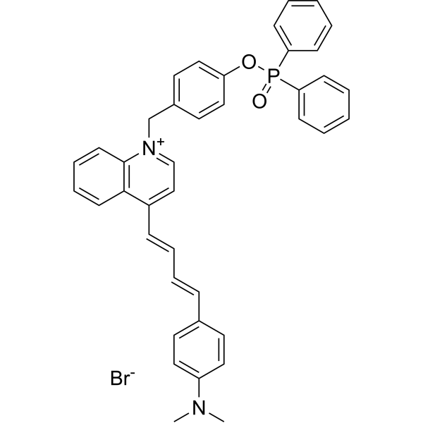
MQA-P
MQA-P is a multifunctional near-infrared (NIR) fluorescent probe for simultaneously detecting ONOO-, viscosity, and polarity within mitochondria. MQA-P exhibits a remarkable turn-on response to ONOO- (λem=645 nm) and is highly sensitive to viscosity/polarity in the NIR channel with λem>704 nm. MQA-P exhibits excited-state intramolecular charge transfer (ESICT) feature that is highly polarity-sensitive by engineering N,N-dimethylamino as the electron donor and a quinoline cationic unit as the electron acceptor. MQA-P is used for ferroptosis or cancer diagnosis in vitro and in vivo via dual-channel images.
商品は「研究用試薬」です。人や動物の医療用・臨床診断用・食品用の製品ではありません。
研究用途以外に使用した場合、当社は一切の責任を負いかねます。
- 分子式: C40H36BrN2O2P
- 分子量:687.60
-
保管条件:
Please store the product under the recommended conditions in the Certificate of Analysis.

To place orders, for customer services and technical support, please contact: MedChemExpress USA
Tel: 609-228-6898 E-mail: [email protected] [email protected]


-
 生物活性
-
化学情報
- プロトコル
- 純度とドキュメンテーション
- 参考文献
-
 Help & FAQs




生物活性
製品説明
体外実験
Guide (Following is our recommended protocol. This protocol only provides a guideline, and should be modified according to your specific needs).
1. MQA-P is dissolved in dimethyl sulfoxide (DMSO) to prepare a stock solution (1.0 mM).
2. For imaging of ONOO- in live cells:
HeLa cells are incubated with MQA-P (5 μM) for 30 min as control; pretreated with SIN-1 (HY-126849; 100 μM) for 30 min and then incubated with MQA-P (5 μM) for another 30 min. The fluorescence images are obtained on a confocal laser scanning microscope with a green channel (λex = 405nm, λem = 550-670 nm).
3. For imaging of viscosity in live cells:
HeLa cells were incubated with MQA-P (5 μM) for 30 min as control; pretreated with Monensin (HY-N4302; 10 μM) for 30 min and then incubated with MQA-P (5 μM) for another 30min. The fluorescence images are obtained on a confocal laser scanning microscope with a red channel (λex = 561 nm, λem = 680-750 nm).
4. For dual-channel imaging of ONOO-, viscosity and polarity during ferroptosis:
HeLa cells are incubated with MQA-P (5 μM) for 30 min as control; pretreated with Erastin (HY-15763; 50 μM) for 30 min and then incubated with MQA-P (5 μM) for another 30 min. The fluorescence images are obtained on a confocal laser scanning microscope with a green channel (λex = 405nm, λem = 550-670 nm) for ONOO- and a red channel (λex = 561 nm, λem = 680-750 nm) for viscosity and polarity[1].
MedChemExpress (MCE) has not independently confirmed the accuracy of these methods. They are for reference only.
体内実験
1. For tissue slices imaging, the normal organs (including heart, liver, spleen, lung, and kidney) and tumor are isolated from the mice, then sectioned as 5 μm thicknesses, respectively.
2. These slices are incubated with MQA-P (20 μM) for 30 min, then washed with PBS (pH 7.4) three times, and finally subjected to in vivo imaging using a confocal laser scanning microscope with a green channel (λex=405nm, λem=550-670 nm) for ONOO- and a red channel(λex=561 nm, λem=680-750 nm) for viscosity and polarity, respectively[1].
MedChemExpress (MCE) has not independently confirmed the accuracy of these methods. They are for reference only.


化学情報
-
分子量 687.60
-
分子式 C40H36BrN2O2P
-
SMILES
CN(C1=CC=C(C=C1)/C=C/C=C/C2=CC=[N+](C3=C2C=CC=C3)CC4=CC=C(C=C4)OP(C5=CC=CC=C5)(C6=CC=CC=C6)=O)C.[Br-]
-
輸送条件
Room temperature in continental US; may vary elsewhere.
-
保管条件
Please store the product under the recommended conditions in the Certificate of Analysis.


純度とドキュメンテーション
参考文献

Calculators
濃度 (開始) × 体積 (開始) = 濃度 (終了) × 体積 (終了)


MQA-P 関連分類
HY-149203 関連分類
 Powered by Bioz