



Retifanlimab

Based on 1 Customer Validation
Retifanlimab is an anti-programmed cell death protein 1 (anti-PD-1) monoclonal antibody (mAb). Retifanlimab can be used for the research of gastroesophageal adenocarcinoma (GEA).
商品は「研究用試薬」です。人や動物の医療用・臨床診断用・食品用の製品ではありません。
研究用途以外に使用した場合、当社は一切の責任を負いかねます。
- 純度 : 96.84%
- CAS 番号: 2079108-44-2
- 分子量:145.35 kDa
-
保管条件:
Please store the product under the recommended conditions in the Certificate of Analysis.


To place orders, for customer services and technical support, please contact: MedChemExpress USA
Tel: 609-228-6898 E-mail: [email protected] [email protected]


-
 生物活性
生物活性
-
化学情報
- プロトコル
- 純度とドキュメンテーション
- 参考文献
-
 Help & FAQs


-

HY-P9903
NivolumabPD-1 Inhibitor -

HY-P9902
PembrolizumabPD-1 Inhibitor -

HY-P99203
CemiplimabPD-1 Inhibitor -
HY-P9904
AtezolizumabAnti-PD-L1 Antibody -

HY-P99345
DostarlimabAnti-PD-1 mAb -
HY-P9919A
Durvalumab (anti-PD-L1)PD-1/PD-L1 Inhibitor -

HY-P9919
DurvalumabPD-L1 Blocking Antibody -

HY-108730
AvelumabAnti-PD-L1 Antibody -

HY-P99833
CosibelimabAnti-PD-L1 Antibody -

HY-P99052
TislelizumabPD-1 Inhibitor -

HY-P9978
ToripalimabPD-1 Inhibitor -

HY-P99048
SintilimabPD-1/PD-L1 Inhibitor


生物活性
製品説明
Isotype
Human IgG4 kappa
Recommend Isotype Controls
Species Reactivity
Human
IC50 & Target
PDCD1/PD-1/CD279
体外実験
Retifanlimab blocks the binding of PD-L1 to PD-1, inhibits the activation of SHP-2, and promotes the secretion of cytokine IFN-γ. Retifanlimab restores the activity of T cells and enhances the immune system's attack on tumor cells[2][3].
MedChemExpress (MCE) has not independently confirmed the accuracy of these methods. They are for reference only. Further protocols information, click here.
体内実験
臨床実験
| NCT Number | Sponsor | Condition | Start Date |
Phase

|
|---|---|---|---|---|
| NCT01329991 | Plexxikon| | 2011-05 | PHASE1 |
遺伝子ID
アクセッション番号
Conjugated
Unconjugated
Reconsititution
The product can be reconstituted/diluted with sterile PBS or saline.
Format
-
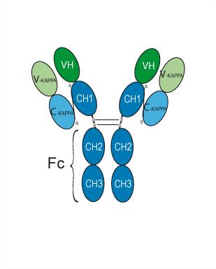
Human IgG4 kappa
アプリケーション
ELISA, FACS, Functional assay
Verified Bioactivity
-
 Immobilized Human PD-1 Protein (ECD, His Tag) can bind Retifanlimab. The EC50 for this effect is 3.71 ng/mL.
 -
 Flow cytometric analysis of 1X10^6 10 ng/mL PMA and 1 μg/mL Ionomycin co-stimulated Jurkat cells(24h) labeling Retifanlimab (HY-P99941, red). Cells were fixed with 4% paraformaldehyde. Then stained with the primary antibody at 1/200 dilution for an hour at 4℃. Alexa Fluor® 488-conjugated AffiniPure Goat Anti-Human IgG H&L (HY-P83776) was used as the secondary antibody at 1/1,000 dilution for 30 minutes at 4℃. Human IgG4 kappa (HY-P99003, blue) was used as the isotype control, cells without incubation with primary antibody were used as the unlabeled control (black).


化学情報
-
CAS 番号 2079108-44-2
-
性状 Liquid
-
分子量 145.35 kDa
-
Color Colorless to light yellow
-
SMILES
[Retifanlimab]
-
別名
INCMGA-00012; MGA-012
-
輸送条件
Shipping with dry ice.
-
Formulation
Please refer to the lot-specific COA for specific buffer information.
-
保管条件
Please store the product under the recommended conditions in the Certificate of Analysis.

プロトコル
-
Apoptosis
Apoptosis, also called programmed cell death, is generally characterized by distinct morphological characteristics.
-
Cell Cytotoxicity Assay
Cytotoxicity assays are usually based on the assessment of cell membrane damage, which can also be indirectly detected by measuring cell viability. Detection methods include MTT assay, CKK-8 assay, LDH assay and ATP assay, etc.
-
Mammalian live/dead viability and cytotoxicity staining
Live/dead viability and cytotoxicity staining assays are based on the simultaneous detection of intracellular esterase activity in metabolically active (viable) cells and membrane integrity loss in non-viable cells. In commonly used dual-staining approaches, membrane-permeant fluorogenic substrates are converted by intracellular esterases into fluorescent products in live cells, while impermeant DNA-binding dyes selectively enter cells with compromised plasma membranes and label nucleic acids in dead or dying cells, enabling discrimination between viable and non-viable populations by fluorescence microscopy or flow cytometry.
-
Apoptosis Solutions
Apoptosis is a regulated, generally non-lytic cell-death pathway that removes unwanted, damaged, infected, or abnormal cells through coordinated morphological changes, caspase activation, DNA fragmentation, and membrane remodeling. The intrinsic apoptosis pathway is controlled mainly by mitochondrial outer membrane permeabilization, BCL-2 family proteins, cytochrome c release, apoptosome formation, caspase-9 activation, and downstream executioner caspase-3/7 activation. The extrinsic apoptosis pathway is initiated by death receptors such as Fas, TNFR, and TRAIL receptors, which recruit adaptor proteins and activate caspase-8 before engaging executioner caspases or mitochondrial amplification through BID cleavage. Apoptosis is linked to many phenotypes, including cancer cell killing, tissue homeostasis, immune regulation, neurodegeneration, infection response, and treatment-induced cytotoxicity; unresolved questions include how apoptosis interacts with necroptosis, pyroptosis, ferroptos


純度とドキュメンテーション
-
データシート (257 KB)
-
SDS (251 KB)
- English - EN (251 KB)
- Français - FR (251 KB)
- Deutsch - DE (251 KB)
- Norwegian - NO (251 KB)
- Español - ES (251 KB)
- Swedish - SV (251 KB)
- Italian - IT (251 KB)
- Korean - KR (251 KB)
- Portuguese - PT (251 KB)
-
Inhibitory Antibodies User Guide (603 KB)
参考文献

Calculators
濃度 (開始) × 体積 (開始) = 濃度 (終了) × 体積 (終了)


Retifanlimab 関連分類
HY-P99941 関連分類
 Powered by Bioz