



RH 795
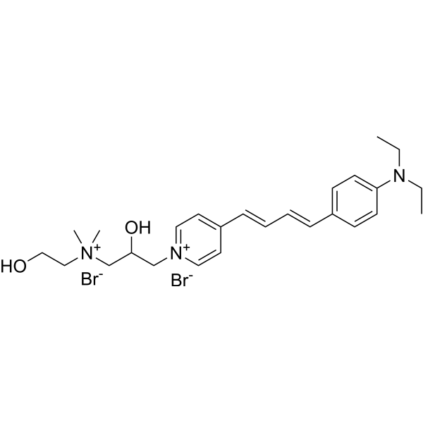
RH 795 is a lipophilic potentiometric styryl fluorescent dye (Ex/Em = 530 nm/712 nm). RH 795 stains the cell membranes of sensory cells and nerve fibers in living cells that maintain membrane potential, while it shows weak staining effect on supporting cells. RH 795 supports in vivo and in vitro confocal microscopy imaging of intact living inner ear cells and neuronal components. RH 795 can be used in studies related to hearing loss.
For research use only. We do not sell to patients.
- CAS No.: 172807-13-5
- Formula: C26H39Br2N3O2
- Molecular Weight:585.41
-
Storage:
Please store the product under the recommended conditions in the Certificate of Analysis.

To place orders, for customer services and technical support, please contact: MedChemExpress USA
Tel: 609-228-6898 E-mail: [email protected] [email protected]


-
 Biological Activity
-
Chemical Information
- Protocols
- Purity & Documentation
- References
-
 Help & FAQs




Biological Activity
Description
In Vitro
RH 795 (7-25 µg/mL) potently labels the cell membranes of inner hair cells, outer hair cells, and associated nerve fibers in unfixed ex vivo cochlear specimens from Balb/c mice, enabling high-resolution visualization via confocal microscopy[2].
Guide (The following is our recommended protocol. This protocol serves only as a guideline and should be modified according to your specific needs).
Ex vivo Live Mouse Temporal Bone[2]
1. Reagent Preparation
1.1 Stock solution: Weigh RH 795 powder and dissolve it in anhydrous DMSO with gentle vortexing to prepare a 1-2 mg/mL (approximately 2-4 mM) stock solution.
1.2 Working solution: Dilute the stock solution with tissue culture medium to a final concentration of approximately 7-25 μg/mL.
Co-staining with Calcein AM (HY-D0041) (for double labeling if needed): Use a final concentration of 20-60 µg/mL for Calcein AM.
2. Procedure
2.1 Preparation: Dissect out the left temporal bone of a mouse, fix it on a plastic holder, open the bony structure of the apical turn to expose the organ of Corti, and insert a perfusion tube into the scala tympani.
2.2 Perfusion system: Connect to a medium-containing reservoir bottle, and perform continuous perfusion of the scala tympani via hydrostatic pressure difference, allowing the medium to flow out through the opening of the apical turn.
2.3 Pre-staining with Calcein (optional for co-labeling): Perfuse with medium containing Calcein (20-30 µg/mL) for several minutes to ~15-30 min to label the cytoplasm of live cells green.
2.4 RH 795 staining: Switch the perfusion solution to fresh medium containing RH 795 (7-25 µg/mL) and perfuse for 15-30 min
Alternatively, add both dyes to the medium simultaneously for double-labeling perfusion
2.5 Washing/Equilibration: Briefly (for several minutes) switch to dye-free medium or continue perfusion to maintain oxygenation, followed immediately by confocal observation
2.6 Imaging parameters
RH 795: Excitation at 568 nm/ long-pass filter ≥ 560 nm (red channel)
Calcein: Excitation at 488 nm/ band-pass filter at 515 nm (green channel)
Reduce laser intensity appropriately to avoid phototoxicity (the membrane-bound dye RH 795 causes relatively strong photodamage).
MedChemExpress (MCE) has not independently confirmed the accuracy of these methods. They are for reference only. Further protocols information, click here.
In Vivo
Guide (The following is our recommended protocol. This protocol serves only as a guideline and should be modified according to your specific needs).
In Vivo Guinea Pig Cochlea[1]
1. Reagent Preparation
1.1 Stock solution: Weigh RH 795 powder and dissolve it in anhydrous DMSO with gentle vortexing to prepare a 1-2 mg/mL (approximately 2-4 mM) stock solution.
1.2 Working solution: Dilute the stock solution with tissue culture medium to a final concentration of 40 μg/mL.
2. Procedure
2.1 Surgical exposure: Anesthetize guinea pigs, perform tracheotomy, open the middle ear bulla to expose the cochlear basal turn, drill a hole in the basal turn scala tympani and insert a perfusion tube; make a small window in the apical turn for optical observation.
2.2 Calcein AM perfusion: Inject medium containing Calcein AM (60 µg/mL) through the basal turn perfusion tube and perfuse for 30 min.
2.3 RH 795 perfusion: Replace the perfusion solution with medium containing RH 795 (40 µg/mL) and perfuse for 15 min.
2.4 Maintenance perfusion: Continuously perfuse with dye-free medium or medium at a low flow rate throughout the experiment to maintain fluid coupling and oxygenation.
2.5 Confocal imaging: Perform simultaneous dual excitation at 488 nm (Calcein) & 543 nm (RH 795), and collect green/red channels respectively.
MedChemExpress (MCE) has not independently confirmed the accuracy of these methods. They are for reference only.
-
Animal Model:Pigmented guinea pigs (250-500 g)[1]
-
Dosage:40 μg/mL
-
Administration:scala tympani perfusion; 15 minutes
-
Result:Preferentially labeled the cell membranes of sensory inner and outer hair cells and cochlear nerve fibers with red fluorescence (emission peak 630 nm), while staining of supporting cells was much weaker.
Enabled clear visualization of individual hair bundles protruding from hair cells, nerve fibers projecting to inner hair cells, and the cytoarchitecture of the hearing organ.
Revealed distinct reorganization of outer hair cell cytoarchitecture, swelling of outer hair cell bodies, and an increase in outer hair cell diameter from ~9-10 μm pre-stimulation to ~15-19 μm post-stimulation following acoustic overstimulation.


Chemical Information
-
CAS No. 172807-13-5
-
Molecular Weight 585.41
-
Formula C26H39Br2N3O2
-
SMILES
OCC[N+](C)(CC(C[N+]1=CC=C(C=C1)/C=C/C=C/C2=CC=C(N(CC)CC)C=C2)O)C.[Br-].[Br-]
-
Shipping
Room temperature in continental US; may vary elsewhere.
-
Storage
Please store the product under the recommended conditions in the Certificate of Analysis.

Protocols
-
Fluorescent plasma-membrane potential dye assay
Fluorescent plasma-membrane potential dye assays measure changes in cell membrane potential using voltage-sensitive dyes whose fluorescence changes when cells depolarize or hyperpolarize. Anionic bis-oxonol dyes such as DiBAC4(3) enter depolarized cells more readily and show increased fluorescence after intracellular binding, while hyperpolarization reduces dye accumulation and fluorescence. FMP/FLIPR membrane-potential dyes are used for faster, homogeneous microplate assays of ion-channel or receptor-mediated membrane-potential changes.


Purity & Documentation
References
[1]. Tomo I, et al. Imaging the living inner ear using intravital confocal microscopy. Neuroimage. 2007;35(4):1393-1400. [Content Brief]
[2]. Le Calvez S, et al. An in vitro preparation to access cellular and neuronal components in the mouse inner ear. J Neurocytol. 2000;29(9):645-652. [Content Brief]

Calculators
Concentration (start) × Volume (start) = Concentration (final) × Volume (final)


RH 795 Related Classifications
HY-D1439 Related Classifications
 Powered by Bioz