



DB1976
Based on 8 publication(s) in Google Scholar
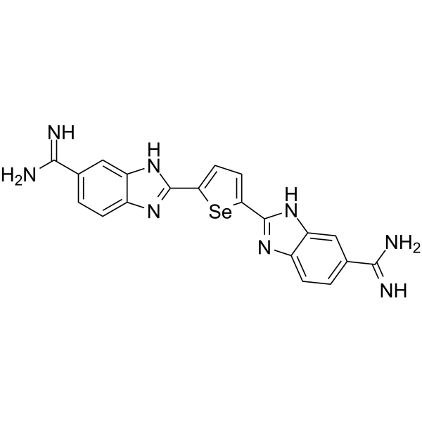
DB1976 is a selenophene analog of DB270 and a potent and cell-permeable fully efficacious transcription factor PU.1 inhibitor. DB1976 potently inhibits PU.1 binding (IC50 of 10 nM) and strongly inhibits the PU.1/DNA complex (with high DB1976-λB affinity, KD of 12 nM) in vitro. DB1976 has apoptosis-inducing effect.
연구목적의 판매만을 진행합니다. 환자를 대상으로 한 판매는 하지 않습니다.
- CAS No.: 1557397-51-9
- 화학식: C20H16N8Se
- 분자량:447.35
-
보관:
Please store the product under the recommended conditions in the Certificate of Analysis.

To place orders, for customer services and technical support, please contact: MedChemExpress USA
Tel: 609-228-6898 E-mail: [email protected] [email protected]


-
 Biological Activity
-
Chemical Information
- Protocol
- 순도&문서
- References
-
 Help & FAQs


Publications Citing Use of MedChemExpress (MCE) DB1976
More- Signal Transduct Target Ther. 2025 Feb 10;10(1):59. [Abstract]
- Nat Immunol. 2023 Nov;24(11):1839-1853. [Abstract]
- J Nanobiotechnology. 2025 Dec 5. [Abstract]
- Adv Healthc Mater. 2026 Jan 24:e05502. [Abstract]
- Int J Biol Macromol. 2025 Sep;322(Pt 1):146515. [Abstract]
- Cell Rep. 2024 Mar 26;43(3):113964. [Abstract]
- Respir Res. 2023 Jan 25;24(1):32. [Abstract]
- Neurochem Int. 2024 Feb:173:105674. [Abstract]


Biological Activity
제품 설명
IC50 & Target
IC50: 10 nM (Transcription factor PU.1)[1]
In Vitro
DB1976 is a classic heterocyclic dication (a single heteroatom) with strong affinity and selectivity for AT-rich sequences commonly found in cognate DNA binding sites for PU.1[2].
DB1976 inhibits PU.1-dependent transactivation of the reporter in a dose-dependent manner with an IC50 value of 2.4 μM in PU.1-negative HEK293 cells[3].
DB1976 treatment leads to a profound decrease in the growth of PU.1 URE–/– AML cells (IC50 of 105 μM), while showing little effect on normal hematopoietic cells at similar concentrations (IC50 of 334 μM)[3].
DB1976 treatment leads to a 1.6-fold increase in apoptotic cells in murine PU.1 URE–/– AML cells, and observed similar effects in human MOLM13 cells[3].
DB1976 treatment leads to a significant decrease in the number of viable cells (primary human AML cells) (mean decrease of 81%) and clonogenic capacity (mean decrease of 36%) compared with vehicle-treated cells. The apoptotic cell fraction increased on average by 1.5-fold with DB1976[3].
MedChemExpress (MCE) has not independently confirmed the accuracy of these methods. They are for reference only.


Chemical Information
-
CAS No. 1557397-51-9
-
분자량 447.35
-
화학식 C20H16N8Se
-
SMILES
N=C(C1=CC=C2N=C(C3=CC=C(C4=NC5=CC=C(C(N)=N)C=C5N4)[Se]3)NC2=C1)N
-
선적
Room temperature in continental US; may vary elsewhere.
-
보관
Please store the product under the recommended conditions in the Certificate of Analysis.


Publications (8)
-
Journal Impact Factor
-
Most Recent
-
Signal Transduct Target Ther
Spatiotemporal transcriptomics elucidates the pathogenesis of fulminant viral myocarditis. [Abstract]2025 Feb 10;10(1):59. PMID: 39924580 -
Nat Immunol
APOE4 impairs the microglial response in Alzheimer's disease by inducing TGFβ-mediated checkpoints. [Abstract]2023 Nov;24(11):1839-1853. PMID: 37749326 -
J Nanobiotechnology
Folate-modified biomimetic nanovesicles loaded with a PU.1 inhibitor alleviate atherosclerosis by suppressing inflammation. [Abstract]2025 Dec 5. PMID: 41345629 -
Adv Healthc Mater
One Stone, Four Birds: Multiple Modulation of Infarct Microenvironment-Based ROS-Responsive Hydrogel for Cardiac Repair. [Abstract]2026 Jan 24:e05502. PMID: 41580910 -
Int J Biol Macromol
EgG1Y162 protein from Echinococcus granulosus modulates the immune functions of mouse splenic lymphocytes and regulates Th9 cells. [Abstract]2025 Sep;322(Pt 1):146515. PMID: 40769342 -
Cell Rep
Nuclear microRNA-mediated transcriptional control determines adult microglial homeostasis and brain function. [Abstract]2024 Mar 26;43(3):113964. PMID: 38489263 -
Respir Res
2023 Jan 25;24(1):32. PMID: 36698141 -
Neurochem Int
M2a macrophages regulate fibrosis and affect the outcome after stroke via PU.1/mTOR pathway in fibroblasts. [Abstract]2024 Feb:173:105674. PMID: 38184171


순도&문서
References
[1]. Munde M, et al. Structure-dependent inhibition of the ETS-family transcription factor PU.1 by novel heterocyclic diamidines. Nucleic Acids Res. 2014 Jan;42(2):1379-90. [Content Brief]
[2]. Stephens DC, et al. Pharmacologic efficacy of PU.1 inhibition by heterocyclic dications: a mechanistic analysis. Nucleic Acids Res. 2016 May 19;44(9):4005-13. [Content Brief]
[3]. Antony-Debré I, et al. Pharmacological inhibition of the transcription factor PU.1 in leukemia. J Clin Invest. 2017 Dec 1;127(12):4297-4313. [Content Brief]

Calculators
Concentration (start) × Volume (start) = Concentration (final) × Volume (final)


DB1976 Related Classifications
HY-135797 Related Classifications
 Powered by Bioz