



Oxyfluorfen

Based on 1 Customer Validation
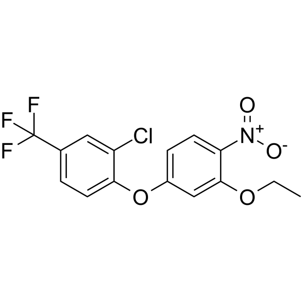
Oxyfluorfen is a pre- and post-emergence diphenyl ether herbicide to control annual broad-leaved and grass weeds. Oxyfluorfen is a protoporphyrinogen oxidase inhibitor and inhibits photosynthesis by blocking chlorophyll synthesis. Oxyfluorfen can inhibit cell growth. Oxyfluorfen induces DNA damage and exhibits toxicity toward aquatic organisms such as Paramisgurnus dabryanus. Oxyfluorfen has genome-level deleterious effects on fish that can lead to stunted skeletal growth. Oxyfluorfen induces transverse limb deficiency or craniosynostosis.
연구목적의 판매만을 진행합니다. 환자를 대상으로 한 판매는 하지 않습니다.
- Purity : 99.69%
- CAS No.: 42874-03-3
- 화학식: C15H11ClF3NO4
- 분자량:361.70
-
보관:Powder -20°C, 3 years , 4°C, 2 years ; In solvent -80°C, 6 months , -20°C, 1 month

To place orders, for customer services and technical support, please contact: MedChemExpress USA
Tel: 609-228-6898 E-mail: [email protected] [email protected]


-
 Biological Activity
-
Chemical Information
-
용액&용해도
- Protocol
- 순도&문서
- References
-
 Help & FAQs




Biological Activity
제품 설명
In Vitro
Oxyfluorfen (10 nM, 12 d) strongly inhibits the growth of normal soybean cells but not resistant cells[2].
Oxyfluorfen (0-100 μM) inhibits the total extractable Protox activity in normal cells but not resistant cells[2].
MedChemExpress (MCE) has not independently confirmed the accuracy of these methods. They are for reference only.
In Vivo
Oxyfluorfen (0-800 ppm, in the diet, 7 or 28 d) exhibits liver toxicity in mice[4].
Oxyfluorfen (40-1600 ppm, in the diet, 3 or 10 d) results in a dose-responsive induction in PPARα genes (Cyp4a10, Acox1, Cte-1 and Pmp-70) in mice model[4].
MedChemExpress (MCE) has not independently confirmed the accuracy of these methods. They are for reference only.
-
Animal Model:Male CD-1 mice (up to 12 weeks) with ad libitum access to feed[4]
-
Dosage:0, 40, 200 and 800 ppm
-
Administration:Added to diet for 7 or 28 days
-
Result:Resulted in significant increases in absolute and relative lever weights with 800 ppm.
Increased the liver weights with no statistical significance at 200 ppm.
Increased peroxisomal ACO activity 1.5- and 5-fold above control levels after 28 days with 200 and 800 ppm, respectively.
Increased cell proliferation after 28 days with 800 ppm.
Led to hypertrophy, vacuolization, single-cell necrosis and additional liver changes in mice given 800 ppm for 28 days.
- Protocol for Cell Counting and Cell Density Analysis
- Protocol for Fluorescence In Situ Hybridization (FISH)
- Somatic Cell Culture
- CCK-8/WST-8 Cell Proliferation Assay
- Cell Counting-Based Growth Curve Assay
- MTT Cell Proliferation Assay
- Genotoxicity/Mutagenicity Study
- Cell Viability Determination by MTT Colorimetric Assay


Chemical Information
-
CAS No. 42874-03-3
-
Appearance Solid
-
분자량 361.70
-
화학식 C15H11ClF3NO4
-
Color Off-white to light yellow
-
SMILES
FC(C1=CC=C(OC2=CC=C([N+]([O-])=O)C(OCC)=C2)C(Cl)=C1)(F)F
-
선적
Room temperature in continental US; may vary elsewhere.
-
보관
Powder -20°C 3 years 4°C 2 years In solvent -80°C 6 months -20°C 1 month


용액&용해도
In Vitro:
DMSO : 100 mg/mL (276.47 mM; Need ultrasonic; Hygroscopic DMSO has a significant impact on the solubility of product, please use newly opened DMSO)
Please refer to the solubility information to select the appropriate solvent. Once prepared, please aliquot and store the solution to prevent product inactivation from repeated freeze-thaw cycles.
Storage method and period of stock solution: -80°C, 6 months; -20°C, 1 month. When stored at -80°C, please use it within 6 months. When stored at -20°C, please use it within 1 month.
Please refer to the solubility information to select the appropriate solvent. Once prepared, please aliquot and store the solution to prevent product inactivation from repeated freeze-thaw cycles.
Storage method and period of stock solution: -80°C, 6 months; -20°C, 1 month. When stored at -80°C, please use it within 6 months. When stored at -20°C, please use it within 1 month.
Concentration (start) × Volume (start) = Concentration (final) × Volume (final)
In Vivo:
Select the appropriate dissolution method based on your experimental animal and administration route.
- For the following dissolution methods, please ensure to first prepare a clear stock solution using an In Vitro approach and then sequentially add co-solvents:
- To ensure reliable experimental results, the clarified stock solution can be appropriately stored based on storage conditions. As for the working solution for In Vivo experiments, it is recommended to prepare freshly and use it on the same day.
- The percentages shown for the solvents indicate their volumetric ratio in the final prepared solution. If precipitation or phase separation occurs during preparation, heat and/or sonication can be used to aid dissolution.
In Vivo Dissolution Calculator
Please enter the basic information of animal experiments:
-
-
-
-
Recommended: Prepare an additional quantity of animals to account for potential losses during experiments.
Please enter your animal formula composition:
-
%DMSO +
Recommended: Keep the proportion of DMSO in working solution below 2% if your animal is weak.
-
%+
-
+%Tween-80 + +
-
%Saline +
The co-solvents required include: DMSO, . All of co-solvents are available by MedChemExpress (MCE). , Tween 80. All of co-solvents are available by MedChemExpress (MCE).
Working solution concentration: 0.22 mg/mL
Method for preparing stock solution: mg drug dissolved in μL DMSO. Stock solution concentration: mg/mL.
1. Take μL DMSO stock solution;
2. Add μL .
μL , mix evenly;
3. Then add μL Tween 80, mix evenly;
4. Then add μL
Please ensure that the stock solution in the first step is dissolved to a clear state, and add co-solvents in sequence. You can use ultrasonic heating (ultrasonic cleaner, recommended frequency 20-40 kHz), vortexing, etc. to assist dissolution.


순도&문서
-
Data Sheet (275 KB)
-
SDS (458 KB)
- English - EN (458 KB)
- Français - FR (458 KB)
- Deutsch - DE (458 KB)
- Norwegian - NO (458 KB)
- Español - ES (458 KB)
- Swedish - SV (458 KB)
- Italian - IT (458 KB)
- Korean - KR (458 KB)
- Portuguese - PT (458 KB)
-
Handling Instructions (2659 KB)
References
[1]. Chi Wu, et al. Sorption, Degradation and Bioavailability of Oxyfluorfen in Biochar-Amended Soils. Sci Total Environ. 2019 Mar 25;658:87-94. [Content Brief]
[2]. E Warabi, et al. Resistance of a Soybean Cell Line to Oxyfluorfen by Overproduction of Mitochondrial Protoporphyrinogen Oxidase. Pest Manag Sci. 2001 Aug;57(8):743-8. [Content Brief]
[4]. Stagg, N. J., et al., (2012). Assessment of possible carcinogenicity of oxyfluorfen to humans using mode of action analysis of rodent liver effects. Toxicological sciences: an official journal of the Society of Toxicology, 128(2), 334–345. [Content Brief]


Complete Stock Solution Preparation Table
Please refer to the solubility information to select the appropriate solvent. Once prepared, please aliquot and store the solution to prevent product inactivation from repeated freeze-thaw cycles.
Storage method and period of stock solution: -80°C, 6 months; -20°C, 1 month. When stored at -80°C, please use it within 6 months. When stored at -20°C, please use it within 1 month.
| Optional Solvent | Concentration Solvent Mass | 1 mg | 5 mg | 10 mg | 25 mg |
|---|---|---|---|---|---|
| DMSO | 1 mM | 2.7647 mL | 13.8236 mL | 27.6472 mL | 69.1181 mL |
| 5 mM | 0.5529 mL | 2.7647 mL | 5.5294 mL | 13.8236 mL | |
| 10 mM | 0.2765 mL | 1.3824 mL | 2.7647 mL | 6.9118 mL | |
| 15 mM | 0.1843 mL | 0.9216 mL | 1.8431 mL | 4.6079 mL | |
| 20 mM | 0.1382 mL | 0.6912 mL | 1.3824 mL | 3.4559 mL | |
| 25 mM | 0.1106 mL | 0.5529 mL | 1.1059 mL | 2.7647 mL | |
| 30 mM | 0.0922 mL | 0.4608 mL | 0.9216 mL | 2.3039 mL | |
| 40 mM | 0.0691 mL | 0.3456 mL | 0.6912 mL | 1.7280 mL | |
| 50 mM | 0.0553 mL | 0.2765 mL | 0.5529 mL | 1.3824 mL | |
| 60 mM | 0.0461 mL | 0.2304 mL | 0.4608 mL | 1.1520 mL | |
| 80 mM | 0.0346 mL | 0.1728 mL | 0.3456 mL | 0.8640 mL | |
| 100 mM | 0.0276 mL | 0.1382 mL | 0.2765 mL | 0.6912 mL |


Oxyfluorfen Related Classifications
HY-119176 Related Classifications
 Powered by Bioz