



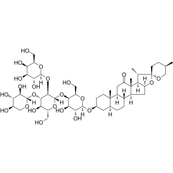
Terrestrosin D

Based on 1 Customer Validation
Terrestrosin D is an orally active apoptosis inducer. Terrestrosin D induces cell cycle arrest at the G1 and S phases, reduces mitochondrial membrane potential, and inhibits the growth of cancer cells and endothelial cells. Terrestrosin D is studied in castration-resistant prostate cancer and pulmonary fibrosis.
연구목적의 판매만을 진행합니다. 환자를 대상으로 한 판매는 하지 않습니다.
- Purity : 99.30%
- CAS No.: 179464-23-4
- 화학식: C50H80O23
- 분자량:1049.16
-
보관:
4°C, protect from light
* In solvent : -80°C, 6 months; -20°C, 1 month (protect from light)

To place orders, for customer services and technical support, please contact: MedChemExpress USA
Tel: 609-228-6898 E-mail: [email protected] [email protected]


-
 Biological Activity
-
Chemical Information
-
용액&용해도
- Protocol
- 순도&문서
- References
-
 Help & FAQs



All VEGFR Isoforms
More

Biological Activity
제품 설명
In Vitro
Terrestrosin D (2.6-41.3 μM; 24 h) exerts significant cytotoxicity in human normal liver LO2 cells (IC50 = 16.88 μM) and human embryonic kidney 293T cells (IC50 = 21.80 μM) following 24 h of treatment[1].
Terrestrosin D (1-5 μM; 24 h) potently inhibits the growth of PC-3, PC-3M, DU145, LNCaP, and 22RV1 human prostate cancer cells with an IC50 below 5 μM after 24 h of treatment[2].
Terrestrosin D (2-5 μM; 24 h) induces G1 phase cell cycle arrest in PC-3 human prostate cancer cells after 24 h of treatment with 2 or 5 μM[2].
Terrestrosin D (1-5 μM; 24 h) potently inhibits the growth of HUVECs and bladder-derived normal human microvascular endothelial cells with an IC50 below 3 μM after 24 h of treatment[2].
Terrestrosin D (2-3 μM; 24 h) induces S phase cell cycle arrest in HUVEC human endothelial cells after 24 h of treatment with 2 or 3 μM[2].
Terrestrosin D (2-5 μM; 24 h) induces dose-dependent, caspase-independent apoptosis in PC-3 human prostate cancer cells, with 60.5% of cells undergoing apoptosis after 24 h of treatment with 5 μM[2].
Terrestrosin D (5 μM; 24 h) does not activate caspase-3 and instead reduces its activity in PC-3 human prostate cancer cells after 24 h of treatment with 5 μM, confirming a caspase-independent apoptotic mechanism[2].
Terrestrosin D (2-3 μM; 24 h) induces dose-dependent, caspase-independent apoptosis in HUVEC human endothelial cells, with 34.3% of cells undergoing apoptosis after 24 h of treatment with 3 μM[2].
Terrestrosin D (3 μM; 24 h) does not activate caspase-3 and instead reduces its activity in HUVEC human endothelial cells after 24 h of treatment with 3 μM, confirming a caspase-independent apoptotic mechanism[2].
Terrestrosin D (2-5 μM; 24 h) induces mitochondrial membrane potential depolarization in 53.8% of PC-3 human prostate cancer cells[2].
Terrestrosin D (2-3 μM; 24 h) induces dose-dependent mitochondrial membrane potential depolarization in HUVEC human endothelial cells[2].
Terrestrosin D (1-5 μM; 24 h) dose-dependently increases VEGF secretion in PC-3 human prostate cancer cells, causing a 1.86-fold increase after 24 h of treatment with 5 μM[2].
Terrestrosin D (1-3 μM; 24 h) dose-dependently increases VEGF secretion in HUVEC human endothelial cells, causing an 11.21-fold increase after 24 h of treatment with 3 μM[2].
MedChemExpress (MCE) has not independently confirmed the accuracy of these methods. They are for reference only. Further protocols information, click here.
-
Cell Line:PC-3 human prostate cancer cells
-
Concentration:2, 5 μM
-
Incubation Time:24 h
-
Result:Caused significant increases in G1 phase populations and concomitant decreases in S phase populations at 5 μM.
Altered cell cycle phase distribution relative to control at 2 μM.
-
Cell Line:PC-3 human prostate cancer cells
-
Concentration:2, 5 μM; 5 μM (with 1 h pre-incubation with z-VAD (HY-164388))
-
Incubation Time:24 h; 24 h (with 1 h pre-incubation with z-VAD)
-
Result:Induced apoptosis in 6.1% of PC-3 cells at 2 μM for 24 h.
Induced apoptosis in 60.5% of PC-3 cells at 5 μM for 24 h.
Did not reduce the number of apoptotic cells induced by 5 μM TED when pre-treated with z-VAD.
-
Cell Line:PC-3 human prostate cancer cells
-
Concentration:1, 2, 3, 4, 5 μM
-
Incubation Time:24 h
-
Result:Induced a dose-dependent increase in VEGF secretion.
Caused a 1.86-fold increase in VEGF levels relative to control at 5 μM.
Parmacokinetics
In Vivo
Terrestrosin D (50 mg/kg; i.p.; 3 times weekly; 4 weeks) significantly suppresses PC-3 xenograft tumor growth in BALB/c nude mice, increases tumor cell apoptosis, and reduces tumor angiogenesis, without causing significant body weight loss[2].
Terrestrosin D (10 mg/kg; i.p.; daily for
6 weeks) significantly attenuates Bleomycin (HY-108345)-induced pulmonary inflammation and fibrosis in male KM mice[3].
MedChemExpress (MCE) has not independently confirmed the accuracy of these methods. They are for reference only.
-
Animal Model:Sprague-Dawley (male, 200 g)[1]
-
Dosage:5 mg/kg; 15 mg/kg
-
Administration:p.o.; daily; 28 days
-
Result:Increased serum alanine aminotransferase to 42.8 U/L, alkaline phosphatase to 264.3 U/L, serum creatinine to 25.4 μmol/L, urinary β-N-Acetylglucosaminidase to 32.0 U/L, and Kidney Injury Molecule 1 to 321.3 ng/L at 5 mg/kg after 28 days of treatment.
Increased serum alanine aminotransferase to 48.8 U/L, aspartate aminotransferase to 151.5 U/L, alkaline phosphatase to 313.5 U/L, blood urea nitrogen to 8.3 mmol/L, serum creatinine to 28.7 μmol/L, urinary β-N-Acetylglucosaminidase to 43.2 U/L, and Kidney Injury Molecule 1 to 332.7 ng/L at 15 mg/kg after 28 days of treatment.
Reduced body weight on Day 8 and food consumption at Week 2 for both doses after 28 days of treatment.
Restored body weight and food consumption to control levels for both doses after 14-day recovery period.
-
Animal Model:BALB/c nude (male, 5 weeks of age, subcutaneous PC-3 cell implantation)[2]
-
Dosage:25 mg/kg; 50 mg/kg
-
Administration:i.p.; 3 times weekly; 4 weeks
-
Result:Suppressed tumor growth to an average volume of 127.41 mm3 after 28 days at 50 mg/kg.
Increased tumor apoptosis index to 58.5 at 50 mg/kg.
Reduced microvessel density to 17.6 per 400× field at 50 mg/kg.
Did not significantly affect mouse body weight at 50 mg/kg.
Showed little effect on tumor growth at 25 mg/kg.
-
Animal Model:KM mice (male, 30 g, bleomycin-induced pulmonary fibrosis model)[3]
-
Dosage:10 mg/kg
-
Administration:i.p.; daily; 2 weeks or 6 weeks
-
Result:Reduced bronchoalveolar lavage fluid (BALF) cell density by ~50% at 2 weeks.
Returned BALF macrophage, neutrophil, and lymphocyte percentages to control levels by 6 weeks.
Significantly reduced BALF levels of TNF-α, IL-6, and IL-8 to near-control levels at 2 and 6 weeks.
Preserved alveolar structure, reducing bleomycin-induced alveolar space reduction and fibrotic focal area formation.


Chemical Information
-
CAS No. 179464-23-4
-
Appearance Solid
-
분자량 1049.16
-
화학식 C50H80O23
-
Color White to off-white
-
SMILES
C[C@@]12[C@]3([H])[C@](O[C@]4(CC[C@@H](C)CO4)[C@H]3C)([H])C[C@@]1([H])[C@@]5([H])[C@]([C@@]6([C@@](C[C@@H](O[C@]7([H])O[C@@H]([C@H](O[C@@]8([H])[C@@H]([C@H]([C@H](O)[C@@H](CO)O8)O[C@@]9([H])[C@@H]([C@H]([C@H](O)CO9)O)O)O[C@]%10([H])O[C@@H]([C@H](O)[C@H](O)[C@H]%10O)CO)[C@H](O)[C@H]7O)CO)CC6)([H])CC5)C)([H])CC2=O
-
Structure Classification
-
Initial Source
-
선적
Room temperature in continental US; may vary elsewhere.
-
보관
4°C, protect from light
* In solvent : -80°C, 6 months; -20°C, 1 month (protect from light)


용액&용해도
In Vitro:
DMSO : 100 mg/mL (95.31 mM; Need ultrasonic; Hygroscopic DMSO has a significant impact on the solubility of product, please use newly opened DMSO)
Please refer to the solubility information to select the appropriate solvent. Once prepared, please aliquot and store the solution to prevent product inactivation from repeated freeze-thaw cycles.
Storage method and period of stock solution: -80°C, 6 months; -20°C, 1 month (protect from light). When stored at -80°C, please use it within 6 months. When stored at -20°C, please use it within 1 month.
Please refer to the solubility information to select the appropriate solvent. Once prepared, please aliquot and store the solution to prevent product inactivation from repeated freeze-thaw cycles.
Storage method and period of stock solution: -80°C, 6 months; -20°C, 1 month (protect from light). When stored at -80°C, please use it within 6 months. When stored at -20°C, please use it within 1 month.
Concentration (start) × Volume (start) = Concentration (final) × Volume (final)
In Vivo:
Select the appropriate dissolution method based on your experimental animal and administration route.
- For the following dissolution methods, please ensure to first prepare a clear stock solution using an In Vitro approach and then sequentially add co-solvents:
- To ensure reliable experimental results, the clarified stock solution can be appropriately stored based on storage conditions. As for the working solution for In Vivo experiments, it is recommended to prepare freshly and use it on the same day.
- The percentages shown for the solvents indicate their volumetric ratio in the final prepared solution. If precipitation or phase separation occurs during preparation, heat and/or sonication can be used to aid dissolution.
Add each solvent one by one: 10% DMSO 40% PEG300 5% Tween-80 45% Saline
Solubility: ≥ 2.5 mg/mL (2.38 mM); Clear solution
This protocol yields a clear solution of ≥ 2.5 mg/mL (saturation unknown).
Taking 1 mL working solution as an example, add 100 μL DMSO stock solution (25.0 mg/mL) to 400 μL PEG300, and mix evenly; then add 50 μL Tween-80 and mix evenly; then add 450 μL Saline to adjust the volume to 1 mL.
Preparation of Saline: Dissolve 0.9 g sodium chloride in ddH₂O and dilute to 100 mL to obtain a clear Saline solution.
Add each solvent one by one: 10% DMSO 90% (20% SBE-β-CD in Saline)
Solubility: ≥ 2.5 mg/mL (2.38 mM); Clear solution
This protocol yields a clear solution of ≥ 2.5 mg/mL (saturation unknown).
Taking 1 mL working solution as an example, add 100 μL DMSO stock solution (25.0 mg/mL) to 900 μL 20% SBE-β-CD in Saline, and mix evenly.
Preparation of 20% SBE-β-CD in Saline (4°C, storage for one week): 2 g SBE-β-CD powder is dissolved in 10 mL Saline, completely dissolve until clear.
In Vivo Dissolution Calculator
Please enter the basic information of animal experiments:
-
-
-
-
Recommended: Prepare an additional quantity of animals to account for potential losses during experiments.
Please enter your animal formula composition:
-
%DMSO +
Recommended: Keep the proportion of DMSO in working solution below 2% if your animal is weak.
-
%+
-
+%Tween-80 + +
-
%Saline +
The co-solvents required include: DMSO, . All of co-solvents are available by MedChemExpress (MCE). , Tween 80. All of co-solvents are available by MedChemExpress (MCE).
Working solution concentration: 0.22 mg/mL
Method for preparing stock solution: mg drug dissolved in μL DMSO. Stock solution concentration: mg/mL. * In solvent : -80°C, 6 months; -20°C, 1 month (protect from light)
1. Take μL DMSO stock solution;
2. Add μL .
μL , mix evenly;
3. Then add μL Tween 80, mix evenly;
4. Then add μL
Please ensure that the stock solution in the first step is dissolved to a clear state, and add co-solvents in sequence. You can use ultrasonic heating (ultrasonic cleaner, recommended frequency 20-40 kHz), vortexing, etc. to assist dissolution.

Protocol
-
Collagen: Sirius Red Staining
Sirius Red or picrosirius red staining is a histochemical method for visualizing collagen-rich extracellular matrix in tissue sections, and collagen fibers are detected as red-stained structures under bright-field microscopy with enhanced birefringence under polarized light. Picrosirius red is useful for assessing total collagen organization, distribution, and fibrosis burden, but polarized color should not be interpreted as a definitive collagen type I versus type III readout because color is affected by fiber orientation, thickness, and packing.
-
Mitochondrial membrane-potential fluorescent assay
Mitochondrial membrane potential fluorescent assays estimate ΔΨm in living cells using lipophilic cationic dyes such as TMRM, TMRE, rhodamine 123, and JC-1, which accumulate in mitochondria according to membrane polarization; loss of signal after FCCP or CCCP treatment is interpreted as mitochondrial depolarization. TMRM/TMRE and rhodamine 123 are commonly used for semi-quantitative live-cell microscopy or flow cytometry, while JC-1 can report a shift from red aggregate fluorescence to green monomer fluorescence during depolarization; interpretation requires controls because dye concentration, quenching mode, cell type, dye efflux, and mitochondrial mass can affect fluorescence independently of ΔΨm.
-
Annexin V plus membrane-impermeant dye apoptosis staining
Annexin V-based apoptosis assays rely on the detection of phosphatidylserine (PS) externalization from the inner leaflet of the plasma membrane to the outer leaflet, an early biochemical hallmark of apoptosis. Fluorescently labeled Annexin V binds PS in a calcium-dependent manner, enabling identification of early apoptotic cells by flow cytometry or fluorescence microscopy. When combined with a membrane-impermeant DNA-binding dye (e. g. , propidium iodide), this approach allows discrimination between viable (Annexin V−/dye−), early apoptotic (Annexin V+/dye−), and late apoptotic or necrotic (Annexin V+/dye+) cell populations by assessing membrane integrity and PS exposure.
-
BrdU Incorporation Assay
Bromodeoxyuridine (BrdU) incorporation assay is based on the principle that BrdU, a thymidine analog, is incorporated into newly synthesized DNA during the S phase of the cell cycle, thereby serving as a marker of DNA replication and cellular proliferation. Incorporated BrdU can be detected using anti-BrdU antibodies following DNA denaturation, enabling visualization or quantification of proliferating cells through immunochemical detection methods such as immunofluorescence or immunohistochemistry.
-
Connective Tissue: Masson's Trichrome/Collagen Trichrome Staining
Masson’s Trichrome (collagen/trichrome staining) is a histological technique that differentially stains tissue compartments using sequential acidic dyes to distinguish collagen from muscle and cytoplasmic components based on dye affinity and tissue permeability differences, enabling visualization of fibrosis and connective tissue architecture in histological sections. The classical formulation typically uses Weigert's iron hematoxylin for nuclear staining, Biebrich scarlet-acid fuchsin for cytoplasm and muscle, and aniline blue (or light green variants) for collagen, producing a characteristic blue/green collagen signal contrasted against red cytoplasm and dark nuclei. The staining principle relies on selective displacement of smaller dye molecules by larger anionic dyes in collagen-rich regions under controlled acidified conditions, which enhances collagen-specific dye retention. This property makes the method widely used for fibrosis assessment in organs such as heart, liver, lung, a
-
Protocol for Cell Cycle
Cell-cycle analysis by flow cytometry measures DNA content in single cells to estimate the fraction of cells in G0/G1, S, and G2/M phases. Propidium iodide intercalates into DNA, and after RNA removal with RNase, fluorescence intensity reflects cellular DNA content: 2N cells are assigned to G0/G1, cells between 2N and 4N to S phase, and 4N cells to G2/M. DNA-content analysis alone cannot reliably separate G0 from G1 or G2 from M. Ki-67 can distinguish quiescent G0 cells from cycling cells, EdU or BrdU incorporation marks active DNA synthesis in S phase, and phospho-histone H3 staining identifies mitotic cells within the 4N population.
-
Fibrosis/Collagen Morphometry
Fibrosis and collagen morphometry is based on the quantitative visualization of fibrillar collagen deposition in tissue sections using histochemical stains such as Sirius Red (Picrosirius Red) or Masson's trichrome, followed by image-based or polarization-enhanced analysis to estimate collagen proportional area as a surrogate of extracellular matrix accumulation during fibrotic remodeling. Sirius Red combined with polarized light microscopy enhances detection of collagen fibers due to birefringence properties, enabling more specific visualization of collagen type I and III fibrils compared to conventional bright-field histology, while whole-section or region-restricted digital morphometry reduces field-selection bias in fibrosis assessment. Alternative quantitative approaches include second harmonic generation (SHG) and two-photon excited fluorescence microscopy, which enable label-free detection of fibrillar collagen and have been validated against histological staining and biochemica
-
Apoptosis
Apoptosis, also called programmed cell death, is generally characterized by distinct morphological characteristics.
-
TUNEL staining for apoptotic DNA fragmentation
TUNEL staining detects DNA strand breaks by using terminal deoxynucleotidyl transferase to add labeled nucleotides to exposed 3′-OH DNA termini, generating either microscopic staining in fixed cells or tissue sections, or fluorescence/cytometric signal in cell suspensions. TUNEL positivity reflects DNA fragmentation but should not be interpreted alone as definitive apoptosis, because TUNEL can also label necrotic, autolytic, mechanically damaged, or DNA-repair-associated DNA breaks.
-
Flow cytometric DNA-content cell-cycle staining
Flow cytometric DNA-content cell-cycle staining measures the fluorescence intensity of DNA-bound fluorochromes in single cells or nuclei to estimate DNA content distributions, allowing assignment of populations to G0/G1, S, and G2/M phases by DNA histogram deconvolution. Propidium iodide (PI) intercalates into DNA, and PI fluorescence is proportional to cellular DNA content when staining is performed under conditions that make DNA accessible and minimize non-DNA signal. Cells with G2/M DNA content are expected to show approximately twice the fluorescence intensity of G0/G1 cells, while S-phase cells occupy intermediate fluorescence values. PI-based DNA-content analysis can also detect cells with fractional DNA content, often reported as sub-G1, when DNA fragmentation and extraction during staining reduce retained DNA signal in apoptotic cells. DAPI is an alternative DNA fluorochrome for univariate DNA-content analysis, while bivariate approaches combining DNA content with proliferation
-
Fluorescent plasma-membrane potential dye assay
Fluorescent plasma-membrane potential dye assays measure changes in cell membrane potential using voltage-sensitive dyes whose fluorescence changes when cells depolarize or hyperpolarize. Anionic bis-oxonol dyes such as DiBAC4(3) enter depolarized cells more readily and show increased fluorescence after intracellular binding, while hyperpolarization reduces dye accumulation and fluorescence. FMP/FLIPR membrane-potential dyes are used for faster, homogeneous microplate assays of ion-channel or receptor-mediated membrane-potential changes.
-
Mitochondrial membrane-potential and mitochondrial mass staining
Mitochondrial membrane potential staining measures the electrochemical polarization across the mitochondrial inner membrane in live cells using lipophilic cationic fluorescent probes; early rhodamine-based work showed that selective mitochondrial dye accumulation is lost when the mitochondrial transmembrane potential is dissipated. JC-1 reports mitochondrial polarization by shifting from green monomer fluorescence to red J-aggregate fluorescence as dye concentration increases within energized mitochondria; therefore, the red/green fluorescence ratio is used as a relative readout of mitochondrial membrane potential. TMRE or TMRM staining provides a single-channel relative readout because these cationic rhodamine esters accumulate in polarized mitochondria, and lower fluorescence indicates reduced mitochondrial polarization when acquisition and dye-loading conditions are controlled. Mitochondrial mass staining is commonly performed with MitoTracker Green FM or related MitoTracker dyes as
-
Endothelial Tube Formation Assay
Endothelial tube formation assay evaluates the ability of endothelial cells to attach, migrate, align, and organize into capillary-like networks when cultured on gelled basement membrane extract or Matrigel; the readout is the morphology and quantity of tube-like networks, which reflects an in vitro endothelial morphogenesis step related to angiogenesis. Basement membrane extract/Matrigel provides laminin-rich extracellular matrix cues that support endothelial differentiation into capillary-like structures, but it can contain biologically active growth factors, so growth-factor-reduced matrix is preferred when testing defined angiogenic stimulators or inhibitors.
-
Apoptosis Solutions
Apoptosis is a regulated, generally non-lytic cell-death pathway that removes unwanted, damaged, infected, or abnormal cells through coordinated morphological changes, caspase activation, DNA fragmentation, and membrane remodeling. The intrinsic apoptosis pathway is controlled mainly by mitochondrial outer membrane permeabilization, BCL-2 family proteins, cytochrome c release, apoptosome formation, caspase-9 activation, and downstream executioner caspase-3/7 activation. The extrinsic apoptosis pathway is initiated by death receptors such as Fas, TNFR, and TRAIL receptors, which recruit adaptor proteins and activate caspase-8 before engaging executioner caspases or mitochondrial amplification through BID cleavage. Apoptosis is linked to many phenotypes, including cancer cell killing, tissue homeostasis, immune regulation, neurodegeneration, infection response, and treatment-induced cytotoxicity; unresolved questions include how apoptosis interacts with necroptosis, pyroptosis, ferroptos


순도&문서
-
Data Sheet (287 KB)
-
SDS (252 KB)
- English - EN (252 KB)
- Français - FR (252 KB)
- Deutsch - DE (252 KB)
- Norwegian - NO (252 KB)
- Español - ES (252 KB)
- Swedish - SV (252 KB)
- Italian - IT (252 KB)
- Korean - KR (252 KB)
- Portuguese - PT (252 KB)
-
Handling Instructions (2659 KB)
References
[1]. Sun XC, et al. Terrestrosin D, a spirostanol saponin from Tribulus terrestris L. with potential hepatorenal toxicity. J Ethnopharmacol. 2022;283:114716. [Content Brief]
[2]. Wei S, et al. Terrestrosin D, a steroidal saponin from Tribulus terrestris L., inhibits growth and angiogenesis of human prostate cancer in vitro and in vivo. Pathobiology. 2014;81(3):123-132. [Content Brief]
[3]. Qiu M, et al. Terrestrosin D from Tribulus terrestris attenuates bleomycin-induced inflammation and suppresses fibrotic changes in the lungs of mice. Pharm Biol. 2019 Dec;57(1):694-700. [Content Brief]


Complete Stock Solution Preparation Table
Please refer to the solubility information to select the appropriate solvent. Once prepared, please aliquot and store the solution to prevent product inactivation from repeated freeze-thaw cycles.
Storage method and period of stock solution: -80°C, 6 months; -20°C, 1 month (protect from light). When stored at -80°C, please use it within 6 months. When stored at -20°C, please use it within 1 month.
| Optional Solvent | Concentration Solvent Mass | 1 mg | 5 mg | 10 mg | 25 mg |
|---|---|---|---|---|---|
| DMSO | 1 mM | 0.9531 mL | 4.7657 mL | 9.5314 mL | 23.8286 mL |
| 5 mM | 0.1906 mL | 0.9531 mL | 1.9063 mL | 4.7657 mL | |
| 10 mM | 0.0953 mL | 0.4766 mL | 0.9531 mL | 2.3829 mL | |
| 15 mM | 0.0635 mL | 0.3177 mL | 0.6354 mL | 1.5886 mL | |
| 20 mM | 0.0477 mL | 0.2383 mL | 0.4766 mL | 1.1914 mL | |
| 25 mM | 0.0381 mL | 0.1906 mL | 0.3813 mL | 0.9531 mL | |
| 30 mM | 0.0318 mL | 0.1589 mL | 0.3177 mL | 0.7943 mL | |
| 40 mM | 0.0238 mL | 0.1191 mL | 0.2383 mL | 0.5957 mL | |
| 50 mM | 0.0191 mL | 0.0953 mL | 0.1906 mL | 0.4766 mL | |
| 60 mM | 0.0159 mL | 0.0794 mL | 0.1589 mL | 0.3971 mL | |
| 80 mM | 0.0119 mL | 0.0596 mL | 0.1191 mL | 0.2979 mL |


Terrestrosin D Related Classifications
HY-N5074 Related Classifications
 Powered by Bioz