



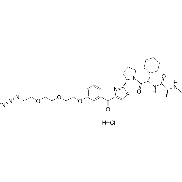
LCL-PEG3-N3 hydrochloride
LCL-PEG3-N3 (hydrochloride) is a decoy oligonucleotide ligand for E3 ligase which can be used for developing chimeric molecules LCL-ER(dec), degrading the estrogen receptor. LCL-PEG3-N3 (hydrochloride) is a click chemistry reagent, it contains an Azide group and can undergo copper-catalyzed azide-alkyne cycloaddition reaction (CuAAc) with molecules containing Alkyne groups. It can also undergo strain-promoted alkyne-azide cycloaddition (SPAAC) reactions with molecules containing DBCO or BCN groups.
For research use only. We do not sell to patients.
- CAS No.: 3033849-91-8
- Formula: C32H46ClN7O6S
- Molecular Weight:692.27
-
Storage:
Please store the product under the recommended conditions in the Certificate of Analysis.

To place orders, for customer services and technical support, please contact: MedChemExpress USA
Tel: 609-228-6898 E-mail: [email protected] [email protected]


-
 Biological Activity
-
Chemical Information
- Protocols
- Purity & Documentation
- References
-
 Help & FAQs


All E3 Ligase Ligand-Linker Conjugates Isoforms
More

Biological Activity
Description
IC50 & Target
|
IAP |


Chemical Information
-
CAS No. 3033849-91-8
-
Molecular Weight 692.27
-
Formula C32H46ClN7O6S
-
SMILES
O=C(C1=CC(OCCOCCOCCN=[N+]=[N-])=CC=C1)C2=CSC([C@H]3N(C([C@H](C4CCCCC4)NC([C@H](C)NC)=O)=O)CCC3)=N2.[H]Cl
-
Shipping
Room temperature in continental US; may vary elsewhere.
-
Storage
Please store the product under the recommended conditions in the Certificate of Analysis.

Protocols
-
EdU Incorporation Assay (Click Chemistry-Based DNA Synthesis Measurement)
The EdU incorporation assay measures DNA synthesis by adding the thymidine analog 5-ethynyl-2′-deoxyuridine to cells or tissues, where it is incorporated into newly synthesized DNA during S phase. Incorporated EdU is detected by copper-catalyzed azide-alkyne cycloaddition, in which a fluorescent azide covalently reacts with the ethynyl group on EdU, allowing S-phase cells to be detected by fluorescence microscopy, flow cytometry, or high-content imaging. EdU detection does not require DNA denaturation or anti-BrdU antibody access, which preserves sample structure and improves compatibility with immunostaining and multiparameter cytometry compared with BrdU-based detection. EdU can be cytotoxic in a cell-type- and exposure-dependent manner, so pulse duration, concentration, and continuous-labeling designs should be validated for each cell type.
-
Research Protocol for Endocrine Diseases
Endocrine diseases often arise from disrupted hormone production, hormone signaling, or target-tissue responsiveness; for diabetes-focused endocrine disease models, insulin signaling regulates glucose uptake, hepatic glucose output, lipid metabolism, and β-cell compensation. Type 2 diabetes develops through interacting defects in insulin resistance, β-cell dysfunction, adipose inflammation, hepatic glucose overproduction, altered incretin signaling, and ectopic lipid metabolism. A major unresolved question is whether endocrine dysfunction is driven primarily by target-tissue insulin resistance, intrinsic β-cell failure, immune/inflammatory stress, or combined multi-organ failure that differs by disease stage.


Purity & Documentation
References

Calculators
Concentration (start) × Volume (start) = Concentration (final) × Volume (final)


LCL-PEG3-N3 hydrochloride Related Classifications
HY-139999A Related Classifications
 Powered by Bioz