



LysoFP-NO2

Based on 1 Customer Validation
LysoFP-NO2 is a turn-on fluorescent probe for carbon monoxide (CO) that localizes to the lysosome. In the presence of lysosomal CO, lysoFP-NO2 is transformed into lysoFP-NH2, which is highly fluorescent. LysoFP-NO2 is selective for CO over various reactive nitrogen, oxygen, and sulfur species. It displays excitation/emission maxima of 440/528 nm, respectively, and is not cytotoxic to HepG2 cells for up to five hours when used at a concentration of 30 μM.
For research use only. We do not sell to patients.
- Purity : 98.2%
- CAS No.: 69408-75-9
- Formula: C18H17N3O5
- Molecular Weight:355.34
-
Storage:Powder -20°C, 3 years ; In solvent -80°C, 6 months , -20°C, 1 month


To place orders, for customer services and technical support, please contact: MedChemExpress USA
Tel: 609-228-6898 E-mail: [email protected] [email protected]


-
 Biological Activity
-
Chemical Information
- Protocols
- Purity & Documentation
- References
-
 Help & FAQs


-
HY-130412
FlAsH-EDT2Protein Labeling Reagent -
HY-111330
Hydroxyphenyl FluoresceinROS Probe -
HY-125603
Termitomycamide BNeuroprotective Agent -
HY-129420
WSP-5Fluorescent Probe -
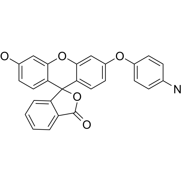
HY-119977
APFFluorescence Probe -
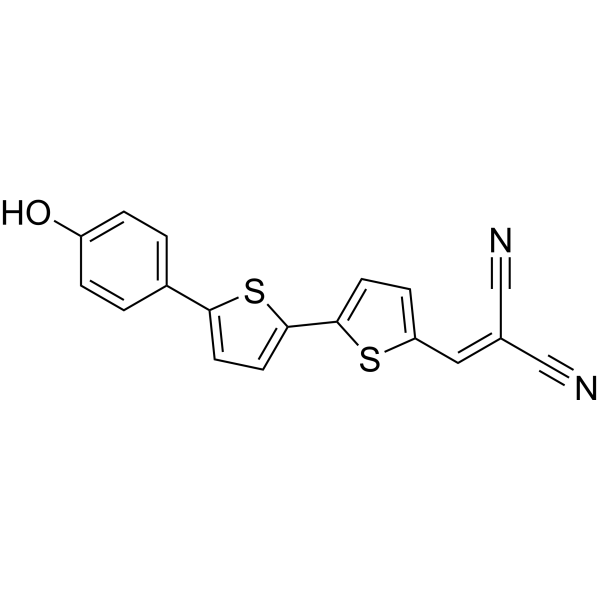
HY-111263
NIAD-4Amyloid-β Fluorophore -
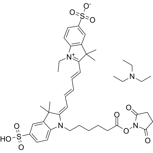
HY-D0819A
CY5-SE triethylamine saltFluorescent Probe -

HY-130785
LysoFP-NH2Lysosomal Probe -

HY-DY1035
Hydroxyphenyl Fluorescein (solution)ROS Probe -
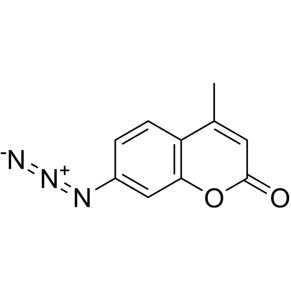
HY-119323
7-Azido-4-methylcoumarinH2S Probe -
HY-118159
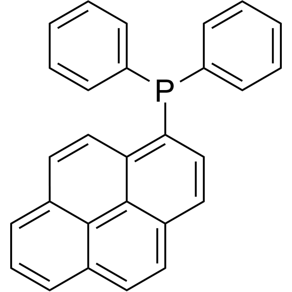
Diphenyl-1-pyrenylphosphinefluorescent probe -
HY-D1275
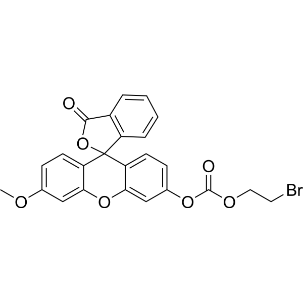
CAY10731H2S Fluorescent Probe


Biological Activity
Description


Chemical Information
-
CAS No. 69408-75-9
-
Appearance Solid
-
Molecular Weight 355.34
-
Formula C18H17N3O5
-
Color Light brown to brown
-
SMILES
O=C1N(CCN2CCOCC2)C(C3=CC([N+]([O-])=O)=CC4=CC=CC1=C34)=O
-
Shipping
Room temperature in continental US; may vary elsewhere.
-
Storage
Powder -20°C 3 years In solvent -80°C 6 months -20°C 1 month

Protocols
-
Lysosome and acidic-vesicle live-cell staining
Lysosome and acidic-vesicle live-cell staining detects acidic intracellular compartments by using membrane-permeant acidotropic probes that accumulate in low-pH vesicles, including lysosomes, late endosomes, autolysosomes, and acidic phagosomes. LysoTracker staining is commonly used as an intensity-based readout of acidic lysosomal compartment abundance or enlargement, while acridine orange produces green fluorescence in less concentrated compartments and red fluorescence after concentration-dependent accumulation in acidic vesicular organelles. Loss or reduction of acridine-orange red signal can be used as a readout of lysosomal membrane permeabilization or reduced acidic-vesicle integrity. This protocol is designed for live cultured cells and can be adapted for fluorescence microscopy, high-content imaging, plate-reader readout, or flow cytometry when the selected literature supports the readout. Because these dyes report acidotropic accumulation rather than lysosome identity alone,
-
Cell Viability Determination by MTT Colorimetric Assay
The following protocol uses the MTT colorimetric assay as a classic literature-established method for assessing cell viability/metabolic activity in cultured mammalian cells. MTT[3-(4,5-dimethylthiazol-2-yl)-2,5-diphenyltetrazolium bromide] is reduced by metabolically active cells to a colored formazan product; the amount of formazan is quantified spectrophotometrically and provides an indirect measure of metabolically active viable cells. Importantly, MTT reduction reflects cellular oxidoreductase/metabolic activity rather than an absolute direct count of living cells, so changes in cellular metabolism can alter the signal independently of cell number.


Purity & Documentation
-
Data Sheet (267 KB)
-
SDS (251 KB)
- English - EN (251 KB)
- Français - FR (251 KB)
- Deutsch - DE (251 KB)
- Norwegian - NO (251 KB)
- Español - ES (251 KB)
- Swedish - SV (251 KB)
- Italian - IT (251 KB)
- Korean - KR (251 KB)
- Portuguese - PT (251 KB)
-
Handling Instructions (2659 KB)
References

Calculators
Concentration (start) × Volume (start) = Concentration (final) × Volume (final)


LysoFP-NO2 Related Classifications
HY-130783 Related Classifications
 Powered by Bioz