



LZWL02003

Based on 1 Customer Validation
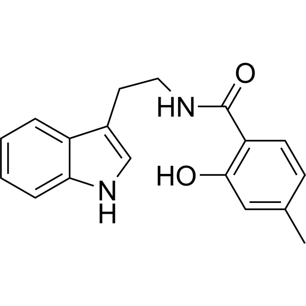
LZWL02003 is an anti-neuroinflammatory agent. LZWL02003 has protective effect on MPP+-induced neuronal damage, and reduces the expression of ROS. LZWL02003 improves cognition, memory, learning, and athletic ability in a Rotenone (HY-B1756)-induced PD rat model. LZWL02003 can be used for research of neurodegenerative disease.
Nur für Forschungszwecke. Wir verkaufen nicht an Patienten.
- Reinheit : 99.56%
- CAS. Nr.: 1371564-45-2
- Formel: C18H18N2O2
- Molecular Weight:294.35
-
Speicherung:Powder -20°C, 3 years , 4°C, 2 years ; In solvent -80°C, 6 months , -20°C, 1 month

To place orders, for customer services and technical support, please contact: MedChemExpress USA
Tel: 609-228-6898 E-mail: [email protected] [email protected]


-
 Biologische Aktivität
-
Chemical Information
-
Lösungsmittel & Löslichkeit
- Protokoll
- Reinheit & Dokumentation
- Verweise
-
 Help & FAQs


-
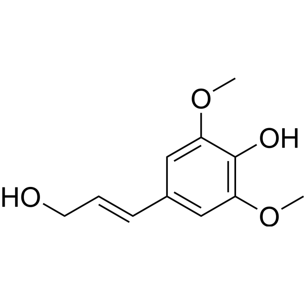
HY-W109812
Sinapyl alcoholAnti-inflammatory/Antinociceptive Agent -
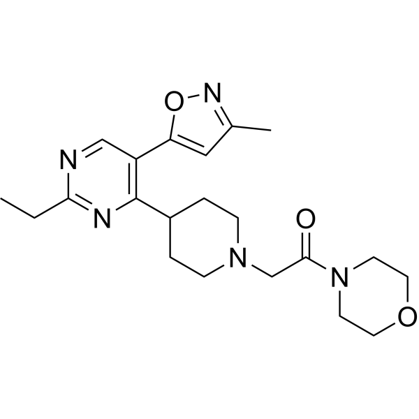
HY-155992
WLB-89462σ2 Receptor Ligand -
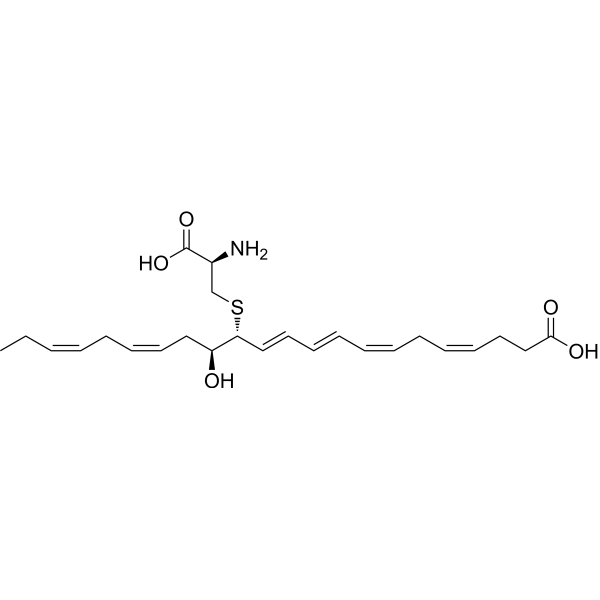
HY-125516
MCTR3Pro-resolving Mediator -
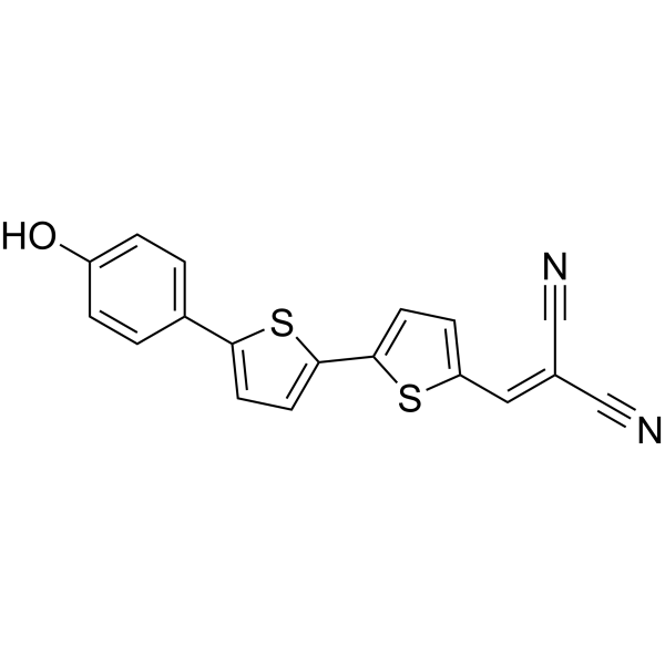
HY-111263
NIAD-4Amyloid-β Fluorophore -
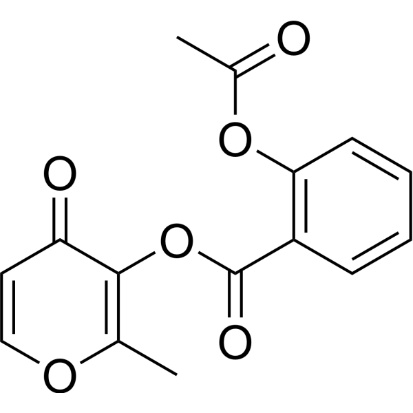
HY-118072
AspalatoneAntiplatelet Aggregant -

HY-W109812R
Sinapyl alcohol (Standard)Anti-inflammatory/Antinociceptive Agent -

HY-139012
RC574GPx1 Activator -

HY-155286
(7S,8R,17S)-RCTR1Resolvin-Sulfido Conjugate -
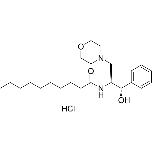
HY-116392A
(−)-L-threo-PDMP hydrochlorideGlucosylceramide Synthase Inhibitor -
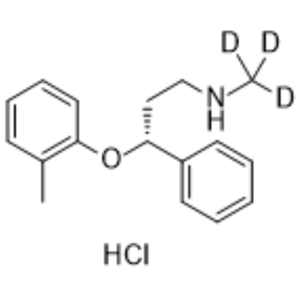
HY-110223
Atomoxetine-d3 hydrochlorideStable Isotope -

HY-170645
Neuroprotectin D1 methyl esterProtectin D1 Intermediate -
HY-120251A
Leteprinim potassiumNeuroprotective Agent


Biologische Aktivität
Beschreibung
Cellular Effect
|
Cell Line

|
Type | Value | Description | References |
|---|---|---|---|---|
| A549 | IC50 |
>100 μM
Compound: E3
|
Antiproliferative activity against human A549 cells after 48 hrs by MTT assay
Antiproliferative activity against human A549 cells after 48 hrs by MTT assay
|
[PMID: 31057737] |
| BV-2 | IC50 |
>100 μM
Compound: 3
|
Cytotoxicity against mouse BV-2 cells assessed as reduction in cell viability after 24 hrs by MTT assay
Cytotoxicity against mouse BV-2 cells assessed as reduction in cell viability after 24 hrs by MTT assay
|
[PMID: 32182488] |
| BV-2 | IC50 |
>100 μM
Compound: L7
|
Cytotoxicity against mouse BV-2 cells assessed as cell viability incubated for 24 hrs by CCK-8 method
Cytotoxicity against mouse BV-2 cells assessed as cell viability incubated for 24 hrs by CCK-8 method
|
[PMID: 39216381] |
| BV-2 | IC50 |
>40 μM
Compound: 3
|
Antiinflammatory activity against LPS-induced mouse BV-2 cells assessed as decrease in COX2 protein expression after 24 hrs by Western blot analysis
Antiinflammatory activity against LPS-induced mouse BV-2 cells assessed as decrease in COX2 protein expression after 24 hrs by Western blot analysis
|
[PMID: 32182488] |
| BV-2 | IC50 |
4.94 μM
Compound: 3
|
Inhibition of LPS-induced NO production in mouse BV-2 cells incubated for 24 hrs by Griess reagent based assay
Inhibition of LPS-induced NO production in mouse BV-2 cells incubated for 24 hrs by Griess reagent based assay
|
[PMID: 32182488] |
| C6 | IC50 |
>100 μM
Compound: 3
|
Cytotoxicity against rat C6 cells assessed as reduction in cell viability after 24 hrs by MTT assay
Cytotoxicity against rat C6 cells assessed as reduction in cell viability after 24 hrs by MTT assay
|
[PMID: 32182488] |
| C6 | IC50 |
7.48 μM
Compound: 3
|
Inhibition of LPS-induced NO production in rat C6 cells incubated for 24 hrs by Griess reagent based assay
Inhibition of LPS-induced NO production in rat C6 cells incubated for 24 hrs by Griess reagent based assay
|
[PMID: 32182488] |
| HeLa | IC50 |
>100 μM
Compound: E3
|
Antiproliferative activity against human HeLa cells after 48 hrs by MTT assay
Antiproliferative activity against human HeLa cells after 48 hrs by MTT assay
|
[PMID: 31057737] |
| HepG2 | IC50 |
>100 μM
Compound: E3
|
Antiproliferative activity against human HepG2 cells after 48 hrs by MTT assay
Antiproliferative activity against human HepG2 cells after 48 hrs by MTT assay
|
[PMID: 31057737] |
| HT-22 | IC50 |
>100 μM
Compound: L7
|
Cytotoxicity against mouse HT-22 cells assessed as cell viability incubated for 24 hrs by CCK-8 method
Cytotoxicity against mouse HT-22 cells assessed as cell viability incubated for 24 hrs by CCK-8 method
|
[PMID: 39216381] |
| MCF7 | IC50 |
>100 μM
Compound: E3
|
Antiproliferative activity against human MCF7 cells after 48 hrs by MTT assay
Antiproliferative activity against human MCF7 cells after 48 hrs by MTT assay
|
[PMID: 31057737] |
| MGC-803 | IC50 |
80 μM
Compound: E3
|
Antiproliferative activity against human MGC803 cells after 48 hrs by MTT assay
Antiproliferative activity against human MGC803 cells after 48 hrs by MTT assay
|
[PMID: 31057737] |


Chemical Information
-
CAS. Nr. 1371564-45-2
-
Appearance Solid
-
Molecular Weight 294.35
-
Formel C18H18N2O2
-
Color White to off-white
-
SMILES
O=C(C1=C(O)C=C(C)C=C1)NCCC2=CNC3=CC=CC=C32
-
Versand
Room temperature in continental US; may vary elsewhere.
-
Speicherung
Powder -20°C 3 years 4°C 2 years In solvent -80°C 6 months -20°C 1 month


Lösungsmittel & Löslichkeit
In Vitro:
DMSO : 100 mg/mL (339.73 mM; Need ultrasonic; Hygroscopic DMSO has a significant impact on the solubility of product, please use newly opened DMSO)
Please refer to the solubility information to select the appropriate solvent. Once prepared, please aliquot and store the solution to prevent product inactivation from repeated freeze-thaw cycles.
Storage method and period of stock solution: -80°C, 6 months; -20°C, 1 month. When stored at -80°C, please use it within 6 months. When stored at -20°C, please use it within 1 month.
Please refer to the solubility information to select the appropriate solvent. Once prepared, please aliquot and store the solution to prevent product inactivation from repeated freeze-thaw cycles.
Storage method and period of stock solution: -80°C, 6 months; -20°C, 1 month. When stored at -80°C, please use it within 6 months. When stored at -20°C, please use it within 1 month.
Konzentration (Stammlösung) × Volumen (Stammlösung) = Konzentration (Ziellösung) × Volumen (Ziellösung)
In Vivo:
Select the appropriate dissolution method based on your experimental animal and administration route.
- For the following dissolution methods, please ensure to first prepare a clear stock solution using an In Vitro approach and then sequentially add co-solvents:
- To ensure reliable experimental results, the clarified stock solution can be appropriately stored based on storage conditions. As for the working solution for In Vivo experiments, it is recommended to prepare freshly and use it on the same day.
- The percentages shown for the solvents indicate their volumetric ratio in the final prepared solution. If precipitation or phase separation occurs during preparation, heat and/or sonication can be used to aid dissolution.
Add each solvent one by one: 10% DMSO 40% PEG300 5% Tween-80 45% Saline
Solubility: ≥ 2.5 mg/mL (8.49 mM); Clear solution
This protocol yields a clear solution of ≥ 2.5 mg/mL (saturation unknown).
Taking 1 mL working solution as an example, add 100 μL DMSO stock solution (25.0 mg/mL) to 400 μL PEG300, and mix evenly; then add 50 μL Tween-80 and mix evenly; then add 450 μL Saline to adjust the volume to 1 mL.
Preparation of Saline: Dissolve 0.9 g sodium chloride in ddH₂O and dilute to 100 mL to obtain a clear Saline solution.
Add each solvent one by one: 10% DMSO 90% (20% SBE-β-CD in Saline)
Solubility: ≥ 2.5 mg/mL (8.49 mM); Clear solution
This protocol yields a clear solution of ≥ 2.5 mg/mL (saturation unknown).
Taking 1 mL working solution as an example, add 100 μL DMSO stock solution (25.0 mg/mL) to 900 μL 20% SBE-β-CD in Saline, and mix evenly.
Preparation of 20% SBE-β-CD in Saline (4°C, storage for one week): 2 g SBE-β-CD powder is dissolved in 10 mL Saline, completely dissolve until clear.
In Vivo Dissolution Calculator
Please enter the basic information of animal experiments:
-
-
-
-
Recommended: Prepare an additional quantity of animals to account for potential losses during experiments.
Please enter your animal formula composition:
-
%DMSO +
Recommended: Keep the proportion of DMSO in working solution below 2% if your animal is weak.
-
%+
-
+%Tween-80 + +
-
%Saline +
The co-solvents required include: DMSO, . All of co-solvents are available by MedChemExpress (MCE). , Tween 80. All of co-solvents are available by MedChemExpress (MCE).
Working solution concentration: 0.22 mg/mL
Method for preparing stock solution: mg drug dissolved in μL DMSO. Stock solution concentration: mg/mL.
1. Take μL DMSO stock solution;
2. Add μL .
μL , mix evenly;
3. Then add μL Tween 80, mix evenly;
4. Then add μL
Please ensure that the stock solution in the first step is dissolved to a clear state, and add co-solvents in sequence. You can use ultrasonic heating (ultrasonic cleaner, recommended frequency 20-40 kHz), vortexing, etc. to assist dissolution.


Reinheit & Dokumentation
-
Data Sheet (275 KB)
-
SDS (251 KB)
- English - EN (251 KB)
- Français - FR (251 KB)
- Deutsch - DE (251 KB)
- Norwegian - NO (251 KB)
- Español - ES (251 KB)
- Swedish - SV (251 KB)
- Italian - IT (251 KB)
- Korean - KR (251 KB)
- Portuguese - PT (251 KB)
-
Handling Instructions (2659 KB)
Verweise


Complete Stock Solution Preparation Table
Please refer to the solubility information to select the appropriate solvent. Once prepared, please aliquot and store the solution to prevent product inactivation from repeated freeze-thaw cycles.
Storage method and period of stock solution: -80°C, 6 months; -20°C, 1 month. When stored at -80°C, please use it within 6 months. When stored at -20°C, please use it within 1 month.
| Optional Solvent | Concentration Solvent Mass | 1 mg | 5 mg | 10 mg | 25 mg |
|---|---|---|---|---|---|
| DMSO | 1 mM | 3.3973 mL | 16.9866 mL | 33.9732 mL | 84.9329 mL |
| 5 mM | 0.6795 mL | 3.3973 mL | 6.7946 mL | 16.9866 mL | |
| 10 mM | 0.3397 mL | 1.6987 mL | 3.3973 mL | 8.4933 mL | |
| 15 mM | 0.2265 mL | 1.1324 mL | 2.2649 mL | 5.6622 mL | |
| 20 mM | 0.1699 mL | 0.8493 mL | 1.6987 mL | 4.2466 mL | |
| 25 mM | 0.1359 mL | 0.6795 mL | 1.3589 mL | 3.3973 mL | |
| 30 mM | 0.1132 mL | 0.5662 mL | 1.1324 mL | 2.8311 mL | |
| 40 mM | 0.0849 mL | 0.4247 mL | 0.8493 mL | 2.1233 mL | |
| 50 mM | 0.0679 mL | 0.3397 mL | 0.6795 mL | 1.6987 mL | |
| 60 mM | 0.0566 mL | 0.2831 mL | 0.5662 mL | 1.4155 mL | |
| 80 mM | 0.0425 mL | 0.2123 mL | 0.4247 mL | 1.0617 mL | |
| 100 mM | 0.0340 mL | 0.1699 mL | 0.3397 mL | 0.8493 mL |


LZWL02003 Related Classifications
HY-155126 Related Classifications
 Powered by Bioz