



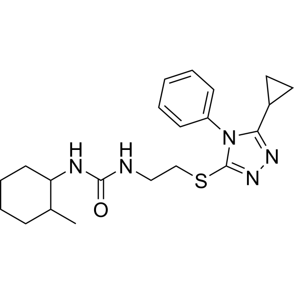
MFN2 agonist-1

Based on 1 Customer Validation
MFN2 agonist-1 is an allosteric agonist of mitofusin 2 (MFN2) with an EC50 of 3 nM. MFN2 agonist-1 mimics the MFN2 HR1 peptide to compete for HR2, disrupt the HR1-HR2 autoinhibitory interaction, stabilize the open conformation of MFN2, and promote GTP hydrolysis and mitochondrial fusion. MFN2 agonist-1 acts in a manner dependent on endogenous MFN1 or MFN2, and does not impair cell viability. MFN2 agonist-1 reverses mitochondrial fragmentation, depolarization, aggregation and axonal transport defects, restores bidirectional movement and improves enhanced autophagy. MFN2 agonist-1 is used in studies related to Charcot-Marie-Tooth disease type 2A, Alzheimer's disease, Parkinson's disease and Huntington's disease.
For research use only. We do not sell to patients.
- Purity : 98.84%
- CAS No.: 2230047-87-5
- Formula: C21H29N5OS
- Molecular Weight:399.55
-
Storage:Powder -20°C, 3 years , 4°C, 2 years ; In solvent -80°C, 6 months , -20°C, 1 month

To place orders, for customer services and technical support, please contact: MedChemExpress USA
Tel: 609-228-6898 E-mail: [email protected] [email protected]


-
 Biological Activity
-
Chemical Information
-
Solvent & Solubility
- Protocols
- Purity & Documentation
- References
-
 Help & FAQs




Biological Activity
Description
IC50 & Target
[1]|
MFN2 3 nM (EC50) |
In Vitro
MFN2 agonist-1 (B-A/l) (0.1 nM-10 μM) potently stimulates mitochondrial fusion in MFN1-/- MFN2-/- mouse embryonic fibroblasts (MEFs), with an EC50 of 3 nM[1].
MFN2 agonist-1 (0.01 nM-10 μM) displaces the MFN2 HR1 peptide (374-384) from the MFN2 HR2 domain, with an IC50 of 150 nM[1].
MFN2 agonist-1 restores mitochondrial fusion in MFN2-/- MEFs expressing the CMT2A mutant MFN2 T105M[1].
MFN2 agonist-1 induces MFN2 to form an open, fusion-permissive conformation in isolated mitochondria and intact cells, which is confirmed by reduced FRET signals[1].
MFN2 agonist-1 corrects mitochondrial fragmentation and depolarization in MFN1+/+ MFN2-/- MEFs expressing the CMT2A mutants MFN2R94Q or MFN2K109A via activation of endogenous MFN1[1].
MFN2 agonist-1 reverses mitochondrial fragmentation and decreased mitochondrial membrane potential in primary cultured neonatal mouse neurons expressing the CMT2A mutant MFN2R94Q[1].
MFN2 agonist-1 (10-60 min post-administration) restores axonal mitochondrial motility and velocity in ex vivo sciatic nerves of MFN2T105M mice[1].
MFN2 agonist-1 restores mitochondrial transport, function and morphology in cultured neonatal mouse neurons expressing the CMT2A mutant MFN2T105M[1].
MedChemExpress (MCE) has not independently confirmed the accuracy of these methods. They are for reference only.
- Cell Cytotoxicity Assay
- Mammalian live/dead viability and cytotoxicity staining
- Human pluripotent stem cell midbrain dopaminergic neuron differentiation
- Autophagy
- CCK-8/WST-8 Cell Proliferation Assay
- Lysosome and acidic-vesicle live-cell staining
- MTT Cell Proliferation Assay
- Macroautophagy Solutions
- Cell Viability Determination by MTT Colorimetric Assay
- Alzheimer’s Disease Modeling


Chemical Information
-
CAS No. 2230047-87-5
-
Appearance Solid
-
Molecular Weight 399.55
-
Formula C21H29N5OS
-
Color White to off-white
-
SMILES
O=C(NC1C(C)CCCC1)NCCSC2=NN=C(C3CC3)N2C4=CC=CC=C4
-
Shipping
Room temperature in continental US; may vary elsewhere.
-
Storage
Powder -20°C 3 years 4°C 2 years In solvent -80°C 6 months -20°C 1 month


Solvent & Solubility
In Vitro:
DMSO : 50 mg/mL (125.14 mM; ultrasonic and warming and heat to 60°C; Hygroscopic DMSO has a significant impact on the solubility of product, please use newly opened DMSO)
Please refer to the solubility information to select the appropriate solvent. Once prepared, please aliquot and store the solution to prevent product inactivation from repeated freeze-thaw cycles.
Storage method and period of stock solution: -80°C, 6 months; -20°C, 1 month. When stored at -80°C, please use it within 6 months. When stored at -20°C, please use it within 1 month.
Please refer to the solubility information to select the appropriate solvent. Once prepared, please aliquot and store the solution to prevent product inactivation from repeated freeze-thaw cycles.
Storage method and period of stock solution: -80°C, 6 months; -20°C, 1 month. When stored at -80°C, please use it within 6 months. When stored at -20°C, please use it within 1 month.
Concentration (start) × Volume (start) = Concentration (final) × Volume (final)


Purity & Documentation
-
Data Sheet (285 KB)
-
SDS (393 KB)
- English - EN (393 KB)
- Français - FR (393 KB)
- Deutsch - DE (393 KB)
- Norwegian - NO (393 KB)
- Español - ES (393 KB)
- Swedish - SV (393 KB)
- Italian - IT (393 KB)
- Korean - KR (393 KB)
- Portuguese - PT (393 KB)
-
Handling Instructions (2659 KB)
References


Complete Stock Solution Preparation Table
Please refer to the solubility information to select the appropriate solvent. Once prepared, please aliquot and store the solution to prevent product inactivation from repeated freeze-thaw cycles.
Storage method and period of stock solution: -80°C, 6 months; -20°C, 1 month. When stored at -80°C, please use it within 6 months. When stored at -20°C, please use it within 1 month.
| Optional Solvent | Concentration Solvent Mass | 1 mg | 5 mg | 10 mg | 25 mg |
|---|---|---|---|---|---|
| DMSO | 1 mM | 2.5028 mL | 12.5141 mL | 25.0282 mL | 62.5704 mL |
| 5 mM | 0.5006 mL | 2.5028 mL | 5.0056 mL | 12.5141 mL | |
| 10 mM | 0.2503 mL | 1.2514 mL | 2.5028 mL | 6.2570 mL | |
| 15 mM | 0.1669 mL | 0.8343 mL | 1.6685 mL | 4.1714 mL | |
| 20 mM | 0.1251 mL | 0.6257 mL | 1.2514 mL | 3.1285 mL | |
| 25 mM | 0.1001 mL | 0.5006 mL | 1.0011 mL | 2.5028 mL | |
| 30 mM | 0.0834 mL | 0.4171 mL | 0.8343 mL | 2.0857 mL | |
| 40 mM | 0.0626 mL | 0.3129 mL | 0.6257 mL | 1.5643 mL | |
| 50 mM | 0.0501 mL | 0.2503 mL | 0.5006 mL | 1.2514 mL | |
| 60 mM | 0.0417 mL | 0.2086 mL | 0.4171 mL | 1.0428 mL | |
| 80 mM | 0.0313 mL | 0.1564 mL | 0.3129 mL | 0.7821 mL | |
| 100 mM | 0.0250 mL | 0.1251 mL | 0.2503 mL | 0.6257 mL |


MFN2 agonist-1 Related Classifications
HY-123985 Related Classifications
 Powered by Bioz