



Minodronic acid
Based on 1 publication(s) in Google Scholar
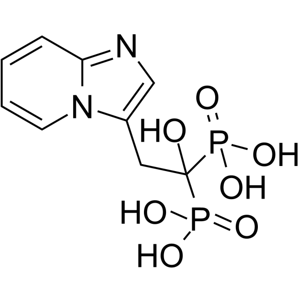
Minodronic acid (YM-529) is an FPP synthase inhibitor with an IC50 of 3 nM, and also an antagonist of P2X2/3 receptors with an IC50 of 62.7 μM. Minodronic acid induces tumor cell apoptosis and inhibits cell growth. Minodronic acid also suppresses bone resorption. Minodronic acid can be used in research related to osteoporosis and cancer.
For research use only. We do not sell to patients.
- Purity : 99.67%
- CAS No.: 180064-38-4
- Formula: C9H12N2O7P2
- Molecular Weight:322.15
-
Storage:Powder -20°C, 3 years , 4°C, 2 years ; In solvent -80°C, 2 years , -20°C, 1 year

To place orders, for customer services and technical support, please contact: MedChemExpress USA
Tel: 609-228-6898 E-mail: [email protected] [email protected]


-
 Biological Activity
-
Chemical Information
-
Solvent & Solubility
- Protocols
- Purity & Documentation
- References
-
 Help & FAQs


-
HY-13062
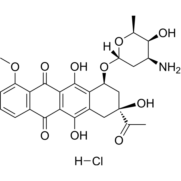
Daunorubicin hydrochlorideTopoisomerase II Inhibitor -
HY-B0631
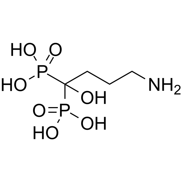
Alendronic acidBisphosphonate -
HY-B0148
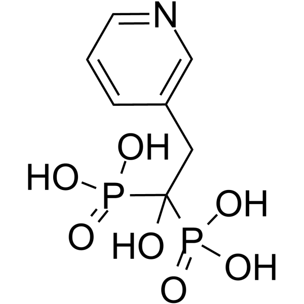
Risedronic acidAnti-resorption Agent -
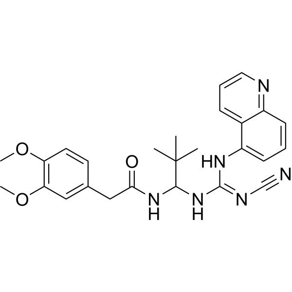
HY-50697
A-740003P2X Receptor Antagonist -
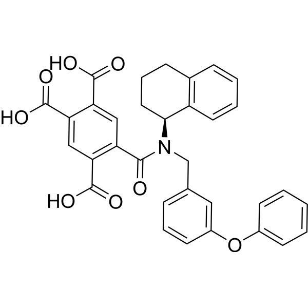
HY-15568
A-317491P2X3/P2X2/3 Antagonist -
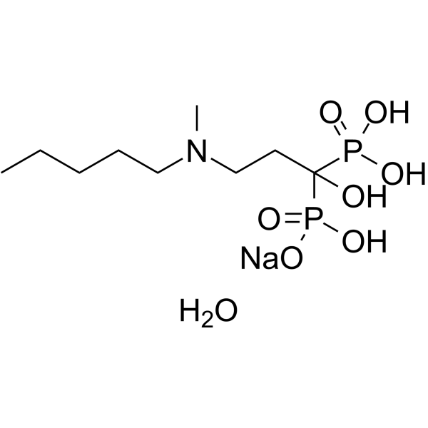
HY-B0515
Ibandronate Sodium MonohydrateFPP synthase Inhibitor -
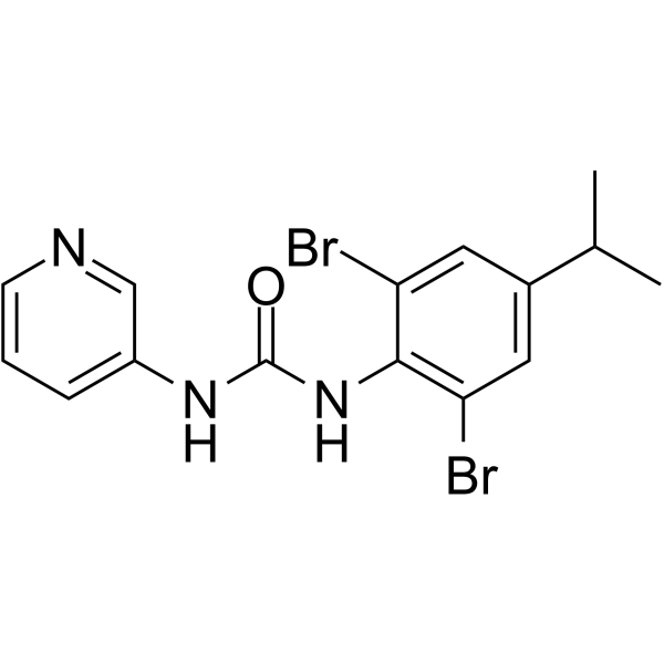
HY-110237
BX430P2X4 Receptor Antagonist -
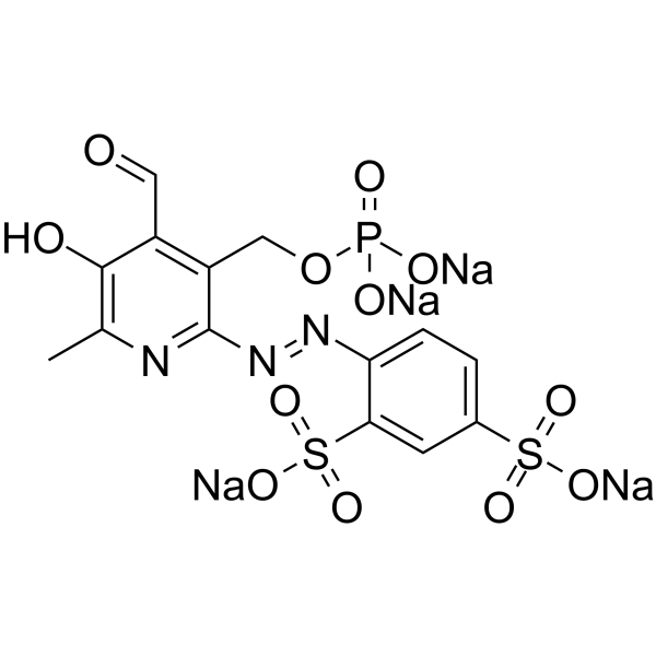
HY-101044
PPADS tetrasodiumP2X Receptor Antagonist -
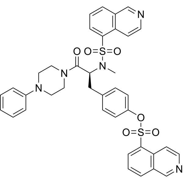
HY-13290
KN-62CaMKII Inhibitor -
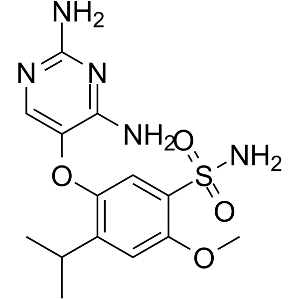
HY-101588
GefapixantP2X3 Antagonist -
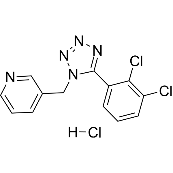
HY-15488A
A 438079 hydrochlorideP2X7 Receptor Antagonist -
HY-B0879A
Suramin sodium saltPTPases Inhibitor
Publications Citing Use of MedChemExpress (MCE) Minodronic acid
MoreAll P2X Receptor Isoforms
More

Biological Activity
Description
IC50 & Target
|
P2X2 Receptor |
P2X3 Receptor |
Cellular Effect
|
Cell Line

|
Type | Value | Description | References |
|---|---|---|---|---|
| HFF | IC50 |
1.88 μM
Compound: 4
|
In vitro inhibitory concentration against the growth of Toxoplasma gondii in human foreskin fibroblast monolayer cells (HFF cells)
In vitro inhibitory concentration against the growth of Toxoplasma gondii in human foreskin fibroblast monolayer cells (HFF cells)
|
[PMID: 15857119] |
| J774 | IC50 |
3 μM
Compound: 4
|
Cytotoxicity against mouse J774 cells assessed as cell viability after 48 hrs by alamar blue assay
Cytotoxicity against mouse J774 cells assessed as cell viability after 48 hrs by alamar blue assay
|
[PMID: 20394422] |
| T-cell | IC50 |
4.1 μM
Compound: 3
|
Inhibitory activity, for stimulation of TNF-alpha release in gamma-delta T cells, using individual observed maximum TNF-alpha release
Inhibitory activity, for stimulation of TNF-alpha release in gamma-delta T cells, using individual observed maximum TNF-alpha release
|
[PMID: 14711309] |
| T-cell | IC50 |
8 μM
Compound: 3
|
Inhibitory activity, for stimulation of TNF-alpha release in gamma-delta T cells, using a constrained maximum TNF-alpha release of 2700 pg/mL
Inhibitory activity, for stimulation of TNF-alpha release in gamma-delta T cells, using a constrained maximum TNF-alpha release of 2700 pg/mL
|
[PMID: 14711309] |
In Vitro
Minodronic acid (5-100 μM; 72 h) inhibits the proliferation of human bladder cancer cell lines 253J, 5637, RT4, RT112, TCCSUP, KU-7 and UM-UC-3 in a dose-dependent manner[1].
Minodronic acid (30 μM; 48 h) induces apoptosis in human bladder cancer cells[1].
Minodronic acid (0.25-2.0 × IC50; 72 h) exhibits synergistic or additive antiproliferative effects when combined with Cisplatin (HY-17394) or Paclitaxel (HY-B0015) in human bladder cancer cell lines KU-7, RT112 and UM-UC-3[1].
Minodronic acid (0.1-1 μM; 3 days) potently inhibits osteoclast-mediated bone resorption in rabbits and induces dose-dependent morphological changes; at 1 μM, it almost completely inhibits bone resorption and causes osteoclast detachment, while at 0.1 μM, it only exerts partial inhibitory effects accompanied by the disappearance of the ruffled border[2].
MedChemExpress (MCE) has not independently confirmed the accuracy of these methods. They are for reference only. Further protocols information, click here.
-
Cell Line:253J, 5637, RT4, RT112, TCCSUP, KU-7, UM-UC-3 human bladder cancer cells
-
Concentration:5, 10, 25, 50, 100 μM
-
Incubation Time:72 h
-
Result:Inhibited cell growth in a dose-dependent manner.
Achieved IC50 values of 12.2 μM (253J), 7.1 μM (5637), 12.2 μM (RT4), 15.4 μM (RT112), 21.8 μM (TCCSUP), 24.8 μM (KU-7), and 20.8 μM (UM-UC-3).
In Vivo
Minodronic acid (30-100 μM; transurethral administration; 5 days) inhibits the growth of orthotopic bladder cancer in SCID mice [1].
Minodronic acid (0.05-5 mg/kg; subcutaneous injection; single administration) accumulates in the calcified and ossified regions of rat bones, concentrates in osteoclast-containing bone tissue sites, and exhibits dose-dependent bone tissue localization characteristics[2].
MedChemExpress (MCE) has not independently confirmed the accuracy of these methods. They are for reference only.
-
Animal Model:BALB/c nu/nu (female, 6-8 weeks old, intracardiac injection of 1×10⁶ UM-UC-3^LUC cells to induce bone metastases)[1]
-
Dosage:80 μg/kg
-
Administration:s.c.; once weekly; 3 weeks
-
Result:Significantly reduced growth of bone metastatic lesions compared to untreated mice.
Did not significantly inhibit visceral metastases.
Showed no significant differences in body weight or serum components (AST, ALT, LDH, TP, BUN, Cre, Ca) compared to untreated mice.
-
Animal Model:SCID (female, 6-8 weeks old, orthotopic implantation of 2×10⁶ UM-UC-3^LUC cells into bladder)[1]
-
Dosage:30 μM; 100 μM
-
Administration:transurethral; daily for 5 consecutive days (days 5-9)
-
Result:Significantly suppressed orthotopic bladder tumor growth, no significant systemic toxicity was observed, and occasional deaths were attributed to mechanical renal injury rather than drug-related toxicity.
-
Animal Model:F334/DuCrj rats (female, 4 days postpartum, 5-7 g)[2]
-
Dosage:0.05 mg/kg (0.311 MBq/kg); 0.5 mg/kg (3.11 MBq/kg); 5 mg/kg (31.1 MBq/kg)
-
Administration:s.c.; single dose
-
Result:Localized approximately 2% of total administered radioactivity to right humeri at 24 hours post-administration, with radioactivity increasing linearly with dose.
Showed radioactive silver grains distributed in the calcified zone, ossification zone, and spongiosa of femurs, but absent in the hypertrophic cartilage zone.
Detected condensed silver grains in spongiosa under and surrounding osteoclasts, with low levels of intracellular silver grains present in osteoclasts.


Chemical Information
-
CAS No. 180064-38-4
-
Appearance Solid
-
Molecular Weight 322.15
-
Formula C9H12N2O7P2
-
Color White to off-white
-
SMILES
OC(P(O)(O)=O)(P(O)(O)=O)CC1=CN=C2C=CC=CN21
-
Synonyms
YM-529
-
Shipping
Room temperature in continental US; may vary elsewhere.
-
Storage
Powder -20°C 3 years 4°C 2 years In solvent -80°C 2 years -20°C 1 year


Publications (1)
-
Journal Impact Factor
-
Most Recent
-
Int J Biol Macromol
Reticuline isomerase AKR1B1 with aldo-keto reductase activity and detoxification function from the insect Blaps rhynchopetera. [Abstract]2026 Mar:352:151224. PMID: 41791543


Solvent & Solubility
In Vitro:
H2O : 5 mg/mL (15.52 mM; Need ultrasonic and warming)
H2O : 5 mg/mL (15.52 mM; ultrasonic and warming and adjust pH to 10 with NaOH and heat to 80°C)
DMSO : < 1 mg/mL (insoluble or slightly soluble)
Please refer to the solubility information to select the appropriate solvent. Once prepared, please aliquot and store the solution to prevent product inactivation from repeated freeze-thaw cycles.
Storage method and period of stock solution: -80°C, 2 years; -20°C, 1 year. When stored at -80°C, please use it within 2 years. When stored at -20°C, please use it within 1 year.
* Note: If you choose water as the stock solution, please dilute it to the working solution, then filter and sterilize it with a 0.22 μm filter before use.
Please refer to the solubility information to select the appropriate solvent. Once prepared, please aliquot and store the solution to prevent product inactivation from repeated freeze-thaw cycles.
Storage method and period of stock solution: -80°C, 2 years; -20°C, 1 year. When stored at -80°C, please use it within 2 years. When stored at -20°C, please use it within 1 year.
* Note: If you choose water as the stock solution, please dilute it to the working solution, then filter and sterilize it with a 0.22 μm filter before use.
Concentration (start) × Volume (start) = Concentration (final) × Volume (final)
In Vivo:
For the following dissolution methods, please prepare the working solution directly:
It is recommended to prepare fresh solutions and use them promptly within a short period of time.
The percentages shown for the solvents indicate their volumetric ratio in the final prepared solution. If precipitation or phase separation occurs during preparation, heat and/or sonication can be used to aid dissolution.
Add each solvent one by one: PBS
Solubility: 2 mg/mL (6.21 mM); Clear solution; Need ultrasonic

Protocols
-
Apoptosis
Apoptosis, also called programmed cell death, is generally characterized by distinct morphological characteristics.
-
TUNEL staining for apoptotic DNA fragmentation
TUNEL staining detects DNA strand breaks by using terminal deoxynucleotidyl transferase to add labeled nucleotides to exposed 3′-OH DNA termini, generating either microscopic staining in fixed cells or tissue sections, or fluorescence/cytometric signal in cell suspensions. TUNEL positivity reflects DNA fragmentation but should not be interpreted alone as definitive apoptosis, because TUNEL can also label necrotic, autolytic, mechanically damaged, or DNA-repair-associated DNA breaks.
-
Cell Cytotoxicity Assay
Cytotoxicity assays are usually based on the assessment of cell membrane damage, which can also be indirectly detected by measuring cell viability. Detection methods include MTT assay, CKK-8 assay, LDH assay and ATP assay, etc.
-
Annexin V plus membrane-impermeant dye apoptosis staining
Annexin V-based apoptosis assays rely on the detection of phosphatidylserine (PS) externalization from the inner leaflet of the plasma membrane to the outer leaflet, an early biochemical hallmark of apoptosis. Fluorescently labeled Annexin V binds PS in a calcium-dependent manner, enabling identification of early apoptotic cells by flow cytometry or fluorescence microscopy. When combined with a membrane-impermeant DNA-binding dye (e. g. , propidium iodide), this approach allows discrimination between viable (Annexin V−/dye−), early apoptotic (Annexin V+/dye−), and late apoptotic or necrotic (Annexin V+/dye+) cell populations by assessing membrane integrity and PS exposure.
-
Protocol for Cell Counting and Cell Density Analysis
Cell counting and cell-density analysis estimate the number of cells in a known volume or field area. Manual hemocytometer counting uses a chamber of defined geometry to convert counted cells into cells/mL, while automated counters and image-analysis workflows detect cell objects from optical, brightfield, fluorescence, impedance, or digital-image features. Trypan blue viability counting is based on dye exclusion: viable cells with intact membranes exclude dye, while non-viable cells with compromised membranes stain blue. The readout is total cell density, viable-cell density, dead-cell density, and percent viability. Cell density can also be estimated from microscopy images by counting objects per image area, from flow cytometry using calibrated volume or reference particles, or from in situ microscopy in bioreactors after calibration against reference methods such as hemocytometer or flow cytometry.
-
Somatic Cell Culture
A method of simulating the in vivo environment in vitro to maintain the cell growth, differentation and main functions.
-
Apoptosis Solutions
Apoptosis is a regulated, generally non-lytic cell-death pathway that removes unwanted, damaged, infected, or abnormal cells through coordinated morphological changes, caspase activation, DNA fragmentation, and membrane remodeling. The intrinsic apoptosis pathway is controlled mainly by mitochondrial outer membrane permeabilization, BCL-2 family proteins, cytochrome c release, apoptosome formation, caspase-9 activation, and downstream executioner caspase-3/7 activation. The extrinsic apoptosis pathway is initiated by death receptors such as Fas, TNFR, and TRAIL receptors, which recruit adaptor proteins and activate caspase-8 before engaging executioner caspases or mitochondrial amplification through BID cleavage. Apoptosis is linked to many phenotypes, including cancer cell killing, tissue homeostasis, immune regulation, neurodegeneration, infection response, and treatment-induced cytotoxicity; unresolved questions include how apoptosis interacts with necroptosis, pyroptosis, ferroptos
-
CCK-8/WST-8 Cell Proliferation Assay
The CCK-8/WST-8 assay is based on the reduction of the water-soluble tetrazolium salt WST-8 to a water-soluble formazan product by cellular dehydrogenases in metabolically active cells, where the generated formazan amount is proportional to the number of living cells and is quantified by measuring absorbance in the visible range, providing a colorimetric readout for cell viability and proliferation assessment. This class of tetrazolium-based assays improves upon earlier MTT-based systems by producing a water-soluble formazan, eliminating the need for organic solubilization steps and enabling direct spectrophotometric measurement in culture medium.
-
Cell Counting-Based Growth Curve Assay
Cell counting-based growth curve assays quantify cell proliferation by directly measuring changes in viable cell number over time using manual or automated counting methods such as hemocytometer-based counting or instrument-assisted cell enumeration, enabling construction of growth curves that reflect population expansion dynamics in response to culture conditions. A widely used approach is trypan blue exclusion with hemocytometer counting, where membrane-compromised (non-viable) cells take up the dye, allowing discrimination between viable and non-viable cells while simultaneously enabling total cell number quantification. Repeated sampling across time points allows estimation of proliferation rate, growth phases, and comparative growth kinetics between experimental conditions.
-
MTT Cell Proliferation Assay
The MTT assay is a colorimetric endpoint assay for estimating viable cell number, cell growth, cytotoxicity, or cell activation in cultured mammalian cells. Living cells reduce the yellow tetrazolium salt MTT into purple/blue formazan, while dead cells do not generate the same signal; the resulting color can be quantified with a multiwell spectrophotometer. MTT reduction is commonly interpreted as a readout of metabolic activity that often correlates with viable cell number, but it should not be treated as a direct cell-counting method unless the assay is optimized for the cell type and experimental condition. Studies show that MTT reduction can involve mitochondrial and non-mitochondrial reducing systems, and formazan may accumulate in intracellular lipid droplets rather than simply marking mitochondria.
-
Cell Viability Determination by MTT Colorimetric Assay
The following protocol uses the MTT colorimetric assay as a classic literature-established method for assessing cell viability/metabolic activity in cultured mammalian cells. MTT[3-(4,5-dimethylthiazol-2-yl)-2,5-diphenyltetrazolium bromide] is reduced by metabolically active cells to a colored formazan product; the amount of formazan is quantified spectrophotometrically and provides an indirect measure of metabolically active viable cells. Importantly, MTT reduction reflects cellular oxidoreductase/metabolic activity rather than an absolute direct count of living cells, so changes in cellular metabolism can alter the signal independently of cell number.
-
Osteoclast differentiation from monocyte/macrophage precursors
Osteoclast differentiation is an in vitro induction assay in which monocyte/macrophage-lineage precursors are exposed to macrophage colony-stimulating factor (M-CSF) and receptor activator of NF-κB ligand (RANKL), generating multinucleated osteoclasts that are commonly identified by tartrate-resistant acid phosphatase (TRAP) staining and functionally confirmed by resorption pits on dentin, bone, or mineralized substrates. M-CSF supports survival and expansion of osteoclast precursors, while RANKL binding to RANK drives osteoclast commitment, fusion, maturation, and resorptive function; osteoprotegerin inhibits this pathway by binding RANKL and preventing RANK activation. The main readouts are the number of TRAP-positive multinucleated cells, formation of F-actin rings, and resorbed surface area; TRAP-positive multinucleated cells indicate osteoclast differentiation, whereas pit formation on dentin, bone, or mineralized coating indicates functional bone-resorbing activity.


Purity & Documentation
-
Data Sheet (316 KB)
-
SDS (393 KB)
- English - EN (393 KB)
- Français - FR (393 KB)
- Deutsch - DE (393 KB)
- Norwegian - NO (393 KB)
- Español - ES (393 KB)
- Swedish - SV (393 KB)
- Italian - IT (393 KB)
- Korean - KR (393 KB)
- Portuguese - PT (393 KB)
-
Handling Instructions (2659 KB)
References
[1]. Sato K, et al. A third-generation bisphosphonate, minodronic acid (YM529), successfully prevented the growth of bladder cancer in vitro and in vivo. Br J Cancer. 2006;95(10):1354-1361. [Content Brief]
[2]. Tanaka M, et al. Minodronic acid induces morphological changes in osteoclasts at bone resorption sites and reaches a level required for antagonism of purinergic P2X2/3 receptors. J Bone Miner Metab. 2018;36(1):54-63. [Content Brief]


Complete Stock Solution Preparation Table
Please refer to the solubility information to select the appropriate solvent. Once prepared, please aliquot and store the solution to prevent product inactivation from repeated freeze-thaw cycles.
Storage method and period of stock solution: -80°C, 2 years; -20°C, 1 year. When stored at -80°C, please use it within 2 years. When stored at -20°C, please use it within 1 year.
| Optional Solvent | Concentration Solvent Mass | 1 mg | 5 mg | 10 mg | 25 mg |
|---|---|---|---|---|---|
| H2O / H2O | 1 mM | 3.1041 mL | 15.5207 mL | 31.0414 mL | 77.6036 mL |
| 5 mM | 0.6208 mL | 3.1041 mL | 6.2083 mL | 15.5207 mL | |
| 10 mM | 0.3104 mL | 1.5521 mL | 3.1041 mL | 7.7604 mL | |
| 15 mM | 0.2069 mL | 1.0347 mL | 2.0694 mL | 5.1736 mL |
* Note: If you choose water as the stock solution, please dilute it to the working solution, then filter and sterilize it with a 0.22 μm filter before use.


Minodronic acid Related Classifications
HY-16322 Related Classifications
 Powered by Bioz