


MK-2206 free base
Based on 483 publication(s) in Google Scholar
MK-2206 free base is an orally active pan-AKT inhibitor, with IC50 values of 8 nM, 12 nM and 65 nM against AKT1, AKT2 and AKT3, respectively. MK-2206 free base inhibits the Akt/mTOR signaling pathway and reduces the levels of downstream GSK3β and Mcl-1 via proteasomal degradation. MK-2206 free base induces G1-phase cell cycle arrest, apoptosis, epithelial-mesenchymal transition, fibroblast activation and extracellular matrix deposition. MK-2206 free base causes transient hyperglycemia and hyperinsulinemia in animals. MK-2206 free base can be used in research related to solid tumors, renal fibrosis and hypercholesterolemia.
For research use only. We do not sell to patients.
- CAS No.: 1032349-93-1
- Formula: C25H21N5O
- Molecular Weight:407.47
-
Storage:
Please store the product under the recommended conditions in the Certificate of Analysis.

To place orders, for customer services and technical support, please contact: MedChemExpress USA
Tel: 609-228-6898 E-mail: [email protected] [email protected]


-
 Biological Activity
-
Chemical Information
- Purity & Documentation
- References
-
 Help & FAQs


Publications Citing Use of MedChemExpress (MCE) MK-2206 free base
More- Signal Transduct Target Ther. 2024 Mar 9;9(1):65. [Abstract]
- Signal Transduct Target Ther. 2021 Jun 18;6(1):234. [Abstract]
- Nature. 2018 Aug;560(7719):499-503. [Abstract]
- Science. 2022 Jul 8;377(6602):eabg9302. [Abstract]
- Cancer Cell. 2025 Jul 15:S1535-6108(25)00271-5. [Abstract]
- Cancer Cell. 2018 Jun 11;33(6):1061-1077.e6. [Abstract]
- Cell. 2014 Feb 13;156(4):771-85. [Abstract]
- Mol Cancer. 2019 Nov 21;18(1):167. [Abstract]
- Nat Cancer. 2025 Dec;6(12):1955-1975. [Abstract]
- Nat Cancer. 2024 Jul;5(7):1082-1101. [Abstract]
- Immunity. 2026 May 29:S1074-7613(26)00180-9. [Abstract]
- Cell Stem Cell. 2019 Dec 5;25(6):754-767.e9. [Abstract]
- Bioact Mater. 2022 Mar 24;18:116-127. [Abstract]
- Cell Mol Immunol. 2023 Feb;20(2):175-188. [Abstract]
- Nat Aging. 2024 Apr;4(4):568-583. [Abstract]
- Cancer Res. 2026 Mar 2. [Abstract]
- Mol Cell. 2025 Feb 20;85(4):843-856.e6. [Abstract]
- Mol Cell. 2025 Mar 19:S1097-2765(25)00186-8. [Abstract]
- Mol Cell. 2022 Jul 21;82(14):2557-2570.e7. [Abstract]
- Cancer Res. 2021 May 1;81(9):2470-2480. [Abstract]
- Mol Cell. 2021 Jan 21;81(2):370-385.e7. [Abstract]
- Nat Commun. 2026 Mar 15;17(1):4002. [Abstract]
- Nat Commun. 2026 Jan 20;17(1):614. [Abstract]
- Nat Commun. 2025 Mar 14;16(1):2528. [Abstract]
- Nat Commun. 2024 Sep 6;15(1):7791. [Abstract]
- Nat Commun. 2024 Jun 17;15(1):5144. [Abstract]
- Nat Commun. 2023 Sep 28;14(1):6069. [Abstract]
- Nat Commun. 2022 Nov 29;13(1):7345. [Abstract]
- Nat Commun. 2022 Aug 2;13(1):4495. [Abstract]
- Nat Commun. 2022 Apr 19;13(1):2136. [Abstract]
- Nat Commun. 2020 Apr 14;11(1):1792. [Abstract]
- Nat Commun. 2021 Jun 30;12(1):4050. [Abstract]
- Nat Commun. 2018 May 8;9(1):1816. [Abstract]
- Cell Death Differ. 2026 Mar 3. [Abstract]
- Cell Death Differ. 2023 Mar;30(3):825-838. [Abstract]
- Cell Death Differ. 2021 Mar;28(3):1026-1040. [Abstract]
- Bone Res. 2025 Feb 24;13(1):25. [Abstract]
- Acta Pharm Sin B. 2021 Jun;11(6):1592-1606. [Abstract]
- Acta Pharm Sin B. 2021 Jan;11(1):71-88. [Abstract]
- Sci Transl Med. 2018 Jul 18;10(450):eaaq1093. [Abstract]
- Autophagy. 2026 Apr 27. [Abstract]
- J Mater Sci Technol. 13 August 2022.
- Adv Sci (Weinh). 2026 Mar;13(13):e15546. [Abstract]
- Adv Sci (Weinh). 2025 Sep 26:e02395. [Abstract]
- Adv Sci (Weinh). 2025 Feb 20:e2408106. [Abstract]
- Adv Sci (Weinh). 2025 Feb 3:e2411719. [Abstract]
- Adv Sci (Weinh). 2024 Jun 3:e2400023. [Abstract]
- Adv Sci (Weinh). 2024 Jul;11(26):e2306348. [Abstract]
- Nat Chem Biol. 2017 Jan;13(1):38-45. [Abstract]
- Leukemia. 2026 May;40(5):955-969. [Abstract]
- Leukemia. 2026 Mar 25. [Abstract]
- Theranostics. 2024 Jul 22;14(11):4481-4498. [Abstract]
- Theranostics. 2024 Jun 17;14(10):3793-3809. [Abstract]
- Theranostics. 2021 Jan 19;11(7):3392-3416. [Abstract]
- Theranostics. 2020 Aug 6;10(21):9899-9912. [Abstract]
- Theranostics. 2020 Feb 3;10(6):2859-2871. [Abstract]
- Theranostics. 2018 Feb 15;8(7):2044-2060. [Abstract]
- Chem Eng J. 2025 Sep 7;522:168177.
- J Adv Res. 2025 May 13:S2090-1232(25)00343-1. [Abstract]
- Biomaterials. 2024 Oct:310:122634. [Abstract]
- Exp Mol Med. 2022 Nov;54(11):2022-2035. [Abstract]
- J Exp Clin Cancer Res. 2023 Aug 10;42(1):204. [Abstract]
- J Exp Clin Cancer Res. 2022 Jan 26;41(1):38. [Abstract]
- J Exp Clin Cancer Res. 2021 Dec 10;40(1):390. [Abstract]
- J Exp Clin Cancer Res. 2021 Aug 20;40(1):262. [Abstract]
- J Exp Clin Cancer Res. 2019 Feb 13;38(1):76. [Abstract]
- J Exp Clin Cancer Res. 2018 Apr 3;37(1):77. [Abstract]
- J Nanobiotechnology. 2022 Sep 24;20(1):422. [Abstract]
- Sci Adv. 2026 Apr 24;12(17):eaed2780. [Abstract]
- Sci Adv. 2024 Dec 13;10(50):eadq4274. [Abstract]
- Carbohydr Polym. 2024 Feb 15:326:121637. [Abstract]
- Sci Adv. 2022 Jan 21;8(3):eabh2635. [Abstract]
- Small. 2025 Mar 19:e2412747. [Abstract]
- J Biomed Sci. 2025 Jan 6;32(1):5. [Abstract]
- Redox Biol. 2025 Sep 16:87:103873. [Abstract]
- Redox Biol. 2022 Feb:49:102217. [Abstract]
- MedComm (2020). 2024 Aug 12;5(8):e684. [Abstract]
- Pharmacol Res. 2025 Dec:222:108027. [Abstract]
- Int J Surg. 2026 Mar 11.
- Nat Struct Mol Biol. 2025 May;32(5):853-863. [Abstract]
- Cancer Lett. 2024 Jul 31:217147. [Abstract]
- J Neuroinflammation. 2024 Aug 2;21(1):192. [Abstract]
- Cancer Lett. 2024 Jun 1:591:216848. [Abstract]
- J Neuroinflammation. 2023 Feb 24;20(1):49. [Abstract]
- Cancer Lett. 2023 Mar 1:556:216063. [Abstract]
- Cancer Lett. 2022 Oct 1:545:215826. [Abstract]
- Cancer Lett. 2021 Jul 28:511:1-14. [Abstract]
- Cancer Lett. 2020 Apr 10;475:53-64. [Abstract]
- Int J Biol Sci. 2024 Oct 14;20(14):5548-5575. [Abstract]
- Int J Biol Sci. 2023 Jan 1;19(1):204-224. [Abstract]
- Int J Biol Sci. 2021 Apr 24;17(7):1821-1836. [Abstract]
- Cell Death Dis. 2025 Dec 22;16(1):922. [Abstract]
- Cell Death Dis. 2025 Oct 6;16(1):706. [Abstract]
- Burns Trauma. 2024 Jun 9:12:tkae035. [Abstract]
- Cell Death Dis. 2022 Sep 29;13(9):838. [Abstract]
- Cell Death Dis. 2022 Apr 19;13(4):370. [Abstract]
- Cell Death Dis. 2020 May 11;11(5):353. [Abstract]
- Cell Death Dis. 2019 Aug 13;10(8):609. [Abstract]
- Cell Death Dis. 2019 May; 10(5): 329. [Abstract]
- Cell Death Dis. 2018 Oct 3;9(10):1015. [Abstract]
- Genes Dis. 2025 Nov 6.
- Genes Dis. 2021 Aug 17;9(2):562-575. [Abstract]
- Angiogenesis. 2025 Feb 3;28(2):13. [Abstract]
- Proc Natl Acad Sci U S A. 2019 Feb 19;116(8):2996-3005. [Abstract]
- Proc Natl Acad Sci U S A. 2016 Jul 26;113(30):E4338-47. [Abstract]
- Cell Commun Signal. 2025 Jun 13;23(1):280. [Abstract]
- Cell Commun Signal. 2025 Apr 23;23(1):194. [Abstract]
- Cell Commun Signal. 2024 Jun 10;22(1):318. [Abstract]
- Int J Biol Macromol. 2026 May:360:151752. [Abstract]
- Int J Biol Macromol. 2025 Dec;333(Pt 2):148750. [Abstract]
- Int J Biol Macromol. 2025 May 15:144179. [Abstract]
- Int J Biol Macromol. 2024 Nov 27:138168. [Abstract]
- Int J Biol Macromol. 2024 Oct 30:137070. [Abstract]
- Int J Biol Macromol. 2024 Aug 26:135109. [Abstract]
- Acta Pharmacol Sin. 2023 Oct;44(10):2004-2018. [Abstract]
- Acta Pharmacol Sin. 2018 Nov;39(11):1787-1796. [Abstract]
- Phytomedicine. 2026 Aug:158:158377. [Abstract]
- Phytomedicine. 2026 Jul:156:158188. [Abstract]
- Phytomedicine. 2026 Jul:156:158199. [Abstract]
- Phytomedicine. 2025 Dec:149:157560. [Abstract]
- Phytomedicine. 2025 Jun:141:156672. [Abstract]
- Phytomedicine. 2024 Jun 27:132:155846. [Abstract]
- Phytomedicine. 2023 Nov:120:155074. [Abstract]
- Phytomedicine. 2023 Sep:118:154933. [Abstract]
- Phytomedicine. 2023 Jul:115:154833. [Abstract]
- Phytomedicine. 2022 Jul;101:154121. [Abstract]
- Phytomedicine. 2021 Nov:92:153687. [Abstract]
- Free Radic Biol Med. 2025 Oct 30:242:275-287. [Abstract]
- Free Radic Biol Med. 2022 Nov 20;193(Pt 1):304-318. [Abstract]
- Food Res Int. 2026 Aug 31:238:119457. [Abstract]
- Sci Total Environ. 2024 May 22:935:173456. [Abstract]
- Sci Total Environ. 2021 Nov 10;794:148732. [Abstract]
- J Orthop Translat. 2026 Feb 11:56:101032. [Abstract]
- Cell Syst. 2020 Jan 22;10(1):66-81.e11. [Abstract]
- J Transl Med. 2025 Sep 24;23(1):1011. [Abstract]
- Biomed Pharmacother. 2025 May 8:187:118136. [Abstract]
- J Transl Med. 2024 Sep 27;22(1):867. [Abstract]
- J Transl Med. 2024 May 14;22(1):457. [Abstract]
- Biomed Pharmacother. 2024 Jan:170:116100. [Abstract]
- J Transl Med. 2023 Feb 3;21(1):74. [Abstract]
- J Transl Med. 2022 Nov 5;20(1):509. [Abstract]
- J Transl Med. 2022 Jul 21;20(1):325. [Abstract]
- Biomed Pharmacother. 2021 Jan:133:111089. [Abstract]
- Biomed Pharmacother. 2019 Dec;120:109498. [Abstract]
- Cell Mol Gastroenterol Hepatol. 2021;11(3):683-696. [Abstract]
- Stem Cell Res Ther. 2025 Feb 28;16(1):100. [Abstract]
- Stem Cell Res Ther. 2022 Jan 10;13(1):13. [Abstract]
- Stem Cell Res Ther. 2021 Jan 26;12(1):88. [Abstract]
- Oncogene. 2020 Oct;39(41):6451-6467. [Abstract]
- Oncogene. 2019 Jun;38(26):5250-5264. [Abstract]
- Oncogene. 2018 Nov;37(45):5997-6009. [Abstract]
- Oncogene. 2017 Aug 10;36(32):4585-4596. [Abstract]
- PLoS Biol. 2025 Sep 11;23(9):e3003362. [Abstract]
- Int Endod J. 2025 Sep 23. [Abstract]
- Blood Adv. 2023 Jul 11;7(13):3199-3212. [Abstract]
- Eur J Cancer. 2022 May 19;170:91-102. [Abstract]
- Cell Death Discov. 2026 Feb 25;12(1):111. [Abstract]
- Cell Death Discov. 2024 Jul 19;10(1):329. [Abstract]
- Cell Death Discov. 2020 Jul 6;6:56. [Abstract]
- Cell Rep. 2023 Oct 17;42(10):113270. [Abstract]
- Cell Rep. 2023 Sep 1;42(9):113048. [Abstract]
- Cell Rep. 2022 Dec 27;41(13):111891. [Abstract]
- Cell Rep. 2023 Jan 31;42(1):111916. [Abstract]
- Cell Rep. 2022 Feb 15;38(7):110392. [Abstract]
- Br J Cancer. 2025 Jun 24. [Abstract]
- Br J Cancer. 2017 Sep 26;117(7):974-983. [Abstract]
- Anal Chem. 2026 Jan 27;98(3):1901-1914. [Abstract]
- Mol Metab. 2024 Sep 19:102032. [Abstract]
- Sci Signal. 2023 May 16;16(785):eade8111. [Abstract]
- Antioxidants (Basel). 2022 Oct 18;11(10):2050. [Abstract]
- Int J Radiat Oncol Biol Phys. 2025 Jun 16:S0360-3016(25)03903-3. [Abstract]
- J Cell Biol. 2021 Oct 4;220(10):e202010118. [Abstract]
- Elife. 2021 Jul 13:10:e66942. [Abstract]
- Phytother Res. 2026 Apr 27. [Abstract]
- Oncoimmunology. 2018 Aug 6;7(10):e1488565. [Abstract]
- J Agric Food Chem. 2024 Jun 5;72(22):12516-12528. [Abstract]
- J Agric Food Chem. 2022 Feb 16;70(6):1911-1922. [Abstract]
- J Agric Food Chem. 2019 Sep 4;67(35):9805-9811. [Abstract]
- JCI Insight. 2025 Sep 23;10(18):e190716. [Abstract]
- Hum Reprod. 2024 Nov 1:deae246. [Abstract]
- Ecotoxicol Environ Saf. 2024 Jul 30:283:116804. [Abstract]
- Ecotoxicol Environ Saf. 2024 Jan 1:269:115816. [Abstract]
- Mol Ther Nucleic Acids. 2022 Jul 20:29:538-549. [Abstract]
- Mol Ther Nucleic Acids. 2022 May 20:29:47-63. [Abstract]
- Cancer Cell Int. 2022 Jan 11;22(1):18. [Abstract]
- Cancer Cell Int. 2021 Apr 26;21(1):235. [Abstract]
- Cell Biol Toxicol. 2026 May 21. [Abstract]
- Transl Res. 2024 Apr:266:16-31. [Abstract]
- J Invest Dermatol. 2019 Jan;139(1):71-80. [Abstract]
- Biochem Pharmacol. 2026 Mar:245:117696. [Abstract]
- Biochem Pharmacol. 2025 Dec 10:245:117629. [Abstract]
- J Ginseng Res. 2025 Dec 29.
- Biochem Pharmacol. 2025 Aug 7;242(Pt 3):117212. [Abstract]
- Biochem Pharmacol. 2025 Jul 26:241:117191. [Abstract]
- Cell Prolif. 2021 Jun;54(6):e13045. [Abstract]
- Neurobiol Dis. 2019 Apr;124:520-530. [Abstract]
- Mol Cancer Ther. 2025 Nov 20. [Abstract]
- Mol Cancer Ther. 2024 Oct 1;23(10):1404-1417. [Abstract]
- J Ethnopharmacol. 2026 Oct 28:369:121888. [Abstract]
- J Ethnopharmacol. 2026 Feb 28:357:120890. [Abstract]
- J Ethnopharmacol. 2025 Aug 27:354:120502. [Abstract]
- J Ethnopharmacol. 2025 Mar 25:119707. [Abstract]
- J Ethnopharmacol. 2024 Sep 13:118829. [Abstract]
- J Ethnopharmacol. 2024 Feb 10:320:117393. [Abstract]
- J Ethnopharmacol. 2022 Apr 6:287:114937. [Abstract]
- J Ethnopharmacol. 2021 Sep 15:277:114224. [Abstract]
- Food Funct. 2021 May 11;12(9):3898-3918. [Abstract]
- Cells. 2025 Dec 23;15(1):29. [Abstract]
- Cells. 2022 Nov 5;11(21):3505. [Abstract]
- Gastric Cancer. 2025 Oct 2. [Abstract]
- Commun Biol. 2025 Mar 31;8(1):529. [Abstract]
- Commun Biol. 2024 Nov 7;7(1):1459. [Abstract]
- Life Sci. 2023 Oct 15:331:122001. [Abstract]
- Life Sci. 2022 Nov 1:308:120907. [Abstract]
- Life Sci. 2020 Nov 1;260:118470. [Abstract]
- Life Sci. 2020 May 15;249:117503. [Abstract]
- Life Sci. 2020 Mar 15;245:117328. [Abstract]
- Nutrients. 2022 Aug 18;14(16):3395. [Abstract]
- Int J Mol Sci. 2026 Jan 22;27(2):1122. [Abstract]
- Int J Mol Sci. 2025 Apr 26;26(9):4133. [Abstract]
- Int J Mol Sci. 2023 Nov 22;24(23):16611. [Abstract]
- Int J Mol Sci. 2023 Jul 6;24(13):11168. [Abstract]
- Int J Mol Sci. 2022 Aug 8;23(15):8798. [Abstract]
- Int J Oncol. 2017 Aug;51(2):625-632. [Abstract]
- Cell Oncol (Dordr). 2021 Oct;44(5):1087-1103. [Abstract]
- Int Immunopharmacol. 2026 Sep 1:184:116961. [Abstract]
- Int Immunopharmacol. 2026 Apr 15:175:116422. [Abstract]
- Int Immunopharmacol. 2026 Apr 1:174:116299. [Abstract]
- Int Immunopharmacol. 2026 Mar 1:172:116189. [Abstract]
- Bioorg Chem. 2026 Jan:168:109303. [Abstract]
- Eur J Pharmacol. 2025 Jun 20:177871. [Abstract]
- Int Immunopharmacol. 2024 Oct 16;143(Pt 2):113399. [Abstract]
- Int Immunopharmacol. 2024 Sep 18;142(Pt B):113186. [Abstract]
- Int Immunopharmacol. 2024 Jul 19:139:112704. [Abstract]
- Mol Cancer Res. 2024 Jul 2;22(7):668-681. [Abstract]
- Eur J Pharmacol. 2024 Apr 5:968:176418. [Abstract]
- Eur J Pharmacol. 2023 Oct 5:956:175957. [Abstract]
- Int Immunopharmacol. 2023 Jun:119:110247. [Abstract]
- Int Immunopharmacol. 2021 Dec;101(Pt A):108264. [Abstract]
- Int Immunopharmacol. 2021 Apr:93:107395. [Abstract]
- Mol Cancer Res. 2020 Mar;18(3):414-423. [Abstract]
- Biosci Rep. 2019 Dec 20;39(12):BSR20191041. [Abstract]
- Eur J Pharmacol. 2019 May 15:851:69-79. [Abstract]
- Cancer Biol Ther. 2025 Dec;26(1):2442556. [Abstract]
- Molecules. 2022 Dec 5;27(23):8568. [Abstract]
- Toxicology. 2022 Oct;480:153326. [Abstract]
- Cancer Biol Ther. 2022 Dec 31;23(1):69-82. [Abstract]
- Endocr Relat Cancer. 2019 Aug;26(8):699-712. [Abstract]
- Mol Oncol. 2023 Sep;17(9):1803-1820. [Abstract]
- Mol Oncol. 2023 Apr;17(4):664-685. [Abstract]
- Front Microbiol. 2022 Sep 12:13:989183. [Abstract]
- Mol Oncol. 2022 Sep;16(17):3192-3209. [Abstract]
- Cancers (Basel). 2023 Dec 12;15(24):5805. [Abstract]
- Transplant Cell Ther. 2023 Aug;29(8):492.e1-492.e10. [Abstract]
- Cancers. 2020 Jul 16;12(7):1918. [Abstract]
- Clin Epigenetics. 2018 May 23:10:69. [Abstract]
- Mol Neurobiol. 2025 Dec 3;63(1):239. [Abstract]
- ACS Omega. 2024 Dec 3;9(50):49739-49748. [Abstract]
- ACS Omega. 2023 Jun 16;8(26):23683-23694. [Abstract]
- Front Cell Dev Biol. 2021 Apr 14:9:649277. [Abstract]
- ACS Omega. 2020 Feb 13;5(7):3709-3716. [Abstract]
- Cancer Sci. 2018 Apr;109(4):944-955. [Abstract]
- FASEB J. 2025 May 31;39(10):e70619. [Abstract]
- J Cell Mol Med. 2025 Jan;29(2):e70352. [Abstract]
- J Cell Mol Med. 2025 Jan;29(1):e70332. [Abstract]
- Lab Invest. 2024 Nov 28:102202. [Abstract]
- Lipids Health Dis. 2024 Dec 27;23(1):424. [Abstract]
- Biochim Biophys Acta Mol Basis Dis. 2024 Sep 3:167496. [Abstract]
- Biochim Biophys Acta Mol Basis Dis. 2024 Mar 2;1870(4):167111. [Abstract]
- J Cell Mol Med. 2024 Apr;28(8):1-11. [Abstract]
- J Cell Mol Med. 2024 Apr;28(7):e18193. [Abstract]
- FASEB J. 2023 Feb;37(2):e22744. [Abstract]
- J Cell Mol Med. 2021 Nov;25(21):10088-10100. [Abstract]
- J Cell Mol Med. 2020 Jun;24(12):6869-6882. [Abstract]
- FASEB J. 2020 Jan;34(1):1481-1496. [Abstract]
- Mediators Inflamm. 2019 Oct 7;2019:6168340. [Abstract]
- FEBS J. 2019 Apr;286(7):1305-1318. [Abstract]
- J Inflamm Res. 2025 Dec 3:18:17025-17044. [Abstract]
- iScience. 2025 Oct 15;28(11):113775. [Abstract]
- iScience. 2025 Oct 27;28(11):113853. [Abstract]
- iScience. 2025 Sep 10;28(9):112919. [Abstract]
- iScience. 2024 Oct 30;27(12):111290. [Abstract]
- Oncol Res. 2024 Nov 13;32(12):1867-1879. [Abstract]
- J Lipid Res. 2024 Nov 15:100701. [Abstract]
- Transl Oncol. 2024 Dec:50:102146. [Abstract]
- iScience. 2024 Mar 5;27(4):109358. [Abstract]
- iScience. 2023 Apr 18;26(5):106688. [Abstract]
- iScience. 2021 Sep 25;24(10):103173. [Abstract]
- Front Mol Biosci. 2021 Jul 8:8:698975. [Abstract]
- J Cell Physiol. 2021 Aug;236(8):5818-5831. [Abstract]
- Fish Shellfish Immunol. 2026 Aug:175:111443. [Abstract]
- J Biol Chem. 2026 Jun;302(6):111461. [Abstract]
- J Biol Chem. 2026 Mar;302(3):111268. [Abstract]
- Sci Rep. 2026 Feb 20. [Abstract]
- Sci Rep. 2025 Dec 3. [Abstract]
- Sci Rep. 2025 Nov 27;15(1):43622. [Abstract]
- Sci Rep. 2025 Nov 18;15(1):40619. [Abstract]
- Sci Rep. 2025 Jul 2;15(1):23103. [Abstract]
- Sci Rep. 2025 Mar 3;15(1):7448. [Abstract]
- Sci Rep. 2024 Dec 30;14(1):31935. [Abstract]
- Sci Rep. 2024 Jun 24;14(1):14552. [Abstract]
- J Biol Chem. 2023 Jan;299(1):102798. [Abstract]
- Sci Rep. 2022 Aug 13;12(1):13796. [Abstract]
- J Biol Chem. 2022 Jun;298(6):102030. [Abstract]
- Aging (Albany NY). 2021 Apr 11;13(7):10672-10687. [Abstract]
- J Biomed Mater Res A. 2021 Jun;109(6):1015-1026. [Abstract]
- Aging. 2020 Jun 12;12(12):11717-11731. [Abstract]
- Sci Rep. 2020 May 7;10(1):7714. [Abstract]
- Oncol Rep. 2019 Aug;42(2):849-856. [Abstract]
- Sci Rep. 2018 Oct 18;8(1):15454 [Abstract]
- J Biol Chem. 2012 Mar 23;287(13):9742-52. [Abstract]
- PNAS Nexus. 2024 Aug 13;3(8):pgae321. [Abstract]
- Cell Signal. 2026 Oct:146:112676. [Abstract]
- Biochim Biophys Acta Mol Cell Res. 2026 Apr;1873(4):120125. [Abstract]
- Cell Signal. 2025 May 17:111877. [Abstract]
- Cell Signal. 2024 Jan:113:110933. [Abstract]
- Mol Cell Biochem. 2024 Sep;479(9):2365-2379. [Abstract]
- Brain Res Bull. 2023 May:196:46-58. [Abstract]
- Reproduction. 2022 Mar 10;163(5):241-250. [Abstract]
- Cell Signal. 2021 Apr:80:109900. [Abstract]
- Brain Res Bull. 2020 Sep;162:166-179. [Abstract]
- Cytokine. 2020 May;129:155046. [Abstract]
- Development. 2024 Nov 1;151(21):dev202899. [Abstract]
- Heliyon. 2024 Oct 11;10(21):e39333. [Abstract]
- Heliyon. 2024 Aug 10;10(16):e36017. [Abstract]
- Biol Trace Elem Res. 2023 Feb;201(2):843-855. [Abstract]
- Bone. 2022 Jan:154:116231. [Abstract]
- Stem Cells. 2019 Dec;37(12):1567-1580. [Abstract]
- Front Med. 2026 May 4:13:1828021. [Abstract]
- Mol Med Rep. 2025 Oct;32(4):284. [Abstract]
- Food Chem Toxicol. 2025 Aug 14:205:115703. [Abstract]
- Mol Med Rep. 2024 Dec;30(6):213. [Abstract]
- Clin Exp Hypertens. 2023 Dec 31;45(1):2208777. [Abstract]
- Mol Med Rep. 2022 Jan;25(1):17. [Abstract]
- Mol Med Rep. 2021 Jul;24(1):508. [Abstract]
- Exp Cell Res. 2020 Dec 1;397(1):112341. [Abstract]
- Food Chem Toxicol. 2018 Oct:120:500-509. [Abstract]
- J Diabetes Res. 2026 Jan 28:2026:5751331. [Abstract]
- BMC Cancer. 2024 Oct 7;24(1):1236. [Abstract]
- J Hepatocell Carcinoma. 2024 Sep 18:11:1763-1775. [Abstract]
- BMC Complement Med Ther. 2023 Oct 10;23(1):358. [Abstract]
- Cell Cycle. 2022 Dec;21(24):2590-2601. [Abstract]
- Toxicol Appl Pharmacol. 2022 Apr 15:441:115969. [Abstract]
- Bosn J Basic Med Sci. 2021 Aug 1;21(4):434-446. [Abstract]
- J Immunol. 2020 Oct 15;205(8):2255-2264. [Abstract]
- Chem Biol Drug Des. 2026 Jan;107(1):e70222. [Abstract]
- Chem Biol Drug Des. 2025 Dec;106(6):e70217. [Abstract]
- Am J Physiol Gastrointest Liver Physiol. 2022 Aug 1;323(2):G102-G113. [Abstract]
- Mol Carcinog. 2025 Jan;64(1):192-208. [Abstract]
- Int J Med Sci. 2025 Jan 1;22(1):209-226. [Abstract]
- Mol Carcinog. 2024 Sep;63(9):1827-1841. [Abstract]
- Cell Stress Chaperones. 2024 Apr;29(2):272-284. [Abstract]
- J Pharm Pharmacol. 2024 Mar 4;76(3):269-282. [Abstract]
- Environ Toxicol. 2024 Jan;39(1):9-22. [Abstract]
- Cytotherapy. 2023 Aug;25(8):858-865. [Abstract]
- Mol Carcinog. 2023 Jul;62(7):963-974. [Abstract]
- J Cancer. 2023 Mar 27;14(5):784-792. [Abstract]
- J Cancer. 2018 Jun 14;9(14):2480-2491. [Abstract]
- Hum Mol Genet. 2017 Sep 15;26(18):3553-3563. [Abstract]
- Naunyn Schmiedebergs Arch Pharmacol. 2025 Oct 24. [Abstract]
- Cancer Med. 2025 Sep;14(18):e71227. [Abstract]
- J Diabetes Complications. 2025 Jun 22;39(9):109111. [Abstract]
- Curr Issues Mol Biol. 2024 Dec 31;47(1):16. [Abstract]
- Arch Biochem Biophys. 2023 May 15:740:109595. [Abstract]
- Mol Pharmacol. 2019 Dec;96(6):862-870. [Abstract]
- Shock. 2023 Jul 1;60(1):100-109. [Abstract]
- J Reprod Immunol. 2023 Jun:157:103928. [Abstract]
- Life Sci Alliance. 2022 Jun 2;5(10):e202101330. [Abstract]
- Cell Immunol. 2022 Feb:372:104475. [Abstract]
- Front Cardiovasc Med. 2021 Sep 3;8:687540. [Abstract]
- Am J Cancer Res. 2021 Apr 15;11(4):1557-1571. [Abstract]
- R Soc Open Sci. 2020 Jul 8;7(7):200635. [Abstract]
- Peptides. 2018 Sep:107:39-44. [Abstract]
- Int J Biochem Cell Biol. 2023 Feb:155:106359. [Abstract]
- Mol Biol Rep. 2023 Jan;50(1):319-329. [Abstract]
- Mol Biol Rep. 2022 Jan;49(1):451-461. [Abstract]
- J Biochem Mol Toxicol. 2019 Nov;33(11):e22391. [Abstract]
- J Cell Biochem. 2019 Oct;120(10):17744-17756. [Abstract]
- Int J Biochem Cell Biol. 2018 Jun:99:43-51. [Abstract]
- J Cell Biochem. 2018 May;119(5):3885-3891. [Abstract]
- Int J Biochem Cell Biol. 2018 Apr:97:16-27. [Abstract]
- Vet Microbiol. 2026 May:316:110992. [Abstract]
- Microbes Infect. 2025 May-Jun;27(4):105481. [Abstract]
- Microvasc Res. 2024 Jul:154:104689. [Abstract]
- J Integr Neurosci. 2024 Jan 16;23(1):14. [Abstract]
- Toxicol In Vitro. 2023 Dec:93:105698. [Abstract]
- Radiat Res. 2018 Aug;190(2):204-215. [Abstract]
- Exp Eye Res. 2018 Jul:172:10-20. [Abstract]
- PLoS One. 2025 Apr 30;20(4):e0320154. [Abstract]
- Platelets. 2023 Dec;34(1):2173505. [Abstract]
- PLoS One. 2016 Jan 28;11(1):e0147682. [Abstract]
- Pulm Circ. 2026 Apr 23;16(2):e70306. [Abstract]
- Chem Biodivers. 2025 Oct 24:e02088. [Abstract]
- Tissue Cell. 2025 Aug 29:97:103112. [Abstract]
- Clin Exp Pharmacol Physiol. 2024 Apr;51(4):e13846. [Abstract]
- J Pain Res. 2020 Dec 1;13:3195-3206. [Abstract]
- Clin Exp Pharmacol Physiol. 2019 Sep;46(9):861-871. [Abstract]
- Medicina (Kaunas). 2026 Feb 27;62(3):456. [Abstract]
- Virology. 2022 Nov:576:83-95. [Abstract]
- PeerJ. 2022 Oct 25:10:e14192. [Abstract]
- Int J Endocrinol. 2024 Nov 12:2024:3494652. [Abstract]
- Thorac Cancer. 2022 Jun;13(12):1772-1782. [Abstract]
- Exp Ther Med. 2022 Jun;23(6):413. [Abstract]
- Exp Ther Med. 2022 Apr;23(4):301. [Abstract]
- Exp Ther Med. 2021 Oct;22(4):1082. [Abstract]
- FEBS Open Bio. 2021 Jun;11(6):1659-1672. [Abstract]
- Exp Ther Med. 2019 May;17(5):3530-3538. [Abstract]
- Biochem Biophys Res Commun. 2026 Apr 30:811:153576. [Abstract]
- Biochem Biophys Res Commun. 2025 May 16:771:152037. [Abstract]
- Biochem Biophys Res Commun. 2025 Jan 16:749:151352. [Abstract]
- Leuk Res. 2024 Jul 7:144:107548. [Abstract]
- Biochem Biophys Res Commun. 2024 Jun 1:723:150200. [Abstract]
- Oncol Lett. 2023 Dec 18;27(2):59. [Abstract]
- Biochem Biophys Res Commun. 2023 Nov 5:680:127-134. [Abstract]
- Biochem Biophys Res Commun. 2023 Jan 1:638:14-22. [Abstract]
- Biochem Biophys Res Commun. 2022 Dec 17:634:152-158. [Abstract]
- Biochem Biophys Res Commun. 2022 Jun 25;610:170-175. [Abstract]
- Biochem Biophys Res Commun. 2021 Jan 1:534:121-127. [Abstract]
- Oncol Lett. 2020 Dec;20(6):398. [Abstract]
- J Mol Histol. 2019 Aug;50(4):369-374. [Abstract]
- Biochem Biophys Res Commun. 2019 Feb 26;510(1):97-103. [Abstract]
- Sex Med. 2025 Aug 6;13(4):qfaf059. [Abstract]
- Cytotechnology. 2026 Jun;78(3):98. [Abstract]
- Cytotechnology. 2025 Apr;77(2):76. [Abstract]
- J Biochem. 2024 Aug 8:mvae057. [Abstract]
- Cell Reprogram. 2021 Dec;23(6):336-348. [Abstract]
- In Vitro Cell Dev Biol Anim. 2021 Sep;57(8):817-824. [Abstract]
- Hematology. 2023 Dec;28(1):2214465. [Abstract]
- Curr Med Sci. 2022 Apr;42(2):387-396. [Abstract]
- Brain Dev. 2024 Feb;46(2):93-102. [Abstract]
- Acta Otolaryngol. 2022 Jan;142(1):6-12. [Abstract]
- Dev Growth Differ. 2016 Apr;58(3):280-92. [Abstract]
- Int J Clin Exp Pathol. 2017;10(3):3033-3042.
- Int J Clin Exp Med. 2016;9(9):18513-18518.
- SSRN. 2026 Apr 28.
- bioRxiv. 2026 Mar 17:2026.03.13.711672. [Abstract]
- bioRxiv. 2025 Nov 3.
- SSRN. 2025 Oct 29.
- SSRN. 2025 Nov 5.
- University of Otago. 2025 Oct 7.
- University of Colorado. 2025.
- SSRN. 2025 Aug 6.
- Mechanobiol Med. 2025 Jul 28;3(3):100145. [Abstract]
- Cornell University. 2025
- bioRxiv. 2025 March 20.
- bioRxiv. 2025 March 03.
- bioRxiv. 2025 Feb 1:2025.01.30.635746. [Abstract]
- SSRN. 2024
- bioRxiv. 2024 August 26.
- bioRxiv. 2020 Jul 15.
- Patent. US20210236501A1.
- bioRxiv. 2024 June 03.
- Research Square Preprint. 2024 Mar 28.
- bioRxiv. 2024 Feb 8.
- Research Square Preprint. 2023 Oct 11.
- Ruperto Carola University Heidelberg. 2023 Jun 1.
- Research Square Preprint. 2023 May 23.
- Research Square Preprint. 2023 May 3.
- bioRxiv. 2023 May 14.
- Research Square Preprint. 2023 May 5.
- Research Square Print. 2023 Mar 9.
- Indian J Pharm Sci. 2022.
- Oxid Med Cell Longev. 2022 Nov 18:2022:7862430. [Abstract]
- Research Square Print. October 11th, 2022.
- Patent. US20220288067A1.
- Research Square Print. August 22nd, 2022.
- Research Square Preprint. 2022 Feb.
- University of Zürich. 2021 Dec.
- Research Square Preprint. 2021 Aug.
- Ann Transl Med. 2021 Apr;9(7):533. [Abstract]
- Research Square Preprint. 2020 Nov.
- bioRxiv. 2020 Mar.
- Radboud University Nijmegen. 2019 Oct.
- Patent. US20190248778A1.
- Oncotarget. 2017 Jul 18;8(29):47642-47654. [Abstract]
- Oncotarget. 2017 Jan 31;8(5):8536-8549. [Abstract]
- Modern Oncology. 2017,25(01):0009-0013.
- Patent. US20160368910A1.
- Harvard University. 2016 Aug.
- Oncotarget. 2016 May 17;7(20):29131-42. [Abstract]
- Okayama University. 2015.
- Patent. US20140309249A1.
- Chem Biol. 2012 Jan 27;19(1):140-54. [Abstract]
 Customer Validation & Images

-
WB
-
WB
-
WB
-
WB
-
IF


Biological Activity
|
Akt1 8 nM (IC50) |
Akt2 12 nM (IC50) |
Akt3 65 nM (IC50) |
|
Cell Line
|
Type | Value | Description | References |
|---|---|---|---|---|
| A549 | IC50 |
8.1 μM
Compound: MK2206
|
Antiproliferative activity against human A549 cells after 48 hrs by MTT assay
Antiproliferative activity against human A549 cells after 48 hrs by MTT assay
|
[PMID: 30777660] |
| AN3-CA | EC50 |
972 nM
Compound: MK-2206
|
Antiproliferative activity against human AN3-CA cells assessed as reduction in cell viability incubated for 5 days by celltiter-glo assay
Antiproliferative activity against human AN3-CA cells assessed as reduction in cell viability incubated for 5 days by celltiter-glo assay
|
[PMID: 30996949] |
| AN3-CA | EC50 |
972 nM
Compound: MK-2206
|
Antiproliferative activity against human AN3-CA cells assessed as reduction of cell growth measured after 5 days by CellTiter-Glo assay
Antiproliferative activity against human AN3-CA cells assessed as reduction of cell growth measured after 5 days by CellTiter-Glo assay
|
[PMID: 31584233] |
| BT-474 | EC50 |
1682 nM
Compound: MK-2206
|
Antiproliferative activity against human BT-474 cells assessed as reduction in cell viability incubated for 5 days by celltiter-glo assay
Antiproliferative activity against human BT-474 cells assessed as reduction in cell viability incubated for 5 days by celltiter-glo assay
|
[PMID: 30996949] |
| HCT-116 | IC50 |
7.5 μM
Compound: MK-2206
|
Growth inhibition of human HCT116 cells after 72 hrs by coulter counter method
Growth inhibition of human HCT116 cells after 72 hrs by coulter counter method
|
[PMID: 24900862] |
| KU-19-19 | EC50 |
7054 nM
Compound: MK-2206
|
Antiproliferative activity against human KU-19-19 cells assessed as reduction in cell viability incubated for 5 days by celltiter-glo assay
Antiproliferative activity against human KU-19-19 cells assessed as reduction in cell viability incubated for 5 days by celltiter-glo assay
|
[PMID: 30996949] |
| MCF7 | EC50 |
571 nM
Compound: MK-2206
|
Antiproliferative activity against human MCF7 cells assessed as reduction in cell viability incubated for 5 days by celltiter-glo assay
Antiproliferative activity against human MCF7 cells assessed as reduction in cell viability incubated for 5 days by celltiter-glo assay
|
[PMID: 30996949] |
| NCI-H1666 | CC50 |
58.5 μM
Compound: 57; MK2206
|
Cytotoxicity against human NCI-H1666 cells
Cytotoxicity against human NCI-H1666 cells
|
[PMID: 33539089] |
| NCI-H358 | IC50 |
<0.1 μM
Compound: A2-NH2; MK2206
|
Antiproliferation activity against human NCI-H358 cells incubated for 3 days in presence of trametinib by MTT assay
Antiproliferation activity against human NCI-H358 cells incubated for 3 days in presence of trametinib by MTT assay
|
[PMID: 38457912] |
| NCI-H460 | IC50 |
5.4 μM
Compound: MK-2206
|
Growth inhibition of human NCI-H460 cells after 72 hrs by coulter counter method
Growth inhibition of human NCI-H460 cells after 72 hrs by coulter counter method
|
[PMID: 24900862] |
| SK-MEL-2 | IC50 |
1 μM
Compound: MK-2206
|
Synergistic cytotoxicity against human SK-MEL-2 cells expressing NRAS mutant assessed as cell growth inhibition measured for 48 hrs in presence of LY3214996 by MTT assay
Synergistic cytotoxicity against human SK-MEL-2 cells expressing NRAS mutant assessed as cell growth inhibition measured for 48 hrs in presence of LY3214996 by MTT assay
|
[PMID: 36961300] |
| SK-MEL-2 | IC50 |
1 μM
Compound: MK-2206
|
Synergistic cytotoxicity against human SK-MEL-2 cells expressing NRAS mutant assessed as cell growth inhibition measured for 48 hrs in presence of MEK-162 by MTT assay
Synergistic cytotoxicity against human SK-MEL-2 cells expressing NRAS mutant assessed as cell growth inhibition measured for 48 hrs in presence of MEK-162 by MTT assay
|
[PMID: 36961300] |
| T47D | EC50 |
411 nM
Compound: MK-2206
|
Antiproliferative activity against human T47D cells assessed as reduction of cell growth measured after 5 days by CellTiter-Glo assay
Antiproliferative activity against human T47D cells assessed as reduction of cell growth measured after 5 days by CellTiter-Glo assay
|
[PMID: 31584233] |
| T47D | EC50 |
583 nM
Compound: MK-2206
|
Antiproliferative activity against human T47D cells assessed as reduction in cell viability incubated for 5 days by celltiter-glo assay
Antiproliferative activity against human T47D cells assessed as reduction in cell viability incubated for 5 days by celltiter-glo assay
|
[PMID: 30996949] |
| ZR-75-1 | EC50 |
571 nM
Compound: MK-2206
|
Antiproliferative activity against human ZR-75-1 cells assessed as reduction of cell growth measured after 5 days by CellTiter-Glo assay
Antiproliferative activity against human ZR-75-1 cells assessed as reduction of cell growth measured after 5 days by CellTiter-Glo assay
|
[PMID: 31584233] |
| ZR-75-1 | EC50 |
63 nM
Compound: MK-2206
|
Antiproliferative activity against human ZR-75-1 cells assessed as reduction in cell viability incubated for 5 days by celltiter-glo assay
Antiproliferative activity against human ZR-75-1 cells assessed as reduction in cell viability incubated for 5 days by celltiter-glo assay
|
[PMID: 30996949] |
MK-2206 free base inhibits Akt1 kinase activity in various human cancer cell lines with an IC50 of approximately 20 nM, blocks the downstream signaling pathway of Akt, and exerts potent antiproliferative effects on cancer cell lines with specific PI3K pathway gene defects, while activation of the Ras pathway predicts no response[1].
MK-2206 free base exerts additive or synergistic antiproliferative and pro-apoptotic sensitizing effects when combined with various chemotherapeutic agents and targeted inhibitors in relevant human cancer cell lines[1].
MK-2206 (72 h) free base potently inhibits the growth of U937, OCI/AML3, MV-4-11 and MOLM-13 acute myeloid leukemia (AML) cell lines, with IC50 values ranging from 0.6 to 2.5 μM, while it exhibits only extremely low cytotoxicity against normal human peripheral blood mononuclear cells (PBMCs)[2].
MK-2206 (1-10 μM; 24 h) free base induces dose-dependent G1 cell cycle arrest in OCI/AML3, MOLM-13 and MV-4-11 AML cell lines[2].
MK-2206 (1-10 μM; 24 h) free base induces dose-dependent apoptosis in OCI/AML3, MOLM-13 and MV-4-11 acute myeloid leukemia (AML) cell lines, with a significant increase in apoptotic cell populations at higher doses[2].
MK-2206 (0.1-10 μM; 2-24 h) free base induces apoptosis in MV-4-11 acute myeloid leukemia (AML) cells via caspase-3 and PARP cleavage, downregulates Mcl-1 protein levels in MV-4-11, OCI/AML3 and U937 AML cells in a dose-dependent manner, and inhibits the phosphorylation of Akt at Ser473 and GSK3β at Ser9 after 2 to 24 h of treatment, respectively[2].
MK-2206 (10 μM; 1-4 h) free base induces downregulation of Mcl-1 in MV-4-11 acute myeloid leukemia (AML) cells via a GSK3β-mediated proteasome-dependent mechanism[2].
Combined administration of MK-2206 (200 nM; 72 h) free base and Cytarabine (HY-13605) synergistically enhances cytotoxicity in MV-4-11, MOLM-13 and OCI/AML3 acute myeloid leukemia (AML) cell lines (ED50 CI value < 1), but exhibits antagonistic effects in U937 AML cells (ED50 CI value = 1.13)[2].
MK-2206 (0.5-5 μM; 48 h) free base inhibits TGF-β1-induced fibrosis, epithelial-mesenchymal transition (EMT), and activation of the Akt/mTOR signaling pathway in HK-2 cells. The effective concentration is 1 μM, which reduces the mRNA expression of Collagen I and Fibronectin, restores E-cadherin levels, and suppresses the expression of mesenchymal markers and phosphorylated Akt/mTOR proteins[3].
MK-2206 (0.5-20 μM; 2-24 h) free base upregulates LDLR protein levels in HepG2 cells treated with sterol feeding or sterol starvation. The maximum induction effect is observed in the sterol starvation group treated with 5 μM for 14 h, while that in the sterol feeding group is achieved with 10 μM treatment for 14 h. Moreover, a significant induction effect occurs within 4 h of treatment with 5 μM[4].
MK-2206 (2.5-5 μM; 2-6 h) free base inhibits the activity of AKT kinase in sterol-fed HepG2 cells. At concentrations ≥2.5 μM, it reduces the phosphorylation levels of AKT and its downstream target PRAS40 within 2 h[4].
MK-2206 (5 μM; 14 h) free base increases cell-surface LDLR expression and stimulates LDL uptake in HepG2 cells under both sterol-fed and sterol-starved conditions[4].
MK-2206 (5-12 μM; 2-24 h) free base induces LDLR mRNA expression in sterol-fed HepG2 cells within 2 h through a mechanism that enhances transcription (rather than mRNA stabilization) and is independent of de novo protein synthesis[4].
MK-2206 (5-10 μM; 14 h) free base upregulates LDLR protein levels in sterol-fed IHH, HeLa, IMH, and Hepac1c7 cells[4].
MK-2206 (5 μM; 24 h) free base inhibits de novo cholesterol biosynthesis in HepG2 cells under sterol starvation conditions[4].
MK-2206 (5 μM; 14 h) free base upregulates LDLR protein levels in CHO cells and HMGCR-deficient UT-2 cells, indicating that this effect is independent of HMGCR activity[4].
MK-2206 (0.5-4 μM; 38 h) free base enhances the LDLR (low-density lipoprotein receptor)-inducing effect of Mevastatin (HY-17408) in sterol-starved HepG2 cells[4].
MK-2206 (5 μM; 2-24 h) free base upregulates the mRNA expression of PCSK9, HMGCR, SREBP-2 and HMGCS1 in sterol-fed HepG2 cells, exerts only minor effects on ACACA, FASN and SCD1, and has no effect on IDOL[4].
MK-2206 (2.5-5 μM; 14-24 h) free base stimulates LDLR promoter activity in sterol-fed HepG2 cells in an SRE-1-dependent manner[4].
MK-2206 (5 μM; 14-24 h) free base upregulates LDLR mRNA levels in sterol-fed HepG2 cells in an SREBP-2-dependent manner[4].
MK-2206 (5 μM; 2-6 h) free base stimulates proteolytic cleavage of FL-SREBP-2 to generate the active NTF-SREBP-2 in sterol-fed HepG2 cells[4].
MK-2206 (2.5-10 μM; 14 h) free base upregulates LDLR protein levels in primary adult hepatocytes[4].
MedChemExpress (MCE) has not independently confirmed the accuracy of these methods. They are for reference only.
-
Cell Line:OCI/AML3, MOLM-13, MV-4-11 human AML cell lines
-
Concentration:1, 5 and 10 μM
-
Incubation Time:24 h
-
Result:Caused a dose-dependent increase in the percentage of cells in the G1 phase and a corresponding decrease in cells in the S and G2/M phases across all three cell lines.
Increased G1 phase cells in OCI/AML3 cells from ~45% (control) to ~75% (10 μM MK-2206).
Increased G1 phase cells in MOLM-13 cells from ~45% to ~75%.
Increased G1 phase cells in MV-4-11 cells from ~60% to ~90%.
-
Cell Line:OCI/AML3, MOLM-13, MV-4-11 human AML cell lines
-
Concentration:1-10 μM
-
Incubation Time:24 h
-
Result:Caused a dose-dependent increase in the percentage of apoptotic cells in all three cell lines.
Increased apoptotic cells in OCI/AML3 cells from ~4% (control) to ~11.5% (10 μM MK-2206).
Increased apoptotic cells in MOLM-13 cells from ~3% to ~9.5%.
Increased apoptotic cells in MV-4-11 cells from ~0.5% to ~14.5%.
-
Cell Line:MV-4-11, OCI/AML3, U937 human AML cell lines
-
Concentration:0.1, 1, 5 and 10 μM (24 h incubation in MV-4-11 cells); 0.1, 1, 5 and 10 μM (2 h incubation in MV-4-11, OCI/AML3, U937 cells)
-
Incubation Time:24 h (MV-4-11 cells); 2 h (MV-4-11, OCI/AML3, U937 cells)
-
Result:Induced dose-dependent cleavage of caspase-3 and PARP, and reduced Mcl-1 protein levels to 51% of control at 10 μM in MV-4-11 cells treated for 24 h, with no effect on Bak, Bcl-2, or Bcl-XL levels.
Reduced Akt phosphorylation at Ser473, reduced GSK3β phosphorylation at Ser9, and reduced Mcl-1 protein levels to 35% of control in MV-4-11, 26% in OCI/AML3, and 33% in U937 at 10 μM in cells treated for 2 h, with no effect on total Akt, total GSK3β, Bcl-2, or Bcl-XL levels.
-
Cell Line:MV-4-11 human AML cell line
-
Concentration:0.1, 1, 5 and 10 μM
-
Incubation Time:24 h
-
Result:Had no significant effect on Mcl-1 mRNA transcript levels in MV-4-11 cells at any tested concentration.
-
Cell Line:MV-4-11 human AML cell line
-
Concentration:10 μM MK-2206 (following 30 min pretreatment with 10 μM MG-132 or 20 mM lithium chloride)
-
Incubation Time:1 h, 2 h, 4 h
-
Result:Pretreatment with MG-132 prevented MK-2206-associated downregulation of Mcl-1, maintaining Mcl-1 levels at near-control values across all incubation times.
Pretreatment with lithium chloride prevented MK-2206-induced Mcl-1 downregulation and maintained GSK3β phosphorylation at Ser9 at levels comparable to control cells.
-
Cell Line:U937, OCI/AML3, MV-4-11, MOLM-13 human AML cell lines
-
Concentration:200 nM MK-2206 (co-administered with varying cytarabine concentrations)
-
Incubation Time:72 h
-
Result:Significantly reduced the IC50 of cytarabine in MV-4-11, OCI/AML3, and MOLM-13 cells, but not in U937 cells.
Produced ED50 CI values of 0.59 (MV-4-11), 0.37 (MOLM-13), 0.38 (OCI/AML3), and 1.13 (U937), indicating synergistic effects in the first three cell lines and an antagonistic effect in U937 cells.
-
Cell Line:human hepatoma HepG2 cells (sterol-fed and sterol-starved conditions)
-
Concentration:0.5, 1, 2.5, 5, 10 and 20 μM (for 14 h); 5 μM (for time-course analysis)
-
Incubation Time:14 h (dose-response); 2-24 h (time-course with 5 μM)
-
Result:Induced LDLR protein expression in a dose-responsive manner, with 5 μM and 10 μM exerting maximal effects in sterol-starved and sterol-fed cells, respectively; concentrations above 10 μM mitigated the LDLR-inducing effect.
Significantly increased LDLR levels within 4 h of 5 μM treatment, reaching maximum levels at 14 h (sterol-starved) and 18 h (sterol-fed), followed by a slight decline by 24 h.
-
Cell Line:sterol-fed human hepatoma HepG2 cells
-
Concentration:2.5, 5 μM (for 2 h); 2.5, 5 μM (for 6 h)
-
Incubation Time:2 h; 6 h
-
Result:Potently inhibited AKT activity, as shown by reduced pAKT and pPRAS40 levels within 2 h of treatment with concentrations as low as 2.5 μM.
-
Cell Line:sterol-fed human hepatoma HepG2 cells
-
Concentration:5 μM
-
Incubation Time:2-24 h; 12 h (followed by actinomycin D treatment for 2-6 h); 5 h (with 1 h cycloheximide preincubation)
-
Result:Induced LDLR mRNA levels in a time-dependent manner, with significant increases observed within 2 h of treatment.
Had no significant stabilizing effect on LDLR mRNA.
Inhibition of protein synthesis with cycloheximide did not reduce MK-2206-mediated induction of LDLR mRNA.
-
Cell Line:sterol-fed immortalized human hepatocytes (IHH), HeLa, immortalized mouse hepatocytes (IMH), and Hepac1c7 cells
-
Concentration:5, 10 μM
-
Incubation Time:14 h
-
Result:Induced LDLR protein expression in all tested cell lines, with significant increases observed at 5 μM and 10 μM.
-
Cell Line:Chinese hamster ovary (CHO) cells and UT-2 cells (HMGCR-deficient CHO cells)
-
Concentration:5 μM
-
Incubation Time:14 h
-
Result:Induced LDLR protein expression in both CHO and UT-2 cells, with significant increases relative to vehicle-treated cells.
-
Cell Line:sterol-starved human hepatoma HepG2 cells treated with mevastatin
-
Concentration:0.5, 1, 2, 4 μM
-
Incubation Time:14 h (following 24 h mevastatin pretreatment)
-
Result:Combination treatment with MK-2206 and mevastatin markedly increased LDLR protein levels relative to treatment with either agent alone.
-
Cell Line:sterol-fed human hepatoma HepG2 cells
-
Concentration:5 μM
-
Incubation Time:2-24 h
-
Result:Induced mRNA expression of PCSK9, HMGCR, SREBP-2, and HMGCS1 in a time-dependent manner, with significant increases observed at multiple time points.
Had no effect on IDOL mRNA levels, and only modestly induced ACACA, FASN, and SCD1 mRNA levels with significant increases at 8 h.
-
Cell Line:sterol-fed human hepatoma HepG2 cells with SREBP-2 knockdown
-
Concentration:5 μM
-
Incubation Time:14 h (24 h post-transfection)
-
Result:Knockdown of SREBP-2 significantly diminished the LDLR-inducing effect of MK-2206 on LDLR mRNA levels.
-
Cell Line:sterol-fed human hepatoma HepG2 cells
-
Concentration:5 μM
-
Incubation Time:2, 4, 6 h
-
Result:Increased levels of CTF-SREBP-2 (membrane fraction) and NTF-SREBP-2 (nuclear extract), with a concomitant reduction in FL-SREBP-2 levels, indicating enhanced proteolytic cleavage of SREBP-2.
MK-2206 (120 mg/kg; p.o.; alternate days; 4 total doses) free base alleviates UUO-induced renal fibrosis in mice by reducing inflammation, inhibiting epithelial-mesenchymal transition, suppressing myofibroblast activation and extracellular matrix deposition, and blocking activation of the Akt/mTOR signaling pathway[3].
MedChemExpress (MCE) has not independently confirmed the accuracy of these methods. They are for reference only.
-
Animal Model:strain not specified (n=6 per group)[3]
-
Dosage:120 mg/kg
-
Administration:p.o.; alternate days; 4 total doses
-
Result:Reduced renal tubular injury and interstitial collagen fiber deposition compared to untreated UUO mice.
Lowered renal mRNA expression of inflammatory factors TGF-β1, IL-1β, and IL-6.
Restored renal E-cadherin expression, reduced expression of myofibroblast markers α-SMA and Vimentin, and downregulated EMT-associated transcription factors Twist and Snail in UUO kidneys.
Decreased renal protein expression of extracellular matrix components Collagen I and Fibronectin.
Inhibited UUO-induced phosphorylation of Akt and mTOR in kidney tissue, reducing the p-Akt/Akt ratio and p-mTOR/mTOR ratio compared to untreated UUO mice.


Chemical Information
-
CAS No. 1032349-93-1
-
Molecular Weight 407.47
-
Formula C25H21N5O
-
SMILES
O=C1N2C(C3=CC(C4=CC=CC=C4)=C(C(C=C5)=CC=C5C6(CCC6)N)N=C3C=C2)=NN1
-
Shipping
Room temperature in continental US; may vary elsewhere.
-
Storage
Please store the product under the recommended conditions in the Certificate of Analysis.


Publications (483)
-
Journal Impact Factor
-
Most Recent
-
Signal Transduct Target Ther
Tet methylcytosine dioxygenase 2 (TET2) deficiency elicits EGFR-TKI (tyrosine kinase inhibitors) resistance in non-small cell lung cancer. [Abstract]2024 Mar 9;9(1):65. PMID: 38461173 -
Signal Transduct Target Ther
M2 macrophages, but not M1 macrophages, support megakaryopoiesis by upregulating PI3K-AKT pathway activity. [Abstract]2021 Jun 18;6(1):234. PMID: 34140465 -
Nature
2018 Aug;560(7719):499-503. PMID: 30051890 -
Science
2022 Jul 8;377(6602):eabg9302. PMID: 35709248 -
Cancer Cell
Macropinocytosis maintains CAF subtype identity under metabolic stress in pancreatic cancer. [Abstract]2025 Jul 15:S1535-6108(25)00271-5. PMID: 40712568 -
Cancer Cell
Liver Cancer Initiation Requires p53 Inhibition by CD44-Enhanced Growth Factor Signaling. [Abstract]2018 Jun 11;33(6):1061-1077.e6. PMID: 29894692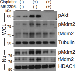
MK-2206 free base purchased from MedChemExpress. Usage Cited in: Cancer Cell. 2018 Jun 11;33(6):1061-1077.e6. [Abstract]
Dih10 cells are treated with CDDP (20 μM) in the presence or absence of the Akt inhibitor MK2206 (5 μM).
-
Cell
Spatial control of the TSC complex integrates insulin and nutrient regulation of mTORC1 at the lysosome. [Abstract]2014 Feb 13;156(4):771-85. PMID: 24529379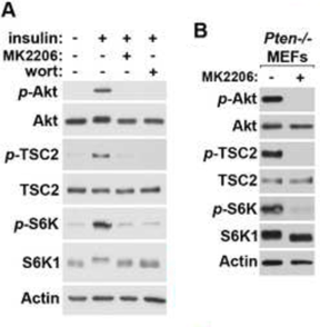
MK-2206 free base purchased from MedChemExpress. Usage Cited in: Cell. 2014 Feb 13;156(4):771-85. [Abstract]
(A) Effects of inhibiting PI3K and Akt in MEFs. Serum starved (16 hr) MEFs are pretreated (30 min) with Wortmannin (100 nM), MK2206 (2 μM) or vehicle (DMSO). Immunoblots of lysates are probed with the indicated antibodies. (B) PTEN null MEFs exhibit constitutive Akt, TSC2 and S6K phosphorylation, which are reversed by the Akt inhibitor MK2206.
-
Mol Cancer
LncRNA-HGBC stabilized by HuR promotes gallbladder cancer progression by regulating miR-502-3p/SET/AKT axis. [Abstract]2019 Nov 21;18(1):167. PMID: 31752906 -
Nat Cancer
MEF2D-expressing cancer precursors reprogram tissue-resident macrophages to support liver tumorigenesis. [Abstract]2025 Dec;6(12):1955-1975. PMID: 41131393 -
Nat Cancer
Targeting a lineage-specific PI3Kɣ-Akt signaling module in acute myeloid leukemia using a heterobifunctional degrader molecule. [Abstract]2024 Jul;5(7):1082-1101. PMID: 38816660
MK-2206 free base purchased from MedChemExpress. Usage Cited in: Nat Cancer. 2024 Jul;5(7):1082-1101. [Abstract]
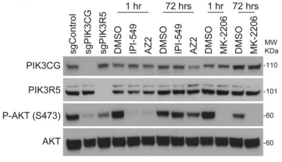
AKT phosphorylation levels by western blot in OCI-AML2 cells either infected with PIK3CG- and PIK3R5-directed sgRNAs for 72 hours or treated with 500nM IPI-549, AZ2, or the AKT inhibitor, MK-2206, for one and 72 hours.
MK-2206 free base purchased from MedChemExpress. Usage Cited in: Nat Cancer. 2024 Jul;5(7):1082-1101. [Abstract]
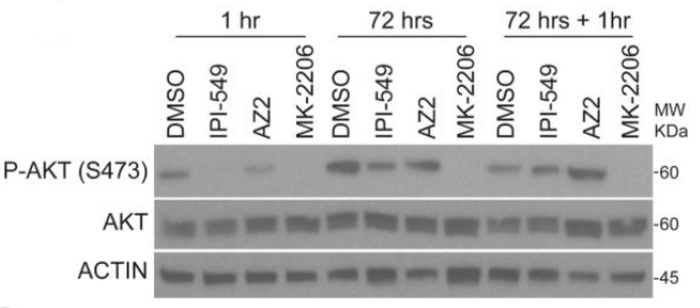
AKT phosphorylation levels by western blot of OCI-AML2 cells treated for one hour, 72 hours, or 72 hours followed by fresh addition of 500nM IPI-549, AZ2, or MK-2206 for one hour.
-
Immunity
Activating an interleukin 4-FLT3-STAT6 axis in multipotent progenitors restores lymphopoiesis in inflammation and aging. [Abstract]2026 May 29:S1074-7613(26)00180-9. PMID: 42214327 -
Cell Stem Cell
Brain Endothelial Cells Maintain Lactate Homeostasis and Control Adult Hippocampal Neurogenesis. [Abstract]2019 Dec 5;25(6):754-767.e9. PMID: 31761722 -
Bioact Mater
Orchestration of energy metabolism and osteogenesis by Mg2+ facilitates low-dose BMP-2-driven regeneration. [Abstract]2022 Mar 24;18:116-127. PMID: 35387176 -
Cell Mol Immunol
β-adrenoreceptor-triggered PKA activation negatively regulates the innate antiviral response. [Abstract]2023 Feb;20(2):175-188. PMID: 36600052 -
Nat Aging
2024 Apr;4(4):568-583. PMID: 38491289 -
Cancer Res
NSD1-Mediated PPARγ Methylation Enhances PTEN Activity to Suppress Glycolysis and Tumor Progression in Endometrial Cancer. [Abstract]2026 Mar 2. PMID: 41770031 -
Mol Cell
2025 Feb 20;85(4):843-856.e6. PMID: 39862868 -
Mol Cell
PI3Kβ functions as a protein kinase to promote cellular protein O-GlcNAcylation and acetyl-CoA production for tumor growth. [Abstract]2025 Mar 19:S1097-2765(25)00186-8. PMID: 40132583 -
Mol Cell
DNA damage promotes HLA class I presentation by stimulating a pioneer round of translation-associated antigen production. [Abstract]2022 Jul 21;82(14):2557-2570.e7. PMID: 35594857 -
Cancer Res
Genomic Alterations in PIK3CA-Mutated Breast Cancer Result in mTORC1 Activation and Limit the Sensitivity to PI3Kα Inhibitors. [Abstract]2021 May 1;81(9):2470-2480. PMID: 33685991 -
Mol Cell
2021 Jan 21;81(2):370-385.e7. PMID: 33271062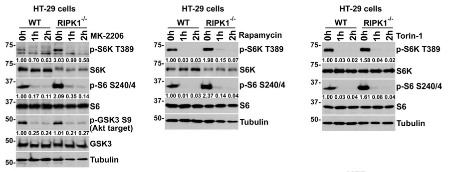
MK-2206 free base purchased from MedChemExpress. Usage Cited in: Mol Cell. 2021 Jan 21;81(2):370-385.e7. [Abstract]
Lack of differential inhibition of mTORC1 signaling in wild-type versus RIPK1−/− HT-29 cells upon inhibition of Akt by MK-2206 and mTORC1 by rapamycin or Torin-1. Cells were treated for indicated time points, and cell lysates were immunoblotted with indicated antibodies.
-
Nat Commun
INSIG1/2 succination mediated by the moonlighting function of ADSL promotes lipogenesis and liver tumorigenesis. [Abstract]2026 Mar 15;17(1):4002. PMID: 41833955 -
Nat Commun
The UFL1-AKT positive feedback loop promotes breast cancer progression by enhancing lipid synthesis. [Abstract]2026 Jan 20;17(1):614. PMID: 41559041 -
Nat Commun
Astrocytic pleiotrophin deficiency in the prefrontal cortex contributes to stress-induced depressive-like responses in male mice. [Abstract]2025 Mar 14;16(1):2528. PMID: 40087317 -
Nat Commun
Astrocyte-derived clusterin disrupts glial physiology to obstruct remyelination in mouse models of demyelinating diseases. [Abstract]2024 Sep 6;15(1):7791. PMID: 39242637 -
Nat Commun
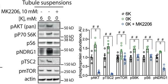
2024 Jun 17;15(1):5144. PMID: 38886379
MK-2206 free base purchased from MedChemExpress. Usage Cited in: Nat Commun. 2024 Jun 17;15(1):5144. [Abstract]
Representative Western blots from isolated tubule suspensions cultured for 30 min in indicated K+ concentrations with or without the AKT inhibitor MK2206 (10 μM).
-
Nat Commun
NLRP6 potentiates PI3K/AKT signalling by promoting autophagic degradation of p85α to drive tumorigenesis. [Abstract]2023 Sep 28;14(1):6069. PMID: 37770465 -
Nat Commun
2022 Nov 29;13(1):7345. PMID: 36446858 -
Nat Commun
Engineering micro oxygen factories to slow tumour progression via hyperoxic microenvironments. [Abstract]2022 Aug 2;13(1):4495. PMID: 35918337 -
Nat Commun
A loss-of-adhesion CRISPR-Cas9 screening platform to identify cell adhesion-regulatory proteins and signaling pathways. [Abstract]2022 Apr 19;13(1):2136. PMID: 35440579 -
Nat Commun
Selective inhibition of cancer cell self-renewal through a Quisinostat-histone H1.0 axis. [Abstract]2020 Apr 14;11(1):1792. PMID: 32286289 -
Nat Commun
SETBP1 accumulation induces P53 inhibition and genotoxic stress in neural progenitors underlying neurodegeneration in Schinzel-Giedion syndrome. [Abstract]2021 Jun 30;12(1):4050. PMID: 34193871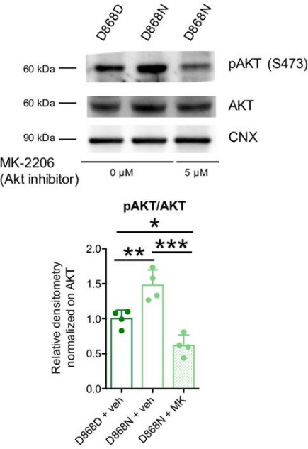
MK-2206 free base purchased from MedChemExpress. Usage Cited in: Nat Commun. 2021 Jun 30;12(1):4050. [Abstract]
Total and phosphorylated AKT (pAKT Ser473) immunoblotting in isogenic control (+vehicle) and mutant NPCs (+vehicle or MK-2206 AKT inhibitor) and quantification as pAKT/AKT ratio.
-
Nat Commun
Recurrent hotspot mutations in HRAS Q61 and PI3K-AKT pathway genes as drivers of breast adenomyoepitheliomas. [Abstract]2018 May 8;9(1):1816. PMID: 29739933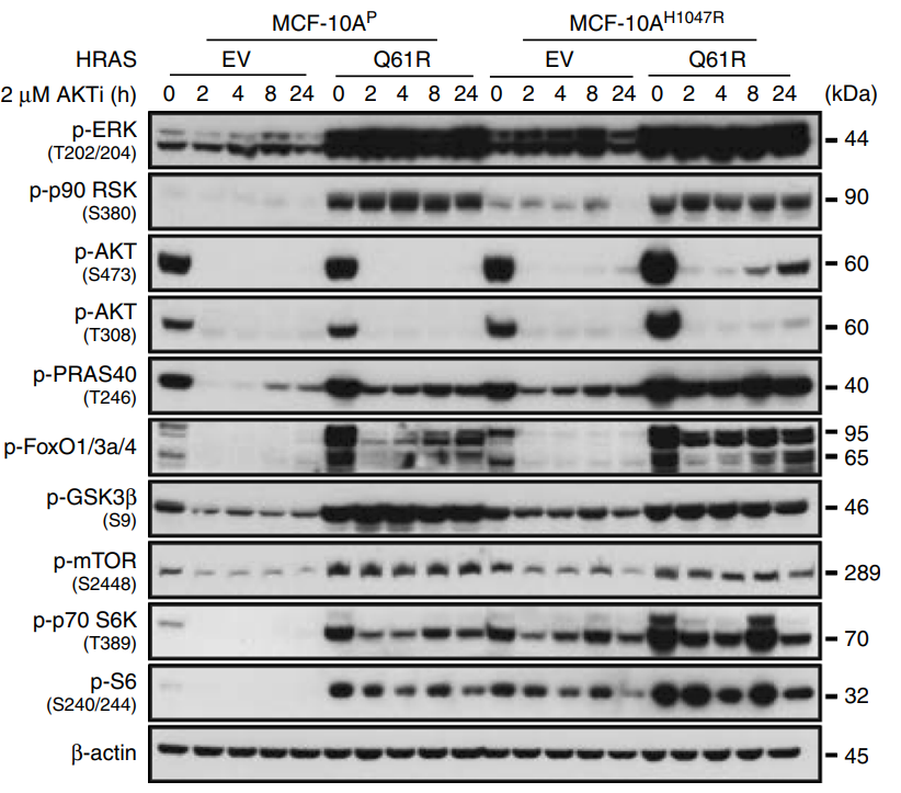
MK-2206 free base purchased from MedChemExpress. Usage Cited in: Nat Commun. 2018 May 8;9(1):1816. [Abstract]
Representative western blot analysis of p-ERK1/2, p-p90 RSK, p-AKT, p-AKT p-PRAS40, p-FOXO1/3a/4, p-GSK3β, p-mTOR, p-p70 S6K, and p-S6 protein in MCF-10AP and MCF-10AH1047R cells stably expressing empty vector (EV) or mutant HRASQ61R treated with 2 µM MK2206 (AKTi) at different time points.
-
Cell Death Differ
Matrix stiffness-induced YEATS2 drives HCC progression via epigenetic activation of the TGFBR2-TAZ-AKT pathway. [Abstract]2026 Mar 3. PMID: 41776086 -
Cell Death Differ
Adaptor protein HIP-55-mediated signalosome protects against ferroptosis in myocardial infarction. [Abstract]2023 Mar;30(3):825-838. PMID: 36639542 -
Cell Death Differ
CHIP-mediated CIB1 ubiquitination regulated epithelial-mesenchymal transition and tumor metastasis in lung adenocarcinoma. [Abstract]2021 Mar;28(3):1026-1040. PMID: 33082516 -
Bone Res
2025 Feb 24;13(1):25. PMID: 39994220 -
Acta Pharm Sin B
HYD-PEP06 suppresses hepatocellular carcinoma metastasis, epithelial-mesenchymal transition and cancer stem cell-like properties by inhibiting PI3K/AKT and WNT/ β-catenin signaling activation. [Abstract]2021 Jun;11(6):1592-1606. PMID: 34221870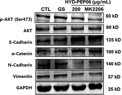
MK-2206 free base purchased from MedChemExpress. Usage Cited in: Acta Pharm Sin B. 2021 Jun;11(6):1592-1606. [Abstract]
HCCLM3 cells were pretreated with or without 10 μmol/L MK2206 (a PI3K/AKT inhibitor) and then with HYD-PEP06 (200 μg/mL) for 24 h. The protein expression of p-AKT, AKT, the epithelial and mesenchymal markers was determined by Western blot.
-
Acta Pharm Sin B
Activated PKB/GSK-3 β synergizes with PKC- δ signaling in attenuating myocardial ischemia/reperfusion injury via potentiation of NRF2 activity: Therapeutic efficacy of dihydrotanshinone-I. [Abstract]2021 Jan;11(1):71-88. PMID: 33532181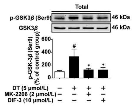
MK-2206 free base purchased from MedChemExpress. Usage Cited in: Acta Pharm Sin B. 2021 Jan;11(1):71-88. [Abstract]
Protein phosphorylation of PKB (Ser473) and GSK-3β (Ser9) in cardiomyocytes treated with DT or MK-2206 or DIF-3, either alone or in combination (n = 5).
-
Sci Transl Med
PP2A inhibition is a druggable MEK inhibitor resistance mechanism in KRAS-mutant lung cancer cells. [Abstract]2018 Jul 18;10(450):eaaq1093. PMID: 30021885 -
Autophagy
The nuclear receptor ESRRA is a crucial regulator of acute kidney injury through inhibition of the lipophagy-ferroptosis axis. [Abstract]2026 Apr 27. PMID: 42041131 -
-
Adv Sci (Weinh)
EGR Proteins Mediate Interferon-Independent Anti-HSV-1 Responses Through Viral and Host Targets. [Abstract]2026 Mar;13(13):e15546. PMID: 41486724 -
Adv Sci (Weinh)
Calhm6 Governs Macrophage Polarization Through Chp1-Camk4-Creb1 Axis and Ectosomal Delivery in Inflammatory Responses. [Abstract]2025 Sep 26:e02395. PMID: 40999918 -
Adv Sci (Weinh)
AKT1 Phosphorylates FDX1 to Promote Cuproptosis Resistance in Triple-Negative Breast Cancer. [Abstract]2025 Feb 20:e2408106. PMID: 39976173 -
Adv Sci (Weinh)
Targeting FDFT1 Reduces Cholesterol and Bile Acid Production and Delays Hepatocellular Carcinoma Progression Through the HNF4A/ALDOB/AKT1 Axis. [Abstract]2025 Feb 3:e2411719. PMID: 39899681 -
Adv Sci (Weinh)
TRIM24 Cooperates with Ras Mutation to Drive Glioma Progression through snoRNA Recruitment of PHAX and DNA-PKcs. [Abstract]2024 Jun 3:e2400023. PMID: 38828688 -
Adv Sci (Weinh)
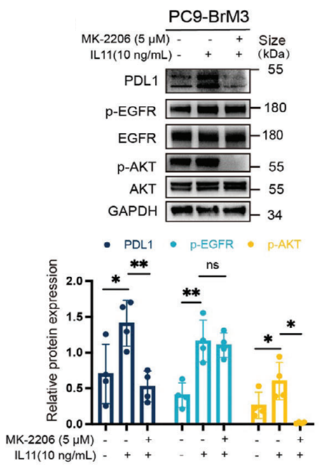
Brain Metastasis from EGFR-Mutated Non-Small Cell Lung Cancer: Secretion of IL11 from Astrocytes Up-Regulates PDL1 and Promotes Immune Escape. [Abstract]2024 Jul;11(26):e2306348. PMID: 38696655
MK-2206 free base purchased from MedChemExpress. Usage Cited in: Adv Sci (Weinh). 2024 Jul;11(26):e2306348. [Abstract]
MK‐2206 (5 µM; 18 h) significantly suppresses IL11‐induced upregulation of PDL1, but did not affect the level of p‐EGFR.
-
Nat Chem Biol
2017 Jan;13(1):38-45. PMID: 27820799 -
Leukemia
BCR::ABL1 tyrosine kinase inhibitors induce ribosome collisions to activate ZAK-dependent ribotoxic stress and apoptosis in chronic myeloid leukemia. [Abstract]2026 May;40(5):955-969. PMID: 41912913 -
Leukemia
A Perturb-seq map of a differentiation hub reveals synergistic vulnerabilities in KMT2A-rearranged acute myeloid leukemia. [Abstract]2026 Mar 25. PMID: 41882099 -
Theranostics
Endosome-microautophagy targeting chimera (eMIATAC) for targeted proteins degradation and enhance CAR-T cell anti-tumor therapy. [Abstract]2024 Jul 22;14(11):4481-4498. PMID: 39113807 -
Theranostics
2024 Jun 17;14(10):3793-3809. PMID: 38994031 -
Theranostics
ILT4 inhibition prevents TAM- and dysfunctional T cell-mediated immunosuppression and enhances the efficacy of anti-PD-L1 therapy in NSCLC with EGFR activation. [Abstract]2021 Jan 19;11(7):3392-3416. PMID: 33537094 -
Theranostics
2020 Aug 6;10(21):9899-9912. PMID: 32863967 -
Theranostics
FGD1 promotes tumor progression and regulates tumor immune response in osteosarcoma via inhibiting PTEN activity. [Abstract]2020 Feb 3;10(6):2859-2871. PMID: 32194840 -
Theranostics
3'-epi-12β-hydroxyfroside, a new cardenolide, induces cytoprotective autophagy via blocking the Hsp90/Akt/mTOR axis in lung cancer cells. [Abstract]2018 Feb 15;8(7):2044-2060. PMID: 29556372 -
-
J Adv Res
An injectable nano-hydroxyapatite-incorporated hydrogel with sustained release of Notoginsenoside R1 enhances bone regeneration by promoting angiogenesis through Notch1/Akt signaling. [Abstract]2025 May 13:S2090-1232(25)00343-1. PMID: 40373960 -
Biomaterials
Nano-sensitizer with self-amplified drug release and hypoxia normalization properties potentiates efficient chemoradiotherapy of pancreatic cancer. [Abstract]2024 Oct:310:122634. PMID: 38823195 -
Exp Mol Med
Protective effect of hepatocyte-enriched lncRNA-Mir122hg by promoting hepatocyte proliferation in acute liver injury. [Abstract]2022 Nov;54(11):2022-2035. PMID: 36424455 -
J Exp Clin Cancer Res
AR antagonists develop drug resistance through TOMM20 autophagic degradation-promoted transformation to neuroendocrine prostate cancer. [Abstract]2023 Aug 10;42(1):204. PMID: 37563661 -
J Exp Clin Cancer Res
A novel FBW7/NFAT1 axis regulates cancer immunity in sunitinib-resistant renal cancer by inducing PD-L1 expression. [Abstract]2022 Jan 26;41(1):38. PMID: 35081978 -
J Exp Clin Cancer Res
The IRF2/CENP-N/AKT signaling axis promotes proliferation, cell cycling and apoptosis resistance in nasopharyngeal carcinoma cells by increasing aerobic glycolysis. [Abstract]2021 Dec 10;40(1):390. PMID: 34893086 -
J Exp Clin Cancer Res
Cooperation between liver-specific mutations of pten and tp53 genetically induces hepatocarcinogenesis in zebrafish. [Abstract]2021 Aug 20;40(1):262. PMID: 34416907 -
J Exp Clin Cancer Res
SKP2 promotes breast cancer tumorigenesis and radiation tolerance through PDCD4 ubiquitination. [Abstract]2019 Feb 13;38(1):76. PMID: 30760284 -
J Exp Clin Cancer Res
Radiotherapy-induced cell death activates paracrine HMGB1-TLR2 signaling and accelerates pancreatic carcinoma metastasis. [Abstract]2018 Apr 3;37(1):77. PMID: 29615080 -
J Nanobiotechnology
Small extracellular vesicles of hypoxic endothelial cells regulate the therapeutic potential of adipose-derived mesenchymal stem cells via miR-486-5p/PTEN in a limb ischemia model. [Abstract]2022 Sep 24;20(1):422. PMID: 36153544 -
Sci Adv
Adissp activates insulin-independent glucose disposal and energy expenditure in white fat to treat diabetes and cardiometabolic disease. [Abstract]2026 Apr 24;12(17):eaed2780. PMID: 42030391 -
Sci Adv
2024 Dec 13;10(50):eadq4274. PMID: 39661665 -
Carbohydr Polym
Inulin-like polysaccharide ABWW may impede CCl4 induced hepatic stellate cell activation through mediating the FAK/PI3K/AKT signaling pathway in vitro & in vivo. [Abstract]2024 Feb 15:326:121637. PMID: 38142102 -
Sci Adv
Fasting improves therapeutic response in hepatocellular carcinoma through p53-dependent metabolic synergism. [Abstract]2022 Jan 21;8(3):eabh2635. PMID: 35061544 -
Small
Tetrahedral DNA Nanostructure-Modified Nanocoating for Improved Bioaffinity and Osseointegration of Titanium. [Abstract]2025 Mar 19:e2412747. PMID: 40103513 -
J Biomed Sci
Fusobacterium nucleatum promotes colorectal cancer liver metastasis via miR-5692a/IL-8 axis by inducing epithelial-mesenchymal transition. [Abstract]2025 Jan 6;32(1):5. PMID: 39757156 -
Redox Biol
Phactr4 promotes oxidative stress and behavioral disorder caused by chronic stress via regulating PP1/GSK3-β pathway. [Abstract]2025 Sep 16:87:103873. PMID: 40967008 -
Redox Biol
2022 Feb:49:102217. PMID: 34942528 -
MedComm (2020)
Ubiquitin-specific protease 22 controls melanoma metastasis and vulnerability to ferroptosis through targeting SIRT1/PTEN/PI3K signaling. [Abstract]2024 Aug 12;5(8):e684. PMID: 39135915 -
Pharmacol Res
CDC7 stabilized by KRAS signaling reactivation impairs chemosensitivity in KRAS-mutant colorectal cancer. [Abstract]2025 Dec:222:108027. PMID: 41207349 -
-
Nat Struct Mol Biol
ERK-USP9X-coupled regulation of thymidine kinase 1 promotes both its enzyme activity-dependent and its enzyme activity-independent functions for tumor growth. [Abstract]2025 May;32(5):853-863. PMID: 39824978 -
Cancer Lett
Forkhead box protein FOXK1 disrupts the circadian rhythm to promote breast tumorigenesis in response to insulin resistance. [Abstract]2024 Jul 31:217147. PMID: 39094826 -
J Neuroinflammation
Inhibition of ANGPTL8 protects against diabetes-associated cognitive dysfunction by reducing synaptic loss via the PirB signaling pathway. [Abstract]2024 Aug 2;21(1):192. PMID: 39095838 -
Cancer Lett
2024 Jun 1:591:216848. PMID: 38604312 -
J Neuroinflammation
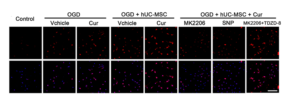
Human umbilical cord-derived mesenchymal stem cell transplantation supplemented with curcumin improves the outcomes of ischemic stroke via AKT/GSK-3β/β-TrCP/Nrf2 axis. [Abstract]2023 Feb 24;20(1):49. PMID: 36829224
MK-2206 free base purchased from MedChemExpress. Usage Cited in: J Neuroinflammation. 2023 Feb 24;20(1):49. [Abstract]
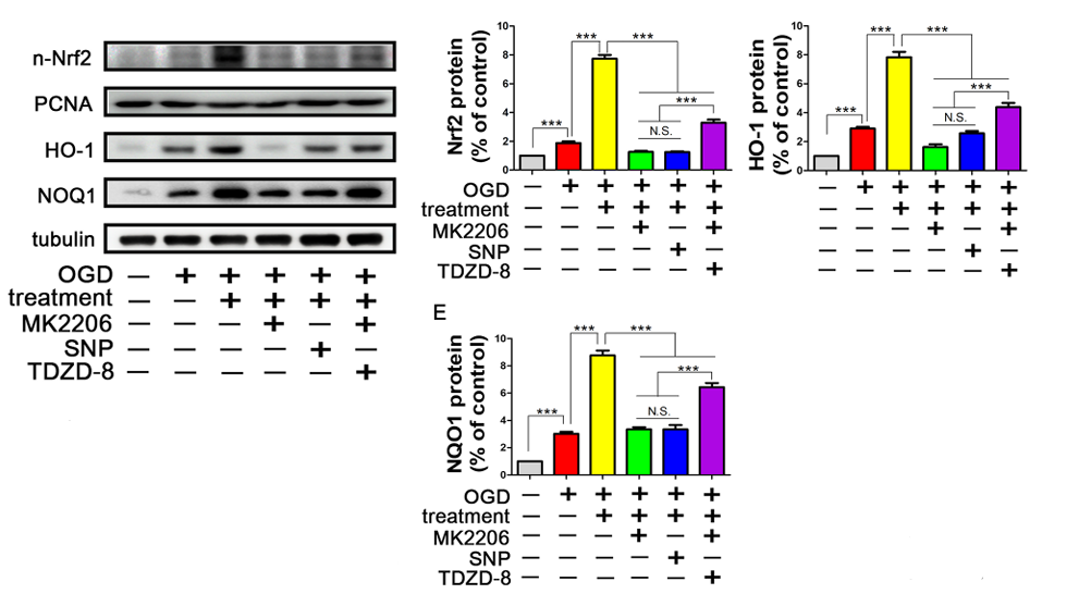
Representative images of immunofluorescence analysis of Nrf2 expression in microglia treated with the AKT inhibitor MK2206 (5 μM,6 h), the GSK3β activator sodium nitroprusside (SNP, 100 μM, 6h), and the GSK3β inhibitor TDZD-8 (5 μM,6 h). Nrf2 (red), nuclei (blue).
MK-2206 free base purchased from MedChemExpress. Usage Cited in: J Neuroinflammation. 2023 Feb 24;20(1):49. [Abstract]
Representative western blots and quantitative data for the expression of Nrf2 in the nucleus and the downstream proteins of Nrf2 pathway, including HO-1 and NQO1, with different treatments (MK2206: 5 μM,6 h; SNP: 100 μM, 6h; TDZD-8: 5 μM,6 h).
MK-2206 free base purchased from MedChemExpress. Usage Cited in: J Neuroinflammation. 2023 Feb 24;20(1):49. [Abstract]
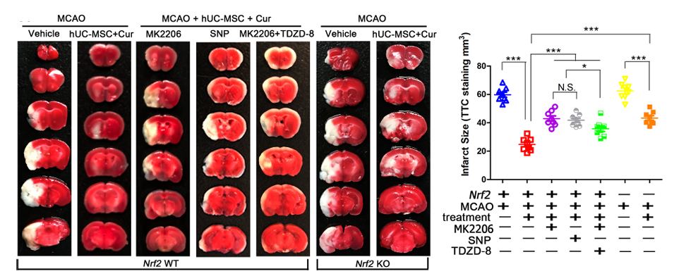
Nrf2 knockout abolished neurological function improvement mediated by combined curcumin-hUC-MSC treatment. TTC-stained brain sections (A) and quantitative analysis (B) showed the decreased infarct volume with combined therapy in MCAO mice (MK2206, SNP or TDZD-8 :5 mg/kg, intraperitoneally,3 consecutive days after MCAO).
-
Cancer Lett
Synergistic efficacy of telomerase-specific oncolytic adenoviral therapy and histone deacetylase inhibition in human hepatocellular carcinoma. [Abstract]2023 Mar 1:556:216063. PMID: 36669725 -
Cancer Lett
Vimentin binds to a novel tumor suppressor protein, GSPT1-238aa, encoded by circGSPT1 with a selective encoding priority to halt autophagy in gastric carcinoma. [Abstract]2022 Oct 1:545:215826. PMID: 35839920 -
Cancer Lett
CXCL13 promotes intestinal tumorigenesis through the activation of epithelial AKT signaling. [Abstract]2021 Jul 28:511:1-14. PMID: 33894331 -
Cancer Lett
Targeting the EphB4 receptor tyrosine kinase sensitizes HER2-positive breast cancer cells to Lapatinib. [Abstract]2020 Apr 10;475:53-64. PMID: 32006616 -
Int J Biol Sci
Targeting PLCG2 Suppresses Tumor Progression, Orchestrates the Tumor Immune Microenvironment and Potentiates Immune Checkpoint Blockade Therapy for Colorectal Cancer. [Abstract]2024 Oct 14;20(14):5548-5575. PMID: 39494327 -
Int J Biol Sci
TGFβ Governs the Pleiotropic Activity of NDRG1 in Triple-Negative Breast Cancer Progression. [Abstract]2023 Jan 1;19(1):204-224. PMID: 36594086 -
Int J Biol Sci
Accelerated Bone Regeneration by Astragaloside IV through Stimulating the Coupling of Osteogenesis and Angiogenesis. [Abstract]2021 Apr 24;17(7):1821-1836. PMID: 33994865 -
Cell Death Dis
mTORC2 inhibition reduces tumor burden via STAT1 activation and enhanced response to anti-PD-L1 therapy. [Abstract]2025 Dec 22;16(1):922. PMID: 41429766 -
Cell Death Dis
Cancer-associated fibroblasts expressing FSTL3 promote vasculogenic mimicry formation and drive colon cancer malignancy. [Abstract]2025 Oct 6;16(1):706. PMID: 41053124 -
Burns Trauma
Neuregulin-1, a member of the epidermal growth factor family, mitigates STING-mediated pyroptosis and necroptosis in ischaemic flaps. [Abstract]2024 Jun 9:12:tkae035. PMID: 38855574 -
Cell Death Dis
Retinoic acid and RARγ maintain satellite cell quiescence through regulation of translation initiation. [Abstract]2022 Sep 29;13(9):838. PMID: 36175396 -
Cell Death Dis
Leukemia inhibitory factor drives glucose metabolic reprogramming to promote breast tumorigenesis. [Abstract]2022 Apr 19;13(4):370. PMID: 35440095 -
Cell Death Dis
2020 May 11;11(5):353. PMID: 32393791 -
Cell Death Dis
Paeonol induces cytoprotective autophagy via blocking the Akt/mTOR pathway in ovarian cancer cells. [Abstract]2019 Aug 13;10(8):609. PMID: 31406198 -
Cell Death Dis
S-nitrosylation of the Peroxiredoxin-2 promotes S-nitrosoglutathione-mediated lung cancer cells apoptosis via AMPK-SIRT1 pathway. [Abstract]2019 May; 10(5): 329. PMID: 30988280 -
Cell Death Dis
Transient receptor potential channel 6 knockdown prevents apoptosis of renal tubular epithelial cells upon oxidative stress via autophagy activation. [Abstract]2018 Oct 3;9(10):1015. PMID: 30282964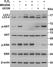
MK-2206 free base purchased from MedChemExpress. Usage Cited in: Cell Death Dis. 2018 Oct 3;9(10):1015. [Abstract]
Representative western blot images are showing the LC3, and the phosphorylated and total protein expression of Akt and ERK1/2 after treatment with H2O2 in the presence and absence of MK2206 (5 μM) and U0126 (25 μM).
-
-
Genes Dis
Protein phosphatase 6 (Pp6) is crucial for regulatory T cell function and stability in autoimmunity. [Abstract]2021 Aug 17;9(2):562-575. PMID: 35224167 -
Angiogenesis
Clioquinol inhibits angiogenesis by promoting VEGFR2 degradation and synergizes with AKT inhibition to suppress triple-negative breast cancer vascularization. [Abstract]2025 Feb 3;28(2):13. PMID: 39899169 -
Proc Natl Acad Sci U S A
2019 Feb 19;116(8):2996-3005. PMID: 30718432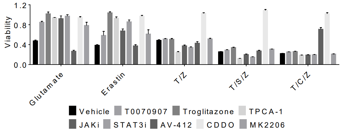
MK-2206 free base purchased from MedChemExpress. Usage Cited in: Proc Natl Acad Sci U S A. 2019 Feb 19;116(8):2996-3005. [Abstract]
HT22 cells were pretreated with the indicated inhibitors (CDDO (Bardoxolone, HY-14909); T0070907 (HY-13202); Troglitazone (HY-50935 ); TPCA-1 (HY-10074); JAKi; STAT3i; AV-412 (HY-10346); and MK2206 (HY-10358)) and then treated with T/Z, T/S/Z, T/C/Z for 9 h or Glutamate, Erastin (T: 20 ng/ml; Z: 50 μM; S: 20 nM; C: 1 μg/ml; Glutamate: 50 mM; Erastin 10 μM) for 12 h. Cell viability was measured by CellTiterGlo assay.
-
Proc Natl Acad Sci U S A
2016 Jul 26;113(30):E4338-47. PMID: 27402769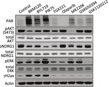
MK-2206 free base purchased from MedChemExpress. Usage Cited in: Proc Natl Acad Sci U S A. 2016 Jul 26;113(30):E4338-47. [Abstract]
HCC1937 cells are treated for 16 h with inhibitors as indicated. Immunoblotting of total cell lysates is performed with antibodies as indicated. Induction of PAR and H2ax phosphorylation (γH2ax) following treatment with inhibitors of pan-PI3K (BKM120, 1.5 μM), PI3Kα (BYL719, 3 μM; PIK75, 0.5 μM), PI3Kβ (TGX221, 30 μM), AZD2281 (5 μM), and inhibitors of AKT (MK2206, 1 μM), SGK (GSK650394, 10 μM), or MAPKK (GSK1120212, 5 nM).
-
Cell Commun Signal
Activation of mechanosensitive ion channel Piezo1 linking metabolic reprogramming and pro-inflammatory responses in hepatocellular carcinoma. [Abstract]2025 Jun 13;23(1):280. PMID: 40514668 -
Cell Commun Signal
CDO1 phosphorylation is required for IL-6-induced tumor cell proliferation through governing cysteine availability. [Abstract]2025 Apr 23;23(1):194. PMID: 40269955 -
Cell Commun Signal
The IL-33-ST2 axis plays a vital role in endometriosis via promoting epithelial-mesenchymal transition by phosphorylating β-catenin. [Abstract]2024 Jun 10;22(1):318. PMID: 38858740 -
Int J Biol Macromol
Medium-molecular weight hyaluronic acid orchestrates hair follicle regeneration via CD44/AKT-driven endogenous ROS activation of β-catenin. [Abstract]2026 May:360:151752. PMID: 41935804 -
Int J Biol Macromol
Exosomal microRNA-20b-5p contributes to cytarabine resistance in acute myeloid leukemia via the microtubule-associated serine/threonine kinase-like-phosphatidylinositol 3-kinase-protein kinase B signaling axis. [Abstract]2025 Dec;333(Pt 2):148750. PMID: 41197693 -
Int J Biol Macromol
Trained immunity driven by Enterococcus faecalis ribosomal protein S11 enhances antigen presentation and boosts influenza vaccine efficacy via nanoparticle delivery. [Abstract]2025 May 15:144179. PMID: 40381785 -
Int J Biol Macromol
Insect chitosan derived from Hermetia illucens larvae suppresses adipogenic signaling and promotes the restoration of gut microbiome balance. [Abstract]2024 Nov 27:138168. PMID: 39613084 -
Int J Biol Macromol
Cepharanthine inhibits African swine fever virus replication by suppressing AKT-associated pathways through disrupting Hsp90-Cdc37 complex. [Abstract]2024 Oct 30:137070. PMID: 39486740 -
Int J Biol Macromol
Sufentanil-induced Nrf2 protein ameliorates cerebral ischemia-reperfusion injury through suppressing neural ferroptosis. [Abstract]2024 Aug 26:135109. PMID: 39197624 -
Acta Pharmacol Sin
FGF10 mitigates doxorubicin-induced myocardial toxicity in mice via activation of FGFR2b/PHLDA1/AKT axis. [Abstract]2023 Oct;44(10):2004-2018. PMID: 37225844 -
Acta Pharmacol Sin
The oncoprotein HBXIP promotes human breast cancer growth through down-regulating p53 via miR-18b/MDM2 and pAKT/MDM2 pathways. [Abstract]2018 Nov;39(11):1787-1796. PMID: 30181579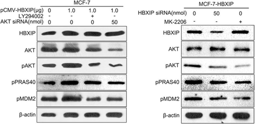
MK-2206 free base purchased from MedChemExpress. Usage Cited in: Acta Pharmacol Sin. 2018 Nov;39(11):1787-1796. [Abstract]
Effects of LY294002 or AKT siRNA on the levels of total AKT, pAKT, phosphorylated PRAS40 and pMDM2 are examined by Western blot analysis in MCF-7 cells when HBXIP was overexpressed. Effects of MK-2206 or HBXIP siRNA on the levels of total AKT, pAKT, pPRAS40, and pMDM2 are examined by Western blot analysis in MCF-7-HBXIP cells.
-
Phytomedicine
Yulin Yangchao formula improves premature ovarian insufficiency by inhibiting granulosa cell apoptosis through the PI3K/AKT/p53 signaling axis. [Abstract]2026 Aug:158:158377. PMID: 42284639 -
Phytomedicine
Juyuanjian attenuates sarcopenia through dual regulation of the Akt/FoxO1 and SIRT1/PGC-1α pathways. [Abstract]2026 Jul:156:158188. PMID: 42068872 -
Phytomedicine
Chlorella pyrenoidosa-derived extracellular vesicles ameliorate ulcerative colitis through microbiota-mediated AKT/mTOR/ferroptosis pathway. [Abstract]2026 Jul:156:158199. PMID: 42030800 -
Phytomedicine
Atraric acid and atranorin inhibit breast cancer energy metabolism and immune evasion through PI3K/AKT/Bcl-xL. [Abstract]2025 Dec:149:157560. PMID: 41297322 -
Phytomedicine
Schisandrin B regulates mitochondrial dynamics via AKT1 activation and mitochondrial targeting to ameliorate renal ischemia-reperfusion injury. [Abstract]2025 Jun:141:156672. PMID: 40220406 -
Phytomedicine
Sophoricoside ameliorates methicillin-resistant Staphylococcus aureus-induced acute lung injury by inhibiting Bach1/Akt pathway. [Abstract]2024 Jun 27:132:155846. PMID: 38964155 -
Phytomedicine
The kava chalcone flavokawain B exerts inhibitory activity and synergizes with BCL-2 inhibition in malignant B-cell lymphoma. [Abstract]2023 Nov:120:155074. PMID: 37716033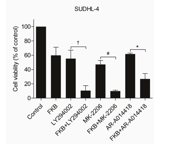
MK-2206 free base purchased from MedChemExpress. Usage Cited in: Phytomedicine. 2023 Nov:120:155074. [Abstract]
SUDHL-4 cells were pretreated with the pan-PI3K inhibitor LY294002 (20 µM), the Akt inhibitor MK2206 (HY-108232; 4 µM), or the GSK3β inhibitor AR-A014418 (HY-10512; 10 µM) for 1 h followed by treating with FKB (Flavokawain B; HY-N2132) at 2.5 µg/ml for 24 h. Cell viability was measured using the MTS assay and results were shown as the percentage relative to the vehicle-treated control. *p < 0.05, #p < 0.01, and †p < 0.001.
MK-2206 free base purchased from MedChemExpress. Usage Cited in: Phytomedicine. 2023 Nov:120:155074. [Abstract]
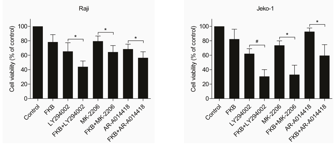
Raji and Jeko-1 cells were pretreated with the pan-PI3K inhibitor LY294002 (20 µM), the Akt inhibitor MK2206 (HY-108232; 4 µM), or the GSK3β inhibitor AR-A014418 (HY-10512; 10 µM) for 1 h followed by treating with FKB (Flavokawain B; HY-N2132) at 2.5 µg/ml for 24 h. Cell viability was measured using the MTS assay and results were shown as the percentage relative to the vehicle-treated control. *p < 0.05, #p < 0.01, and †p < 0.001.
-
Phytomedicine
Quercetin induces MGMT+ glioblastoma cells apoptosis via dual inhibition of Wnt3a/β-Catenin and Akt/NF-κB signaling pathways. [Abstract]2023 Sep:118:154933. PMID: 37451151 -
Phytomedicine
RNA-binding domain 2 of nucleolin is important for the autophagy induction of curcumol in nasopharyngeal carcinoma cells. [Abstract]2023 Jul:115:154833. PMID: 37137203 -
Phytomedicine
Ginsenoside Rh2 attenuates CDAHFD-induced liver fibrosis in mice by improving intestinal microbial composition and regulating LPS-mediated autophagy. [Abstract]2022 Jul;101:154121. PMID: 35489327 -
Phytomedicine
Aesculin suppresses the NLRP3 inflammasome-mediated pyroptosis via the Akt/GSK3β/NF-κB pathway to mitigate myocardial ischemia/reperfusion injury. [Abstract]2021 Nov:92:153687. PMID: 34482222 -
Free Radic Biol Med
7,8-Dihydroxyflavone protects acetaminophen induced liver injury through activating PI3K/Akt/NRF2/GPX4 mediated ferroptosis suppression. [Abstract]2025 Oct 30:242:275-287. PMID: 41173313 -
Free Radic Biol Med
BP5 alleviates endotoxemia-induced acute lung injury by activating Nrf2 via dual regulation of the Keap1-Nrf2 interaction and the Akt (Ser473)/GSK3β (Ser9)/Fyn pathway. [Abstract]2022 Nov 20;193(Pt 1):304-318. PMID: 36272670 -
Food Res Int
δ-Tocotrienol re-sensitizes vemurafenib-resistant melanoma cells to BRAF inhibition via modulation of AKT signaling. [Abstract]2026 Aug 31:238:119457. PMID: 42215124 -
Sci Total Environ
Parental exposure to Cypermethrin causes intergenerational toxicity in zebrafish offspring. [Abstract]2024 May 22:935:173456. PMID: 38788937 -
Sci Total Environ
Chronic MC-LR exposure promoted Aβ and p-tau accumulation via regulating Akt/GSK-3β signal pathway. [Abstract]2021 Nov 10;794:148732. PMID: 34323745 -
J Orthop Translat
Profibrotic macrophage-derived CXCL4 promotes pericyte-to-myofibroblast transition after spinal cord injury. [Abstract]2026 Feb 11:56:101032. PMID: 41836554 -
Cell Syst
Torin2 Exploits Replication and Checkpoint Vulnerabilities to Cause Death of PI3K-Activated Triple-Negative Breast Cancer Cells. [Abstract]2020 Jan 22;10(1):66-81.e11. PMID: 31812693 -
J Transl Med
Inhibition of DDR1 potentiates carbon ion radiotherapy by promoting ferroptosis and immunogenic death in head and neck squamous cell carcinoma. [Abstract]2025 Sep 24;23(1):1011. PMID: 40993737 -
Biomed Pharmacother
Inhibition of the PI3K/AKT signaling pathway contributes to the anti-renal cell carcinoma effects of deoxyelephantopin. [Abstract]2025 May 8:187:118136. PMID: 40344699 -
J Transl Med
NL101 synergizes with the BCL-2 inhibitor venetoclax through PI3K-dependent suppression of c-Myc in acute myeloid leukaemia. [Abstract]2024 Sep 27;22(1):867. PMID: 39334157 -
J Transl Med
Resveratrol attenuates inflammation and fibrosis in rheumatoid arthritis-associated interstitial lung disease via the AKT/TMEM175 pathway. [Abstract]2024 May 14;22(1):457. PMID: 38745204 -
Biomed Pharmacother
Hyaluronan delays human amniotic epithelial stem cell senescence by regulating CD44 isoform switch to activate AKT/mTOR signals. [Abstract]2024 Jan:170:116100. PMID: 38159379 -
J Transl Med
VEGF-A enhances the cytotoxic function of CD4+ cytotoxic T cells via the VEGF-receptor 1/VEGF-receptor 2/AKT/mTOR pathway. [Abstract]2023 Feb 3;21(1):74. PMID: 36737819 -
J Transl Med
Chemotherapy induces ACE2 expression in breast cancer via the ROS-AKT-HIF-1α signaling pathway: a potential prognostic marker for breast cancer patients receiving chemotherapy. [Abstract]2022 Nov 5;20(1):509. PMID: 36335375 -
J Transl Med
NCAPG promotes the proliferation of hepatocellular carcinoma through the CKII-dependent regulation of PTEN. [Abstract]2022 Jul 21;20(1):325. PMID: 35864529 -
Biomed Pharmacother
USP13 mediates PTEN to ameliorate osteoarthritis by restraining oxidative stress, apoptosis and inflammation via AKT-dependent manner. [Abstract]2021 Jan:133:111089. PMID: 33378983 -
Biomed Pharmacother
High glucose inhibits myogenesis and induces insulin resistance by down-regulating AKT signaling. [Abstract]2019 Dec;120:109498. PMID: 31634780 -
Cell Mol Gastroenterol Hepatol
2021;11(3):683-696. PMID: 33075564 -
Stem Cell Res Ther
Mesenchymal stem cells overexpressing neuropeptide S promote the recovery of rats with spinal cord injury by activating the PI3K/AKT/GSK3β signaling pathway. [Abstract]2025 Feb 28;16(1):100. PMID: 40022159 -
Stem Cell Res Ther
Thymosin beta-4 improves endothelial function and reparative potency of diabetic endothelial cells differentiated from patient induced pluripotent stem cells. [Abstract]2022 Jan 10;13(1):13. PMID: 35012642 -
Stem Cell Res Ther
Akt signaling is activated by TGFβ2 and impacts tenogenic induction of mesenchymal stem cells. [Abstract]2021 Jan 26;12(1):88. PMID: 33499914 -
Oncogene
Aberrant JmjC domain-containing protein 8 (JMJD8) expression promotes activation of AKT and tumor epithelial-mesenchymal transition. [Abstract]2020 Oct;39(41):6451-6467. PMID: 32879443 -
Oncogene
AKT drives SOX2 overexpression and cancer cell stemness in esophageal cancer by protecting SOX2 from UBR5-mediated degradation. [Abstract]2019 Jun;38(26):5250-5264. PMID: 30894683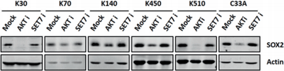
MK-2206 free base purchased from MedChemExpress. Usage Cited in: Oncogene. 2019 Jun;38(26):5250-5264. [Abstract]
Western blotting analysis shows the effect of AKT inhibitor MK2206 (AKT i) and SET7 inhibitor (R)-PFI-2 on the levels of SOX2 proteins. The cells are treated with 1 μM MK2206 for 24 h and 10 μM (R)-PFI-2 for 12 h before being harvested for analysis.
-
Oncogene
PIM2-mediated phosphorylation of hexokinase 2 is critical for tumor growth and paclitaxel resistance in breast cancer. [Abstract]2018 Nov;37(45):5997-6009. PMID: 29985480 -
Oncogene
2017 Aug 10;36(32):4585-4596. PMID: 28368422 -
PLoS Biol
The mTORC2 subunit RICTOR drives breast cancer progression by promoting ganglioside biosynthesis through transcriptional and epigenetic mechanisms. [Abstract]2025 Sep 11;23(9):e3003362. PMID: 40934285 -
Int Endod J
Fibroblast Dysregulation in Multiple Idiopathic External Cervical Resorption: Laboratory Investigation From a Rare Case of 13 Affected Teeth. [Abstract]2025 Sep 23. PMID: 40988149 -
Blood Adv
Melanocortin/MC5R axis regulates the proliferation of hematopoietic stem cells in mice after ionizing radiation injury. [Abstract]2023 Jul 11;7(13):3199-3212. PMID: 36920787 -
Eur J Cancer
2022 May 19;170:91-102. PMID: 35598361 -
Cell Death Discov
The protective up-regulation of metallothionein-2A in intervertebral disc degeneration inhibits nucleus pulposus cell ferroptosis through activation of the PI3K/AKT/mTOR pathway. [Abstract]2026 Feb 25;12(1):111. PMID: 41741409 -
Cell Death Discov
2024 Jul 19;10(1):329. PMID: 39030174 -
Cell Death Discov
TGR5-HNF4α axis contributes to bile acid-induced gastric intestinal metaplasia markers expression. [Abstract]2020 Jul 6;6:56. PMID: 32655894 -
Cell Rep
Collagen 1-mediated CXCL1 secretion in tumor cells activates fibroblasts to promote radioresistance of esophageal cancer. [Abstract]2023 Oct 17;42(10):113270. PMID: 37851572 -
Cell Rep
2023 Sep 1;42(9):113048. PMID: 37659078 -
Cell Rep
2022 Dec 27;41(13):111891. PMID: 36577384 -
Cell Rep
The deubiquitinase OTUD1 noncanonically suppresses Akt activation through its N-terminal intrinsically disordered region. [Abstract]2023 Jan 31;42(1):111916. PMID: 36640312 -
Cell Rep
Renal Klotho and inorganic phosphate are extrinsic factors that antagonistically regulate hematopoietic stem cell maintenance. [Abstract]2022 Feb 15;38(7):110392. PMID: 35172146 -
Br J Cancer
Deciphering the role of IGF2BP2 and PRMT5 in gallbladder cancer progression: insights from multi-omics analysis. [Abstract]2025 Jun 24. PMID: 40555776 -
Br J Cancer
Activation of an AKT/FOXM1/STMN1 pathway drives resistance to tyrosine kinase inhibitors in lung cancer. [Abstract]2017 Sep 26;117(7):974-983. PMID: 28850563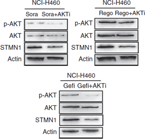
MK-2206 free base purchased from MedChemExpress. Usage Cited in: Br J Cancer. 2017 Sep 26;117(7):974-983. [Abstract]
The effect of the AKT inhibitor MK2206 (10 μM) on the expression levels of phosphor-AKT, AKT, and STMN1 in TKI-pretreated NCI-H460 cells. β-actin is used as a loading control.
-
Anal Chem
TROP2 Promotes Tumor Cell Migration through Downregulation of DSG2 Revealed by Super-Resolution Fluorescence Imaging. [Abstract]2026 Jan 27;98(3):1901-1914. PMID: 41531208 -
Mol Metab
Linking metabolism and histone acetylation dynamics by integrated metabolic flux analysis of Acetyl-CoA and histone acetylation sites. [Abstract]2024 Sep 19:102032. PMID: 39305948 -
Sci Signal
The Bacillus cereus toxin alveolysin disrupts the intestinal epithelial barrier by inducing microtubule disorganization through CFAP100. [Abstract]2023 May 16;16(785):eade8111. PMID: 37192300 -
Antioxidants (Basel)
Sphingosine-1-Phosphate Alleviates Irradiation Induced Salivary Gland Hypofunction through Preserving Endothelial Cells and Resident Macrophages. [Abstract]2022 Oct 18;11(10):2050. PMID: 36290773 -
Int J Radiat Oncol Biol Phys
2025 Jun 16:S0360-3016(25)03903-3. PMID: 40532892 -
J Cell Biol
Membrane-bound SCF and VCAM-1 synergistically regulate the morphology of hematopoietic stem cells. [Abstract]2021 Oct 4;220(10):e202010118. PMID: 34402812 -
Elife
2021 Jul 13:10:e66942. PMID: 34253290 -
Phytother Res
(+)-Matairesinol, Derived From Olive Oil, Enhanced T-Cell Anti-Tumor Immune via JAK3-STAT1 Signal and Synergized With Anti-PD-1 Efficacy. [Abstract]2026 Apr 27. PMID: 42037203 -
Oncoimmunology
Ex vivo AKT-inhibition facilitates generation of polyfunctional stem cell memory-like CD8+ T cells for adoptive immunotherapy. [Abstract]2018 Aug 6;7(10):e1488565. PMID: 30288356 -
J Agric Food Chem
Platycodin D Ameliorates Cognitive Impairment in Type 2 Diabetes Mellitus Mice via Regulating PI3K/Akt/GSK3β Signaling Pathway. [Abstract]2024 Jun 5;72(22):12516-12528. PMID: 38491972 -
J Agric Food Chem
Ginsenoside Rg2 Ameliorating CDAHFD-Induced Hepatic Fibrosis by Regulating AKT/mTOR-Mediated Autophagy. [Abstract]2022 Feb 16;70(6):1911-1922. PMID: 35104139 -
J Agric Food Chem
Stachydrine Mediates Rapid Vascular Relaxation: Activation of Endothelial Nitric Oxide Synthase Involving AMP-Activated Protein Kinase and Akt Phosphorylation in Vascular Endothelial Cells. [Abstract]2019 Sep 4;67(35):9805-9811. PMID: 31407895 -
JCI Insight
FXR shapes an immunosuppressive microenvironment in PD-L1lo/- non-small cell lung cancer by upregulating HVEM. [Abstract]2025 Sep 23;10(18):e190716. PMID: 40985894 -
Hum Reprod
CDC42 deficiency leads to endometrial stromal cell senescence in recurrent implantation failure. [Abstract]2024 Nov 1:deae246. PMID: 39487595 -
Ecotoxicol Environ Saf
Neohesperidin alleviates the inhibitory effect of bisphenol A on the myogenic differentiation of umbilical cord mesenchymal stem cells via the IGF1R/AKT1/RHOA signaling pathway. [Abstract]2024 Jul 30:283:116804. PMID: 39083871 -
Ecotoxicol Environ Saf
Interleukin-37 relieves PM2.5-triggered lung injury by inhibiting autophagy through the AKT/mTOR signaling pathway in vivo and in vitro. [Abstract]2024 Jan 1:269:115816. PMID: 38091678 -
Mol Ther Nucleic Acids
2022 Jul 20:29:538-549. PMID: 36035756 -
Mol Ther Nucleic Acids
Upregulation of miR-520c-3p via hepatitis B virus drives hepatocellular migration and invasion by the PTEN/AKT/NF-κB axis. [Abstract]2022 May 20:29:47-63. PMID: 35795482 -
Cancer Cell Int
Short-term starvation synergistically enhances cytotoxicity of Niraparib via Akt/mTOR signaling pathway in ovarian cancer therapy. [Abstract]2022 Jan 11;22(1):18. PMID: 35016681 -
Cancer Cell Int
Silencing of PSMC2 inhibits development and metastasis of prostate cancer through regulating proliferation, apoptosis and migration. [Abstract]2021 Apr 26;21(1):235. PMID: 33902600 -
Cell Biol Toxicol
ARF6 promotes the nuclear translocation of β-catenin to facilitate AFP deficiency-mediated metastasis in CTNNB1S33Y-mutant hepatocellular carcinoma. [Abstract]2026 May 21. PMID: 42165897 -
Transl Res
TRPV4 in adipose tissue ameliorates diet-induced obesity by promoting white adipocyte browning. [Abstract]2024 Apr:266:16-31. PMID: 37926276 -
J Invest Dermatol
Cornulin Is Induced in Psoriasis Lesions and Promotes Keratinocyte Proliferation via Phosphoinositide 3-Kinase/Akt Pathways. [Abstract]2019 Jan;139(1):71-80. PMID: 30009832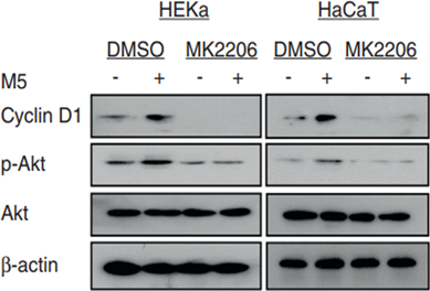
MK-2206 free base purchased from MedChemExpress. Usage Cited in: J Invest Dermatol. 2019 Jan;139(1):71-80. [Abstract]
HEKa (left) and HaCaT (right) cells are seeded in six-cell plates and pretreated with MK2206 for 6 hours, and then stimulated with M5 (2.5 ng/mL) for 48 hours before measuring the protein levels of cyclin D1, Akt, and phosphoAkt by Western blot analysis.
-
Biochem Pharmacol
Dopamine receptor D2 regulates inflammation and fibrosis in metabolic dysfunction-associated steatohepatitis disease. [Abstract]2026 Mar:245:117696. PMID: 41519398 -
Biochem Pharmacol
Celastrol synergizes with MEK1 inhibitor nedometinib to overcome resistance in bladder cancer via dual suppression of CDK1/CDC5L and feedback-activated Akt/STAT3. [Abstract]2025 Dec 10:245:117629. PMID: 41386559 -
-
Biochem Pharmacol
Lasiokaurin suppresses breast cancer growth by blocking autophagic flux and regulating cellular energy homeostasis. [Abstract]2025 Aug 7;242(Pt 3):117212. PMID: 40782951 -
Biochem Pharmacol
2025 Jul 26:241:117191. PMID: 40721007 -
Cell Prolif
SETD2 epidermal deficiency promotes cutaneous wound healing via activation of AKT/mTOR Signalling. [Abstract]2021 Jun;54(6):e13045. PMID: 33949020 -
Neurobiol Dis
Rho-kinase inhibition has antidepressant-like efficacy and expedites dendritic spine pruning in adolescent mice. [Abstract]2019 Apr;124:520-530. PMID: 30593834 -
Mol Cancer Ther
AXL kinase inhibition promotes cytosolic DNA sensor cGAS activity and sensitizes poorly immunogenic tumors to chemo-immunotherapy. [Abstract]2025 Nov 20. PMID: 41263056 -
Mol Cancer Ther
AKT Inhibition Sensitizes to Polo-Like Kinase 1 Inhibitor Onvansertib in Prostate Cancer. [Abstract]2024 Oct 1;23(10):1404-1417. PMID: 38894678 -
J Ethnopharmacol
"Shihu" promote T-cell immunity against breast cancer through bioactive constituent THMB enhancing JAK3-STAT1 signal. [Abstract]2026 Oct 28:369:121888. PMID: 42202911 -
J Ethnopharmacol
Paris polyphylla Smith var. yunnanensis-derived saponins potentiate the antitumor activity of GPX4 inhibitors. [Abstract]2026 Feb 28:357:120890. PMID: 41232632 -
J Ethnopharmacol
Targeting GRB2-Akt-SREBP1 signaling axis by saponins from Paris polyphylla Smith var. yunnanensis (Franch.) Hand.-Mazz to overcome cisplatin resistance in bladder cancer. [Abstract]2025 Aug 27:354:120502. PMID: 40882789 -
J Ethnopharmacol
Mechanism and molecular targets of Euphorbia fischeriana Steud root extract in hepatocellular carcinoma. [Abstract]2025 Mar 25:119707. PMID: 40147679 -
J Ethnopharmacol
Network pharmacology and phytochemical composition combined with validation in vivo and in vitro reveal the mechanism of platycodonis radix ameliorating PM2.5-induced acute lung injury. [Abstract]2024 Sep 13:118829. PMID: 39278295 -
J Ethnopharmacol
Moluodan promotes DSS-induced intestinal inflammation involving the reprogram of macrophage function and polarization. [Abstract]2024 Feb 10:320:117393. PMID: 37952735 -
J Ethnopharmacol
Bolbostemma paniculatum (Maxim.) Franquet extract suppresses the development of colorectal cancer through downregulation of PI3K/Akt pathway. [Abstract]2022 Apr 6:287:114937. PMID: 34958876 -
J Ethnopharmacol
Astragalus saponins improves stroke by promoting the proliferation of neural stem cells through phosphorylation of Akt. [Abstract]2021 Sep 15:277:114224. PMID: 34044075 -
Food Funct
Hesperetin ameliorates hepatic oxidative stress and inflammation via the PI3K/AKT-Nrf2-ARE pathway in oleic acid-induced HepG2 cells and a rat model of high-fat diet-induced NAFLD. [Abstract]2021 May 11;12(9):3898-3918. PMID: 33977953 -
Cells
Integrin-Mediated TIMP1 Signaling Reprograms Liver Macrophages and Accelerates Colorectal Cancer Metastasis. [Abstract]2025 Dec 23;15(1):29. PMID: 41511313 -
Cells
MK-2206 Alleviates Renal Fibrosis by Suppressing the Akt/mTOR Signaling Pathway In Vivo and In Vitro. [Abstract]2022 Nov 5;11(21):3505. PMID: 36359901 -
Gastric Cancer
COPS5 inhibition synergizes with the antitumor effect of trastuzumab by PTEN upregulation in HER2-amplified gastric cancer. [Abstract]2025 Oct 2. PMID: 41039054 -
Commun Biol
2025 Mar 31;8(1):529. PMID: 40164822 -
Commun Biol
PI3K-AKT-mediated phosphorylation of Thr260 in CgCaspase-3/6/7 regulates heat-induced activation in oysters. [Abstract]2024 Nov 7;7(1):1459. PMID: 39511363 -
Life Sci
Lactobacillus rhamnosus GG aggravates vascular calcification in chronic kidney disease: A potential role for extracellular vesicles. [Abstract]2023 Oct 15:331:122001. PMID: 37625519 -
Life Sci
A bovine milk-derived peptide ameliorates alloxan-injured pancreatic β cells through IRS2/PI3K/Akt signaling. [Abstract]2022 Nov 1:308:120907. PMID: 36037870 -
Life Sci
DHCR24 overexpression modulates microglia polarization and inflammatory response via Akt/GSK3β signaling in Aβ25-35 treated BV-2 cells. [Abstract]2020 Nov 1;260:118470. PMID: 32950573 -
Life Sci
IGF-1 promotes multiple myeloma progression through PI3K/Akt-mediated epithelial-mesenchymal transition. [Abstract]2020 May 15;249:117503. PMID: 32142767 -
Life Sci
Osteopontin induces atrial fibrosis by activating Akt/GSK-3β/β-catenin pathway and suppressing autophagy. [Abstract]2020 Mar 15;245:117328. PMID: 31954162 -
Nutrients
Phosphate Metabolic Inhibition Contributes to Irradiation-Induced Myelosuppression through Dampening Hematopoietic Stem Cell Survival. [Abstract]2022 Aug 18;14(16):3395. PMID: 36014901 -
Int J Mol Sci
Traditional Medicinal Plant Dahlia pinnata Selectively Suppresses TNF-α Expression Through Modulation of NF-κB and p38 Signaling. [Abstract]2026 Jan 22;27(2):1122. PMID: 41596763 -
Int J Mol Sci
Hypercapnia Increases Influenza A Virus Infection of Bronchial Epithelial Cells by Augmenting Cellular Cholesterol via mTOR and Akt. [Abstract]2025 Apr 26;26(9):4133. PMID: 40362373 -
Int J Mol Sci
Metformin Suppresses Stemness of Non-Small-Cell Lung Cancer Induced by Paclitaxel through FOXO3a. [Abstract]2023 Nov 22;24(23):16611. PMID: 38068934 -
Int J Mol Sci
Involvement of the p38/MK2 Pathway in MCLR Hepatotoxicity Revealed through MAPK Pharmacological Inhibition and Phosphoproteomics in HepaRG Cells. [Abstract]2023 Jul 6;24(13):11168. PMID: 37446360 -
Int J Mol Sci
PEBP4 Directs the Malignant Behavior of Hepatocellular Carcinoma Cells via Regulating mTORC1 and mTORC2. [Abstract]2022 Aug 8;23(15):8798. PMID: 35955931 -
Int J Oncol
2017 Aug;51(2):625-632. PMID: 28656278 -
Cell Oncol (Dordr)
Blocking MMP-12-modulated epithelial-mesenchymal transition by repurposing penfluridol restrains lung adenocarcinoma metastasis via uPA/uPAR/TGF-β/Akt pathway. [Abstract]2021 Oct;44(5):1087-1103. PMID: 34319576 -
Int Immunopharmacol
Rheumatoid arthritis synovial fibroblasts promote the glycolysis of myeloid-derived suppressor cells via TREM1/mTOR axis. [Abstract]2026 Sep 1:184:116961. PMID: 42242136 -
Int Immunopharmacol
IL-5 promotes airway remodeling in asthma by mediating epithelial-mesenchymal transition via the AKT/NLRP3 pathway. [Abstract]2026 Apr 15:175:116422. PMID: 41759267 -
Int Immunopharmacol
The mechanism study of isoorientin regulating neuroinflammation after subarachnoid hemorrhage through AKT/GSK3β. [Abstract]2026 Apr 1:174:116299. PMID: 41671619 -
Int Immunopharmacol
Gasdermin D-dependent macrophage pyroptosis mediates polystyrene microplastics-induced pulmonary fibrosis. [Abstract]2026 Mar 1:172:116189. PMID: 41539003 -
Bioorg Chem
Gypenoside L inhibits oral squamous cell carcinoma progression by targeting the AKT signaling pathway. [Abstract]2026 Jan:168:109303. PMID: 41343873 -
Eur J Pharmacol
Investigating the effects of dendrobine on fibrosis and inflammation in orbital fibroblasts of thyroid-associated ophthalmopathy via network pharmacology, molecular docking, and transcriptomics. [Abstract]2025 Jun 20:177871. PMID: 40544935 -
Int Immunopharmacol
CARD9 promotes cholangiocarcinoma by regulating the IL-17A/Hedgehog and the THEM4/AKT/mTOR signaling pathways. [Abstract]2024 Oct 16;143(Pt 2):113399. PMID: 39418733 -
Int Immunopharmacol
The role of the PI3K/AKT/mTOR pathway in mediating PD-L1 upregulation during fibroblast transdifferentiation. [Abstract]2024 Sep 18;142(Pt B):113186. PMID: 39298817 -
Int Immunopharmacol
Role of glucose metabolism reprogramming in keratinocytes in the link between psoriasis and metabolic syndrome. [Abstract]2024 Jul 19:139:112704. PMID: 39032466 -
Mol Cancer Res
Vasorin exocytosed from glioma cells facilitates angiogenesis via VEGFR2/AKT signaling pathway. [Abstract]2024 Jul 2;22(7):668-681. PMID: 38488456 -
Eur J Pharmacol
Licochalcone A induces mitochondria-dependent apoptosis and interacts with venetoclax in acute myeloid leukemia. [Abstract]2024 Apr 5:968:176418. PMID: 38350590 -
Eur J Pharmacol
Platycodin D induces apoptotic cell death through PI3K/AKT and MAPK/ERK pathways and synergizes with venetoclax in acute myeloid leukemia. [Abstract]2023 Oct 5:956:175957. PMID: 37541375 -
Int Immunopharmacol
Paeoniflorin promotes intestinal stem cell-mediated epithelial regeneration and repair via PI3K-AKT-mTOR signalling in ulcerative colitis. [Abstract]2023 Jun:119:110247. PMID: 37159966 -
Int Immunopharmacol
Topoisomerase 2 inhibitor etoposide promotes interleukin-10 production in LPS-induced macrophages via upregulating transcription factor Maf and activating PI3K/Akt pathway. [Abstract]2021 Dec;101(Pt A):108264. PMID: 34715493 -
Int Immunopharmacol
Z-guggulsterone induces PD-L1 upregulation partly mediated by FXR, Akt and Erk1/2 signaling pathways in non-small cell lung cancer. [Abstract]2021 Apr:93:107395. PMID: 33529916 -
Mol Cancer Res
2020 Mar;18(3):414-423. PMID: 31932471 -
Biosci Rep
PD-L1 regulates tumorigenesis and autophagy of ovarian cancer by activating mTORC signaling. [Abstract]2019 Dec 20;39(12):BSR20191041. PMID: 31799599 -
Eur J Pharmacol
AS1949490, an inhibitor of 5'-lipid phosphatase SHIP2, promotes protein kinase C-dependent stabilization of brain-derived neurotrophic factor mRNA in cultured cortical neurons. [Abstract]2019 May 15:851:69-79. PMID: 30753865 -
Cancer Biol Ther
2025 Dec;26(1):2442556. PMID: 39699276 -
Molecules
Geniposidic Acid from Eucommia ulmoides Oliver Staminate Flower Tea Mitigates Cellular Oxidative Stress via Activating AKT/NRF2 Signaling. [Abstract]2022 Dec 5;27(23):8568. PMID: 36500666 -
Toxicology
Environmental exposure to microcystin-LR increases the risks of urinary bladder proliferation and carcinogenesis: Evidence from case control, animal, and in vitro studies. [Abstract]2022 Oct;480:153326. PMID: 36115647 -
Cancer Biol Ther
Repeated treatments of Capan-1 cells with PARP1 and Chk1 inhibitors promote drug resistance, migration and invasion. [Abstract]2022 Dec 31;23(1):69-82. PMID: 35000525 -
Endocr Relat Cancer
2019 Aug;26(8):699-712. PMID: 31146260 -
Mol Oncol
PP2A-based triple-strike therapy overcomes mitochondrial apoptosis resistance in brain cancer cells. [Abstract]2023 Sep;17(9):1803-1820. PMID: 37458534 -
Mol Oncol
L1CAM promotes vasculogenic mimicry formation by miR-143-3p-induced expression of hexokinase 2 in glioma. [Abstract]2023 Apr;17(4):664-685. PMID: 36708044 -
Front Microbiol
Toll-like receptor 2 signaling pathway activation contributes to a highly efficient inflammatory response in Japanese encephalitis virus-infected mouse microglial cells by proteomics. [Abstract]2022 Sep 12:13:989183. PMID: 36171749 -
Mol Oncol
Kaiso phosphorylation at threonine 606 leads to its accumulation in the cytoplasm, reducing its transcriptional repression of the tumour suppressor CDH1. [Abstract]2022 Sep;16(17):3192-3209. PMID: 35851744 -
Cancers (Basel)
The Targeted Degradation of BRAF V600E Reveals the Mechanisms of Resistance to BRAF-Targeted Treatments in Colorectal Cancer Cells. [Abstract]2023 Dec 12;15(24):5805. PMID: 38136350 -
Transplant Cell Ther
Thymosin β4 exerts cytoprotective function and attenuates liver injury in murine hepatic sinusoidal obstruction syndrome after hematopoietic stem cell transplantation. [Abstract]2023 Aug;29(8):492.e1-492.e10. PMID: 37192732 -
Cancers
Expression of MALAT1 Promotes Trastuzumab Resistance in HER2 Overexpressing Breast Cancers. [Abstract]2020 Jul 16;12(7):1918. PMID: 32708561 -
Clin Epigenetics
Retinoic acid-induced 2 (RAI2) is a novel tumor suppressor, and promoter region methylation of RAI2 is a poor prognostic marker in colorectal cancer. [Abstract]2018 May 23:10:69. PMID: 29796120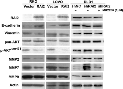
MK-2206 free base purchased from MedChemExpress. Usage Cited in: Clin Epigenetics. 2018 May 23:10:69. [Abstract]
The expression levels of E-cadherin, vimentin, pan-AKT, p-AKTser473, MMP-2, MMP-7, MMP-9, and actin (control) are detected by Western blot in RAI2 un-expressed and re-expressed RKO and LOVO cells as well as in the control group, shRAI2 knockdown group, and shRAI2 knockdown plus MK2206 treated group.
-
Mol Neurobiol
Regulation of Melatonin on AQP4 Expression in Brain of SD Newborn Rats with Bilirubin Encephalopathy and Protection Mechanism. [Abstract]2025 Dec 3;63(1):239. PMID: 41335355 -
ACS Omega
Dynamic Detection of the E3-PROTAC-Target Protein Ternary Complex In Vitro and In Vivo via Bimolecular Fluorescence Complementation. [Abstract]2024 Dec 3;9(50):49739-49748. PMID: 39713624 -
ACS Omega
MGST1 Expression Is Associated with Poor Prognosis, Enhancing the Wnt/β-Catenin Pathway via Regulating AKT and Inhibiting Ferroptosis in Gastric Cancer. [Abstract]2023 Jun 16;8(26):23683-23694. PMID: 37426275 -
Front Cell Dev Biol
Accelerated Bone Regeneration by Adrenomedullin 2 Through Improving the Coupling of Osteogenesis and Angiogenesis via β-Catenin Signaling. [Abstract]2021 Apr 14:9:649277. PMID: 33937244 -
ACS Omega
2020 Feb 13;5(7):3709-3716. PMID: 32118186 -
Cancer Sci
Reverse of non-small cell lung cancer drug resistance induced by cancer-associated fibroblasts via a paracrine pathway. [Abstract]2018 Apr;109(4):944-955. PMID: 29383798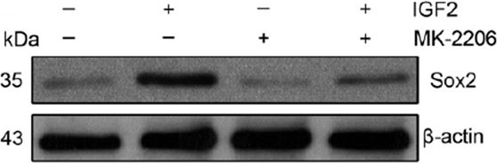
MK-2206 free base purchased from MedChemExpress. Usage Cited in: Cancer Sci. 2018 Apr;109(4):944-955. [Abstract]
Western blotting of Sox2 and b-actin in A549 cells with or without IGF2 treated in the presence or absence of MK-2206.
-
FASEB J
Rhythmic Insulin Induced by Eating During the Active Phase Promotes Skeletal Muscle Growth via Synchronizing Mitochondrial Energy Supply Rhythm and Cell Cycle. [Abstract]2025 May 31;39(10):e70619. PMID: 40354091 -
J Cell Mol Med
Stromal Cell Derived Factor-1 Promotes Hepatic Insulin Resistance via Inhibiting Hepatocyte Lipophagy. [Abstract]2025 Jan;29(2):e70352. PMID: 39855896 -
J Cell Mol Med
Foxk2 Enhances Adipogenic Differentiation by Relying on the Transcriptional Activation of Peroxisome Proliferator-Activated Receptor Gamma. [Abstract]2025 Jan;29(1):e70332. PMID: 39789420 -
Lab Invest
HECW1-Mediated Ubiquitination of HIPK2 Drives Metastasis in Gastric Cancer Through the AKT Signaling Pathway. [Abstract]2024 Nov 28:102202. PMID: 39615883 -
Lipids Health Dis
2024 Dec 27;23(1):424. PMID: 39731125 -
Biochim Biophys Acta Mol Basis Dis
2024 Sep 3:167496. PMID: 39237046 -
Biochim Biophys Acta Mol Basis Dis
Resistance exercise preconditioning prevents disuse muscle atrophy by inhibiting apoptosis and protein degradation via SESN2 in C57BL/6J mice. [Abstract]2024 Mar 2;1870(4):167111. PMID: 38432454 -
J Cell Mol Med
NEDD4L is a promoter for angiogenesis and cell proliferation in human umbilical vein endothelial cells. [Abstract]2024 Apr;28(8):1-11. PMID: 38526036 -
J Cell Mol Med
Reduced chemokine C-C motif ligand 1 expression may negatively regulate colorectal cancer progression at liver metastatic sites. [Abstract]2024 Apr;28(7):e18193. PMID: 38506205 -
FASEB J
LDHA deficiency inhibits trophoblast proliferation via the PI3K/AKT/FOXO1/CyclinD1 signaling pathway in unexplained recurrent spontaneous abortion. [Abstract]2023 Feb;37(2):e22744. PMID: 36583693 -
J Cell Mol Med
Slight up-regulation of Kir2.1 channel promotes endothelial progenitor cells to transdifferentiate into a pericyte phenotype by Akt/mTOR/Snail pathway. [Abstract]2021 Nov;25(21):10088-10100. PMID: 34592781 -
J Cell Mol Med
Aberrant methylation of WD-repeat protein 41 contributes to tumour progression in triple-negative breast cancer. [Abstract]2020 Jun;24(12):6869-6882. PMID: 32394588 -
FASEB J
Par6 regulates cell cycle progression through enhancement of Akt/PI3K/GSK-3β signaling pathway activation in glioma. [Abstract]2020 Jan;34(1):1481-1496. PMID: 31914615 -
Mediators Inflamm
Andrographolide Protects against HG-Induced Inflammation, Apoptosis, Migration, and Impairment of Angiogenesis via PI3K/AKT-eNOS Signalling in HUVECs. [Abstract]2019 Oct 7;2019:6168340. PMID: 31686985 -
FEBS J
Systems analysis identifies potential target genes to overcome cetuximab resistance in colorectal cancer cells. [Abstract]2019 Apr;286(7):1305-1318. PMID: 30719834 -
J Inflamm Res
Huanglian Jiedu Decoction Alleviates Myocardial Ischemia-Reperfusion Injury via AKT-Mediated Regulation of Endoplasmic Reticulum Stress. [Abstract]2025 Dec 3:18:17025-17044. PMID: 41368345 -
iScience
TRIM36 inhibits lung fibroblast activation and pulmonary fibrosis through the degradation of phospho-AKT1. [Abstract]2025 Oct 15;28(11):113775. PMID: 41323265 -
iScience
ATP6V0D2 drives triple-negative breast cancer progression and inhibits cisplatin sensitivity by promoting PHLPP2 lysosomal degradation. [Abstract]2025 Oct 27;28(11):113853. PMID: 41312388 -
iScience
Integrative multi-omics analysis decodes HOXC9-driven malignant transformation and metastasis in OSCC. [Abstract]2025 Sep 10;28(9):112919. PMID: 41054515 -
iScience
Broad and diverse roles of sphingosine-1-phosphate/sphingosine-1-phosphate receptors in the prostate. [Abstract]2024 Oct 30;27(12):111290. PMID: 39618500 -
Oncol Res
LncRNA AFAP1-AS1 exhibits oncogenic characteristics and promotes gemcitabine-resistance of cervical cancer cells through miR-7-5p/EGFR axis. [Abstract]2024 Nov 13;32(12):1867-1879. PMID: 39574469 -
J Lipid Res
Metallothionein 1B attenuates inflammation and hepatic steatosis in MASH by inhibiting the AKT/PI3K pathway. [Abstract]2024 Nov 15:100701. PMID: 39551239 -
Transl Oncol
Unraveling the mechanisms of RECQL4-mediated cervical cancer progression through the PI3K/AKT pathway. [Abstract]2024 Dec:50:102146. PMID: 39378549 -
iScience
SGMS1 facilitates osteogenic differentiation of MSCs and strengthens osteogenesis-angiogenesis coupling by modulating Cer/PP2A/Akt pathway. [Abstract]2024 Mar 5;27(4):109358. PMID: 38544565 -
iScience
Trigeminal nerve-derived substance P regulates limbal stem cells by the PI3K-AKT pathway. [Abstract]2023 Apr 18;26(5):106688. PMID: 37187698 -
iScience
Schlafen 11 expression in human acute leukemia cells with gain-of-function mutations in the interferon-JAK signaling pathway. [Abstract]2021 Sep 25;24(10):103173. PMID: 34693224 -
Front Mol Biosci
The Role of TRPC6 in Renal Ischemia/Reperfusion and Cellular Hypoxia/Reoxygenation Injuries. [Abstract]2021 Jul 8:8:698975. PMID: 34307458 -
J Cell Physiol
BKCa channel participates in insulin-induced lipid deposition in adipocytes by increasing intracellular calcium. [Abstract]2021 Aug;236(8):5818-5831. PMID: 33432604 -
Fish Shellfish Immunol
M1 macrophage polarization induces inflammatory response of adipocytes via MAPK/AKT-PPARγ pathway in large yellow croaker (Larimichthys crocea). [Abstract]2026 Aug:175:111443. PMID: 42162881 -
J Biol Chem
2026 Jun;302(6):111461. PMID: 41999888 -
J Biol Chem
Downregulation of ClC-3 chloride channels in dorsal root ganglia neurons contributes to bone metastasis-induced pain. [Abstract]2026 Mar;302(3):111268. PMID: 41655695 -
Sci Rep
Involvement of AKT/β-catenin in Enhanced Osteogenesis by Cannabidiol in Bone Stromal Cells Harvested from Human Tori. [Abstract]2026 Feb 20. PMID: 41720800 -
Sci Rep
Pseudomonas aeruginosa-derived DnaJ functions as a novel immunomodulator inducing IFNβ via CME-SGK1-IRF3 axis in macrophages. [Abstract]2025 Dec 3. PMID: 41339751 -
Sci Rep
Panax notoginseng saponins suppress the PI3K/AKT pathway to enhance autophagy and apoptosis in pulmonary fibrosis. [Abstract]2025 Nov 27;15(1):43622. PMID: 41309997 -
Sci Rep
Establishment of novel cholangiocarcinoma cell lines with ARID1A deficiency and preclinical validation of synthetic lethality therapies. [Abstract]2025 Nov 18;15(1):40619. PMID: 41254205 -
Sci Rep
SREBP-2 promotes cancer progression through the mevalonate-Akt pathway in non-small cell lung cancer. [Abstract]2025 Jul 2;15(1):23103. PMID: 40595126 -
Sci Rep
AKT activation triggers Rab14-mediated ADAM10 translocation to the cell surface in human aortic endothelial cells. [Abstract]2025 Mar 3;15(1):7448. PMID: 40032916 -
Sci Rep
Arctiin alleviates knee osteoarthritis by suppressing chondrocyte oxidative stress induced by accumulated iron via AKT/NRF2/HO-1 signaling pathway. [Abstract]2024 Dec 30;14(1):31935. PMID: 39738432 -
Sci Rep
Blockage of Akt activation suppresses cadmium-induced renal tubular cellular damages through aggrephagy in HK-2 cells. [Abstract]2024 Jun 24;14(1):14552. PMID: 38914593 -
J Biol Chem
Nuclear factor Nrf2 promotes glycosidase OGG1 expression by activating the AKT pathway to enhance leukemia cell resistance to cytarabine. [Abstract]2023 Jan;299(1):102798. PMID: 36528059 -
Sci Rep
Development of actionable targets of multi-kinase inhibitors (AToMI) screening platform to dissect kinase targets of staurosporines in glioblastoma cells. [Abstract]2022 Aug 13;12(1):13796. PMID: 35963891 -
J Biol Chem
Prolonged deprivation of arginine or leucine induces PI3K/Akt-dependent reactivation of mTORC1. [Abstract]2022 Jun;298(6):102030. PMID: 35577075 -
Aging (Albany NY)
NET1 promotes HCC growth and metastasis in vitro and in vivo via activating the Akt signaling pathway. [Abstract]2021 Apr 11;13(7):10672-10687. PMID: 33839702 -
J Biomed Mater Res A
Immunomodulatory property and its regulatory mechanism of double network hydrogel on dendritic cells. [Abstract]2021 Jun;109(6):1015-1026. PMID: 32856407 -
Aging
Resveratrol inhibits the malignant progression of hepatocellular carcinoma via MARCH1-induced regulation of PTEN/AKT signaling. [Abstract]2020 Jun 12;12(12):11717-11731. PMID: 32530437 -
Sci Rep
Vitamin K2 promotes PI3K/AKT/HIF-1α-mediated glycolysis that leads to AMPK-dependent autophagic cell death in bladder cancer cells. [Abstract]2020 May 7;10(1):7714. PMID: 32382009 -
Oncol Rep
Downregulation of PDCD4 induced by progesterone is mediated by the PI3K/AKT signaling pathway in human endometrial cancer cells. [Abstract]2019 Aug;42(2):849-856. PMID: 31233196 -
Sci Rep
E-cadherin loss induces targetable autocrine activation of growth factor signalling in lobular breast cancer. [Abstract]2018 Oct 18;8(1):15454 PMID: 30337563 -
J Biol Chem
Kinome-wide selectivity profiling of ATP-competitive mammalian target of rapamycin (mTOR) inhibitors and characterization of their binding kinetics. [Abstract]2012 Mar 23;287(13):9742-52. PMID: 22223645 -
PNAS Nexus
Ribosome stalling during c-myc translation presents actionable cancer cell vulnerability. [Abstract]2024 Aug 13;3(8):pgae321. PMID: 39161732 -
Cell Signal
FOLR2+ tumor-associated macrophages drive metastatic microenvironment remodeling and lymphatic dissemination in colorectal cancer. [Abstract]2026 Oct:146:112676. PMID: 42297063 -
Biochim Biophys Acta Mol Cell Res
FBXO32 activates the PI3K/AKT pathway by inhibiting PTEN through ubiquitination of TAL1 in hepatocellular carcinoma. [Abstract]2026 Apr;1873(4):120125. PMID: 41713558 -
Cell Signal
2025 May 17:111877. PMID: 40389045 -
Cell Signal
STEAP4 modulates cell proliferation and oxidative stress in benign prostatic hyperplasia. [Abstract]2024 Jan:113:110933. PMID: 37866665 -
Mol Cell Biochem
Inhibition of STIM1 alleviates high glucose-induced proliferation and fibrosis by inducing autophagy in mesangial cells. [Abstract]2024 Sep;479(9):2365-2379. PMID: 37736800 -
Brain Res Bull
Hypocretin-1 suppresses malignant progression of glioblastoma cells through Notch1 signaling pathway. [Abstract]2023 May:196:46-58. PMID: 36925051 -
Reproduction
WNT16 from decidual stromal cells regulates HTR8/SVneo trophoblastic cell function via AKT/beta-catenin pathway. [Abstract]2022 Mar 10;163(5):241-250. PMID: 35174787 -
Cell Signal
Akt phosphorylation regulated by IKKε in response to low shear stress leads to endothelial inflammation via activating IRF3. [Abstract]2021 Apr:80:109900. PMID: 33370582 -
Brain Res Bull
2020 Sep;162:166-179. PMID: 32599128 -
Cytokine
PI3K mediates tumor necrosis factor induced-necroptosis through initiating RIP1-RIP3-MLKL signaling pathway activation. [Abstract]2020 May;129:155046. PMID: 32114297 -
Development
Axin1 regulates tooth root development by inhibiting AKT1-mTORC1 activation and Shh translation in Hertwig's epithelial root sheath. [Abstract]2024 Nov 1;151(21):dev202899. PMID: 39344774 -
Heliyon
Role of HDAC3 in the epithelial-mesenchymal transition of retinal pigment epithelium cells: Implications for proliferative vitreoretinopathy. [Abstract]2024 Oct 11;10(21):e39333. PMID: 39524785 -
Heliyon
PTOV1 facilitates colorectal cancer cell proliferation through activating AKT1 signaling pathway. [Abstract]2024 Aug 10;10(16):e36017. PMID: 39229496 -
Biol Trace Elem Res
Ortho-silicic Acid Plays a Protective Role in Glucocorticoid-Induced Osteoporosis via the Akt/Bad Signal Pathway In Vitro and In Vivo. [Abstract]2023 Feb;201(2):843-855. PMID: 35314965 -
Bone
PTX3 promotes osteogenic differentiation by triggering HA/CD44/FAK/AKT positive feedback loop in an inflammatory environment. [Abstract]2022 Jan:154:116231. PMID: 34653679 -
Stem Cells
M2 Macrophages Enhance the Cementoblastic Differentiation of Periodontal Ligament Stem Cells via the Akt and JNK Pathways. [Abstract]2019 Dec;37(12):1567-1580. PMID: 31400241 -
Front Med
Wogonoside alleviates hyperosmotic stress-induced inflammation and apoptosis in human corneal epithelial cells via PI3K/AKT signaling. [Abstract]2026 May 4:13:1828021. PMID: 42210954 -
Mol Med Rep
ER‑α36 knockdown is associated with lysosomal dysfunction and proliferation inhibition in liver cancer cells. [Abstract]2025 Oct;32(4):284. PMID: 40808340 -
Food Chem Toxicol
Chronic exposure to polystyrene micro/nanoplastics triggers testicular dysfunction through PI3K/AKT/mTOR signaling-mediated spermatocyte senescence in mice. [Abstract]2025 Aug 14:205:115703. PMID: 40818636 -
Mol Med Rep
MUC1‑ND interacts with TRPV1 to promote corneal epithelial cell proliferation in diabetic dry eye mice by partly activating the AKT signaling pathway. [Abstract]2024 Dec;30(6):213. PMID: 39370807 -
Clin Exp Hypertens
FGFR2 modulates the Akt/Nrf2/ARE signaling pathway to improve angiotensin II-induced hypertension-related endothelial dysfunction. [Abstract]2023 Dec 31;45(1):2208777. PMID: 37154169 -
Mol Med Rep
CMTM5 inhibits the development of prostate cancer via the EGFR/PI3K/AKT signaling pathway. [Abstract]2022 Jan;25(1):17. PMID: 34791506 -
Mol Med Rep
LY‑294002 enhances the chemosensitivity of liver cancer to oxaliplatin by blocking the PI3K/AKT/HIF‑1α pathway. [Abstract]2021 Jul;24(1):508. PMID: 33982772 -
Exp Cell Res
CKIP-1 augments autophagy in steatotic hepatocytes by inhibiting Akt/mTOR signal pathway. [Abstract]2020 Dec 1;397(1):112341. PMID: 33191205 -
Food Chem Toxicol
Diallyl sulfide protects against lipopolysaccharide/d-galactosamine-induced acute liver injury by inhibiting oxidative stress, inflammation and apoptosis in mice. [Abstract]2018 Oct:120:500-509. PMID: 30075314 -
J Diabetes Res
Targeting Oxidative Stress and Apoptosis via PI3K/Akt/Nrf2 Pathway: The Therapeutic Role of Bletilla striata Polysaccharide in Diabetic Wound Repair. [Abstract]2026 Jan 28:2026:5751331. PMID: 41613340 -
BMC Cancer
Aldo-keto reductase family 1 member C3 mediates radioresistance of esophageal cancer cells through suppressing MAPK and AKT signaling. [Abstract]2024 Oct 7;24(1):1236. PMID: 39375680 -
J Hepatocell Carcinoma
ZDHHC20 Activates AKT Signaling Pathway to Promote Cell Proliferation in Hepatocellular Carcinoma. [Abstract]2024 Sep 18:11:1763-1775. PMID: 39309302 -
BMC Complement Med Ther
Astragalus polysaccharide restores insulin secretion impaired by lipopolysaccharides through the protein kinase B /mammalian target of rapamycin/glucose transporter 2 pathway. [Abstract]2023 Oct 10;23(1):358. PMID: 37817130 -
Cell Cycle
Cancer-associated fibroblast-released extracellular vesicles carrying miR-199a-5p induces the progression of gastric cancer through regulation of FKBP5-mediated AKT1/mTORC1 signaling pathway. [Abstract]2022 Dec;21(24):2590-2601. PMID: 36005478 -
Toxicol Appl Pharmacol
Icariin promotes mouse Leydig cell testosterone synthesis via the Esr1/Src/Akt/Creb/Sf-1 pathway. [Abstract]2022 Apr 15:441:115969. PMID: 35259346 -
Bosn J Basic Med Sci
Stomach-specific c-Myc overexpression drives gastric adenoma in mice through AKT/mammalian target of rapamycin signaling. [Abstract]2021 Aug 1;21(4):434-446. PMID: 33259779 -
J Immunol
2020 Oct 15;205(8):2255-2264. PMID: 32929041 -
Chem Biol Drug Des
Exploring the Therapeutic Potential of Oridonin in the Treatment of Laryngeal Cancer: A Comprehensive Strategy Involving Network Pharmacology, Molecular Docking, Dynamic Simulation, and Experimental Verification. [Abstract]2026 Jan;107(1):e70222. PMID: 41525139 -
Chem Biol Drug Des
Therapeutic Potential of 6-Gingerol in Glioma: Mechanistic Insights and Experimental Validation. [Abstract]2025 Dec;106(6):e70217. PMID: 41351348 -
Am J Physiol Gastrointest Liver Physiol
Plant green pigment of chlorophyllin attenuates inflammatory bowel diseases by suppressing autophagy activation in mice. [Abstract]2022 Aug 1;323(2):G102-G113. PMID: 35638642 -
Mol Carcinog
ZNF480 Accelerates Chemotherapy Resistance in Breast Cancer by Competing With TRIM28 and Stabilizing LSD1 to Upregulate the AKT-GSK3β-Snail Pathway. [Abstract]2025 Jan;64(1):192-208. PMID: 39503216 -
Int J Med Sci
Enhanced Bone Regeneration by Schwann Cells through Coupling of Osteogenesis and Angiogenesis via β-catenin signaling in a Preclinical Model of Distraction Osteogenesis. [Abstract]2025 Jan 1;22(1):209-226. PMID: 39744173 -
Mol Carcinog
LAIR1 promotes hepatocellular carcinoma cell metastasis and induces M2-macrophage infiltration through activating AKT-IKKβ-p65 axis. [Abstract]2024 Sep;63(9):1827-1841. PMID: 39016636 -
Cell Stress Chaperones
2024 Apr;29(2):272-284. PMID: 38485044 -
J Pharm Pharmacol
Ethyl 2,2-difluoro-2-(2-oxo-2H-chromen-3-yl) acetate attenuates the malignant biological behaviors of non-small cell lung cancer via suppressing EGFR/PI3K/AKT/mTOR signaling pathway. [Abstract]2024 Mar 4;76(3):269-282. PMID: 38241189 -
Environ Toxicol
MUC1 promotes lymph node metastasis in esophageal squamous cell carcinoma by downregulating DNAJB6 expression. [Abstract]2024 Jan;39(1):9-22. PMID: 37584547 -
Cytotherapy
Mesenchymal stromal cells regulate THP-1-differentiated macrophage cytokine production by activating Akt/mammalian target of rapamycin complex 1 pathway. [Abstract]2023 Aug;25(8):858-865. PMID: 37125989 -
Mol Carcinog
LATS-regulated nuclear-cytoplasmic translocation of SREBP2 inhibits hepatocellular carcinoma cell migration and invasion via epithelial-mesenchymal transition. [Abstract]2023 Jul;62(7):963-974. PMID: 37042569 -
J Cancer
2023 Mar 27;14(5):784-792. PMID: 37056386 -
J Cancer
Cry 1 Regulates the Clock Gene Network and Promotes Proliferation and Migration Via the Akt/P53/P21 Pathway in Human Osteosarcoma Cells. [Abstract]2018 Jun 14;9(14):2480-2491. PMID: 30026846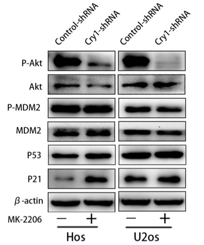
MK-2206 free base purchased from MedChemExpress. Usage Cited in: J Cancer. 2018 Jun 14;9(14):2480-2491. [Abstract]
Analysis of indicated proteins in Cry1 silencing osteosarcoma cells after MK-2206 treatment. Data are expressed as the mean±standard deviation.
-
Hum Mol Genet
BMP4 uses several different effector pathways to regulate proliferation and differentiation in the epithelial and mesenchymal tissue compartments of the developing mouse ureter. [Abstract]2017 Sep 15;26(18):3553-3563. PMID: 28655168
MK-2206 free base purchased from MedChemExpress. Usage Cited in: Hum Mol Genet. 2017 Sep 15;26(18):3553-3563. [Abstract]
Immunofluorescence analysis for expression of the I-cell marker ΔNP63 on proximal sections of ureters explanted from E12.5 wildtype (control) embryos and cultured for 6 d in the presence of solvent (DMSO) (A), the AKT inhibitor (AKT-i) MK2206 (B), the P38 inhibitor (P38-i) SB203580 (C), the ERK1/2 inhibitor (ERK1/2-i) PD98059 (D) or combinations as indicated (E and F).
-
Naunyn Schmiedebergs Arch Pharmacol
Medicarpin reduces cisplatin resistance and causes apoptosis by inhibiting the AKT/Bcl2 pathway in breast cancer. [Abstract]2025 Oct 24. PMID: 41134353 -
Cancer Med
Focal Adhesion Kinase Intersects With the BRD4-MYC Axis and YAP1 to Drive Tumor Cell Growth, Phenotypic Plasticity, Stemness, and Metastatic Potential in Colorectal Cancer. [Abstract]2025 Sep;14(18):e71227. PMID: 40959971 -
J Diabetes Complications
Modulation of the sodium-chloride cotransporter by insulin in auditory cells: A potential link to diabetes-related hearing complications. [Abstract]2025 Jun 22;39(9):109111. PMID: 40561788 -
Curr Issues Mol Biol
2024 Dec 31;47(1):16. PMID: 39852131 -
Arch Biochem Biophys
Carboxypeptidase A6 suppresses the proliferation and invasion of colorectal cancer cells and is negatively regulated by miR-96-3p. [Abstract]2023 May 15:740:109595. PMID: 37011707 -
Mol Pharmacol
Functional RNAi Screens Define Distinct Protein Kinase Vulnerabilities in EGFR-Dependent HNSCC Cell Lines. [Abstract]2019 Dec;96(6):862-870. PMID: 31554698 -
Shock
ATAXIA TELANGIECTASIA MUTATED PROTECTS AGAINST LIPOPOLYSACCARIDE-INDUCED BLOOD-BRAIN BARRIER DISRUPTION BY REGULATING ATK/DRP1-MEDIATED MITOCHONDRIAL HOMEOSTASIS. [Abstract]2023 Jul 1;60(1):100-109. PMID: 37141173 -
J Reprod Immunol
4-vinylcyclohexene diepoxide induces premature ovarian insufficiency in rats by triggering the autophagy of granule cells through regulating miR-144. [Abstract]2023 Jun:157:103928. PMID: 36889083 -
Life Sci Alliance
2022 Jun 2;5(10):e202101330. PMID: 35654587 -
Cell Immunol
2022 Feb:372:104475. PMID: 35063904 -
Front Cardiovasc Med
TANK Promotes Pressure Overload Induced Cardiac Hypertrophy via Activating AKT Signaling Pathway. [Abstract]2021 Sep 3;8:687540. PMID: 34540911 -
Am J Cancer Res
TM2D1 contributes the epithelial-mesenchymal transition of hepatocellular carcinoma via modulating AKT/β-catenin axis. [Abstract]2021 Apr 15;11(4):1557-1571. PMID: 33948373 -
R Soc Open Sci
Ultra-performance liquid chromatography-tandem mass spectrometric method for quantitation of the recently Food and Drug Administration approved combination of vaborbactam and meropenem in human plasma. [Abstract]2020 Jul 8;7(7):200635. PMID: 32874656 -
Peptides
2018 Sep:107:39-44. PMID: 30081043 -
Int J Biochem Cell Biol
2023 Feb:155:106359. PMID: 36586532 -
Mol Biol Rep
Combined treatment with ruxolitinib and MK-2206 inhibits the JAK2/STAT5 and PI3K/AKT pathways via apoptosis in MDA-MB-231 breast cancer cell line. [Abstract]2023 Jan;50(1):319-329. PMID: 36331743 -
Mol Biol Rep
mTOR inhibitor PP242 increases antitumor activity of sulforaphane by blocking Akt/mTOR pathway in esophageal squamous cell carcinoma. [Abstract]2022 Jan;49(1):451-461. PMID: 34731371 -
J Biochem Mol Toxicol
ATGL promotes the proliferation of hepatocellular carcinoma cells via the p-AKT signaling pathway. [Abstract]2019 Nov;33(11):e22391. PMID: 31476254 -
J Cell Biochem
Absent in melanoma 2 suppresses epithelial-mesenchymal transition via Akt and inflammasome pathways in human colorectal cancer cells. [Abstract]2019 Oct;120(10):17744-17756. PMID: 31210372 -
Int J Biochem Cell Biol
NDRG2 suppresses proliferation, migration, invasion and epithelial-mesenchymal transition of esophageal cancer cells through regulating the AKT/XIAP signaling pathway. [Abstract]2018 Jun:99:43-51. PMID: 29530788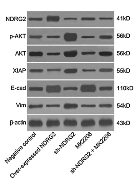
MK-2206 free base purchased from MedChemExpress. Usage Cited in: Int J Biochem Cell Biol. 2018 Jun:99:43-51. [Abstract]
Western blot analysis of NDRG2, p-ATK, XIAP, E-cadherin and Vimentin in TE-13 cells shows NDRG2 negatively regulates the expressions of AKT, XIAP, E-cadherin and Vimentin proteins.
-
J Cell Biochem
Matrine reversed multidrug resistance of breast cancer MCF-7/ADR cells through PI3K/AKT signaling pathway. [Abstract]2018 May;119(5):3885-3891. PMID: 29130495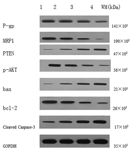
MK-2206 free base purchased from MedChemExpress. Usage Cited in: J Cell Biochem. 2018 May;119(5):3885-3891. [Abstract]
Western blot showed the protein expression of P-gp,MRP1, PTEN, p-AKT, Bax, Bcl-2, and cleaved caspase-3.
-
Int J Biochem Cell Biol
STYK1 promotes cancer cell proliferation and malignant transformation by activating PI3K-AKT pathway in gallbladder carcinoma. [Abstract]2018 Apr:97:16-27. PMID: 29413947 -
Vet Microbiol
The Chinese medicine monomer Schisandrin C inhibits PRRSV infection by regulating the OGT-PI3K/AKT/mTOR signaling pathway. [Abstract]2026 May:316:110992. PMID: 41865607 -
Microbes Infect
Pseudomonas aeruginosa-derived DnaJ induces TLR2 expression through TLR10-mediated activation of the PI3K-SGK1 pathway in macrophages. [Abstract]2025 May-Jun;27(4):105481. PMID: 39978578 -
Microvasc Res
2024 Jul:154:104689. PMID: 38636926 -
J Integr Neurosci
Heliox Protects SH-SY5Y Cells from Oxygen-Glucose Deprivation/Reperfusion-Induced Ferroptosis. [Abstract]2024 Jan 16;23(1):14. PMID: 38287843 -
Toxicol In Vitro
Everolimus prevents doxorubicin-induced apoptosis in H9c2 cardiomyocytes but not in MCF-7 cancer cells: Cardioprotective roles of autophagy, mitophagy, and AKT. [Abstract]2023 Dec:93:105698. PMID: 37739323 -
Radiat Res
Mitochondrial Dysfunctions Regulated Radioresistance through Mitochondria-to-Nucleus Retrograde Signaling Pathway of NF-κB/PI3K/AKT2/mTOR. [Abstract]2018 Aug;190(2):204-215. PMID: 29863983
MK-2206 free base purchased from MedChemExpress. Usage Cited in: Radiat Res. 2018 Aug;190(2):204-215. [Abstract]
When 6 Gy irradiation is combined with AKT2 inhibitor MK-2206 treatment, the protein levels in phosphorylated AKT2, mTOR and IKBa are decreased, and the downstream proapoptotic proteins, caspase 3 and caspase 8, are increased.
-
Exp Eye Res
Expression and regulation of alarmin cytokine IL-1α in human retinal pigment epithelial cells. [Abstract]2018 Jul:172:10-20. PMID: 29551335 -
PLoS One
Exosomes from human umbilical cord mesenchymal stem cells promote the growth of human hair dermal papilla cells. [Abstract]2025 Apr 30;20(4):e0320154. PMID: 40305498 -
Platelets
2023 Dec;34(1):2173505. PMID: 36813739 -
PLoS One
2016 Jan 28;11(1):e0147682. PMID: 26821351 -
Pulm Circ
Effect of Gouteng Jiangya Jieyu Prescription on Neuronal Damage in Rats With Hypertension Complicated by Depression via ROS/AKT/NF-κB Pathway. [Abstract]2026 Apr 23;16(2):e70306. PMID: 42039681 -
Chem Biodivers
Total Flavonoids of Hovenia dulcis Seeds Ameliorate Hepatic Fibrosis via Modulating PI3K/AKT Signaling Pathway In Vitro and In Vivo. [Abstract]2025 Oct 24:e02088. PMID: 41135015 -
Tissue Cell
Nanosphere loaded with curcumin attenuates adipogenesis and lipid metabolism by modulating AKT/mTORC1/PPARγ phosphorylation. [Abstract]2025 Aug 29:97:103112. PMID: 40886657 -
Clin Exp Pharmacol Physiol
Dapagliflozin alleviates high glucose-induced injury of endothelial cells via inducing autophagy. [Abstract]2024 Apr;51(4):e13846. PMID: 38382536 -
J Pain Res
Intravenous Administration of Triptonide Attenuates CFA-Induced Pain Hypersensitivity by Inhibiting DRG AKT Signaling Pathway in Mice. [Abstract]2020 Dec 1;13:3195-3206. PMID: 33293856 -
Clin Exp Pharmacol Physiol
SPNS2 promotes the malignancy of colorectal cancer cells via regulating Akt and ERK pathway. [Abstract]2019 Sep;46(9):861-871. PMID: 31206801 -
Medicina (Kaunas)
2026 Feb 27;62(3):456. PMID: 41901538 -
Virology
The Pan-ErbB tyrosine kinase inhibitor afatinib inhibits multiple steps of the mammarenavirus life cycle. [Abstract]2022 Nov:576:83-95. PMID: 36183499 -
PeerJ
Icariside II induces rapid phosphorylation of endothelial nitric oxide synthase via multiple signaling pathways. [Abstract]2022 Oct 25:10:e14192. PMID: 36312762 -
Int J Endocrinol
Umbilical Cord-Derived Mesenchymal Stem Cells Improve Ornidazole-Induced Asthenozoospermia in Rats via Activation of the AKT/mTOR Pathway. [Abstract]2024 Nov 12:2024:3494652. PMID: 39564353 -
Thorac Cancer
ACT001 inhibits the proliferation of non-small cell lung cancer cells by upregulating NKTR expression. [Abstract]2022 Jun;13(12):1772-1782. PMID: 35537816 -
Exp Ther Med
mTORC1 is a key regulator that mediates OGD- and TGFβ1-induced myofibroblast transformation and chondroitin-4-sulfate expression in cardiac fibroblasts. [Abstract]2022 Jun;23(6):413. PMID: 35601064 -
Exp Ther Med
Assessing the anti-inflammatory effects of quercetin using network pharmacology and in vitro experiments. [Abstract]2022 Apr;23(4):301. PMID: 35340883 -
Exp Ther Med
Benazepril hydrochloride protects against doxorubicin cardiotoxicity by regulating the PI3K/Akt pathway. [Abstract]2021 Oct;22(4):1082. PMID: 34447475 -
FEBS Open Bio
METTL3 mediates bone marrow mesenchymal stem cell adipogenesis to promote chemoresistance in acute myeloid leukaemia. [Abstract]2021 Jun;11(6):1659-1672. PMID: 33932138 -
Exp Ther Med
miRNA-214 suppresses oxidative stress in diabetic nephropathy via the ROS/Akt/mTOR signaling pathway and uncoupling protein 2. [Abstract]2019 May;17(5):3530-3538. PMID: 30988734 -
Biochem Biophys Res Commun
HYAL2-generated low-molecular-weight hyaluronic acid promotes intervertebral disc degeneration via the CD44/AKT signaling axis. [Abstract]2026 Apr 30:811:153576. PMID: 41806573 -
Biochem Biophys Res Commun
Flavonoid derivative FB-15 inhibits the progression of nasopharyngeal carcinoma by targeting the PI3K-Akt signaling pathway. [Abstract]2025 May 16:771:152037. PMID: 40409114 -
Biochem Biophys Res Commun
2025 Jan 16:749:151352. PMID: 39847995 -
Leuk Res
PI3K/AKT confers intrinsic and acquired resistance to pirtobrutinib in chronic lymphocytic leukemia. [Abstract]2024 Jul 7:144:107548. PMID: 39018782 -
Biochem Biophys Res Commun
PCK2 induces gefitinib resistance by suppresses ferroptosis in non-small cell lung cancer. [Abstract]2024 Jun 1:723:150200. PMID: 38850814 -
Oncol Lett
EGFR mediates epithelial‑mesenchymal transition through the Akt/GSK-3β/Snail signaling pathway to promote liver cancer proliferation and migration. [Abstract]2023 Dec 18;27(2):59. PMID: 38192662 -
Biochem Biophys Res Commun
Engrailed 2 triggers the activation of multiple phosphorylation-induced signaling pathways in both transcription-dependent and -independent manners. [Abstract]2023 Nov 5:680:127-134. PMID: 37738902 -
Biochem Biophys Res Commun
Monotropein attenuates doxorubicin-induced oxidative stress, inflammation, and arrhythmia via the AKT signal pathway. [Abstract]2023 Jan 1:638:14-22. PMID: 36436337 -
Biochem Biophys Res Commun
2022 Dec 17:634:152-158. PMID: 36244113 -
Biochem Biophys Res Commun
CaMKII and Kalirin, a Rac1-GEF, regulate Akt phosphorylation involved in contraction-induced glucose uptake in skeletal muscle cells. [Abstract]2022 Jun 25;610:170-175. PMID: 35462099 -
Biochem Biophys Res Commun
2021 Jan 1:534:121-127. PMID: 33321289 -
Oncol Lett
7-dehydrocholesterol suppresses melanoma cell proliferation and invasion via Akt1/NF-κB signaling. [Abstract]2020 Dec;20(6):398. PMID: 33193858 -
J Mol Histol
2019 Aug;50(4):369-374. PMID: 31190160 -
Biochem Biophys Res Commun
SRPIN340 protects heart muscle from oxidative damage via SRPK1/2 inhibition-mediated AKT activation. [Abstract]2019 Feb 26;510(1):97-103. PMID: 30661787 -
Sex Med
Hinokiflavone alleviates high-fat diet-induced erectile dysfunction via the EGFR/PI3K/Akt/eNOS signaling pathway. [Abstract]2025 Aug 6;13(4):qfaf059. PMID: 40808867 -
Cytotechnology
Lysine-proline-valine peptide attenuates hepatic lipid accumulation through ROS-dependent regulation of the PPARγ pathway in HepG2 cells. [Abstract]2026 Jun;78(3):98. PMID: 42064835 -
Cytotechnology
β-sitosterol in Yijing Hugui decoction prevents cyclophosphamide-induced premature ovarian insufficiency via the AKT1/Nrf2 pathway. [Abstract]2025 Apr;77(2):76. PMID: 40078376 -
J Biochem
2024 Aug 8:mvae057. PMID: 39115281 -
Cell Reprogram
Comparison of the Cytokine Profile in Mesenchymal Stem Cells from Human Adipose, Umbilical Cord, and Placental Tissues. [Abstract]2021 Dec;23(6):336-348. PMID: 34677101 -
In Vitro Cell Dev Biol Anim
MicroRNA-7a inhibits Isl1 expression to regulate insulin secretion by targeting Raf1 and Mapkap1 in NIT-1 cells. [Abstract]2021 Sep;57(8):817-824. PMID: 34713362 -
Hematology
2023 Dec;28(1):2214465. PMID: 37222135 -
Curr Med Sci
Celastrol Induces Apoptosis and Autophagy via the AKT/mTOR Signaling Pathway in the Pituitary ACTH-secreting Adenoma Cells. [Abstract]2022 Apr;42(2):387-396. PMID: 35419676 -
Brain Dev
RCOR1 improves neurobehaviors and neuron injury in rat cerebral palsy by Endothelin-1 targeting-induced Akt/GSK-3β pathway upregulation. [Abstract]2024 Feb;46(2):93-102. PMID: 37978036 -
Acta Otolaryngol
Insulin regulates Nedd4-2 via a PKB-dependent mechanism in HEI-OC1 auditory cells-crosstalks with sphingolipid and cAMP signaling. [Abstract]2022 Jan;142(1):6-12. PMID: 34962430 -
Dev Growth Differ
Inhibition of phosphorylated Ser473-Akt from translocating into the nucleus contributes to 2-cell arrest and defective zygotic genome activation in mouse preimplantation embryogenesis. [Abstract]2016 Apr;58(3):280-92. PMID: 27004788 -
-
-
-
bioRxiv
2026 Mar 17:2026.03.13.711672. PMID: 41890037 -
-
-
-
-
-
-
Mechanobiol Med
Piezo1 activates nitric oxide synthase in red blood cells via protein kinase C with increased activity in diabetes. [Abstract]2025 Jul 28;3(3):100145. PMID: 40809083 -
-
-
-
bioRxiv
2025 Feb 1:2025.01.30.635746. PMID: 39975205 -
-
-
-
-
-
-
-
-
-
-
-
-
-
-
-
Oxid Med Cell Longev
Hypoxia Enhances Glioma Resistance to Sulfasalazine-Induced Ferroptosis by Upregulating SLC7A11 via PI3K/AKT/HIF-1 α Axis. [Abstract]2022 Nov 18:2022:7862430. PMID: 36439690 -
-
-
-
-
-
-
Ann Transl Med
ESM1 promotes triple-negative breast cancer cell proliferation through activating AKT/NF-κB/Cyclin D1 pathway. [Abstract]2021 Apr;9(7):533. PMID: 33987231 -
-
-
-
-
Oncotarget
αMSH inhibits adipose inflammation via reducing FoxOs transcription and blocking Akt/JNK pathway in mice. [Abstract]2017 Jul 18;8(29):47642-47654. PMID: 28514752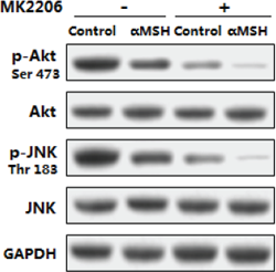
MK-2206 free base purchased from MedChemExpress. Usage Cited in: Oncotarget. 2017 Jul 18;8(29):47642-47654. [Abstract]
Akt/JNK signal pathway is impaired in the inhibition of αMSH on adipocyte inflammation and FoxOs expressions. Mouse adipocytes are pretreated with αMSH and MK-2206, respectively. Relative protein levels of Akt, p-Aktser473, JNK, p-JNKThr183 with or without MK-2206 (n=3).
-
Oncotarget
The angiotensin II type 1 receptor antagonist telmisartan inhibits cell proliferation and tumor growth of esophageal adenocarcinoma via the AMPKα/mTOR pathway in vitro and in vivo. [Abstract]2017 Jan 31;8(5):8536-8549. PMID: 28052030
MK-2206 free base purchased from MedChemExpress. Usage Cited in: Oncotarget. 2017 Jan 31;8(5):8536-8549. [Abstract]
Western blot analysis of cyclin D1 and cyclin E in OE19 cells treated with the control, BIBR 277 alone, MK-2206 alone, or BIBR 277 combined with MK-2206 for 48 h.
-
MK-2206 free base purchased from MedChemExpress. Usage Cited in: Modern Oncology. 2017,25(01):0009-0013.
The related protein expression of Bcl-2, Bax, Caspase-3, PARP, GSK-3β, p65 gene in each intervention group. Western blot assay:1:Control group; 2:MK2206 positive group; 3:0.3g/L Mat; 4: 0.6g/L Mat; 5:1.2g/LMat.
-
-
MK-2206 free base purchased from MedChemExpress. Usage Cited in: Harvard University. 2016 Aug.
Mouse embryonic fibroblasts (MEFs) are deprived of Arginine and treated with 2 µM MK2206 for up to 6 hours.
-
Oncotarget
Dual inhibition of AKT/FLT3-ITD by A674563 overcomes FLT3 ligand-induced drug resistance in FLT3-ITD positive AML. [Abstract]2016 May 17;7(20):29131-42. PMID: 27074558 -
MK-2206 free base purchased from MedChemExpress. Usage Cited in: Okayama University. 2015.
Expression of Sema4D and PlexinB1 in various oral squamous cell carcinoma cell lines following the addition of IGF-I. Notably, the addition of IGF-I resulted in increased expression of Sema4D, particularly in HSC-2 and SAS cells. This increase was suppressed when IGF-I was administered in the presence of inhibitors targeting its receptor or downstream signaling pathways. No changes in PlexinB1 expression were observed following the addition of IGF-I.
-
-
Chem Biol
2012 Jan 27;19(1):140-54. PMID: 22284361


Purity & Documentation
References
[2]. Lu JW, et al. MK-2206 induces apoptosis of AML cells and enhances the cytotoxicity of cytarabine. Med Oncol. 2015;32(7):206. [Content Brief]
[3]. Chen M, et al. MK-2206 Alleviates Renal Fibrosis by Suppressing the Akt/mTOR Signaling Pathway In Vivo and In Vitro. Cells. 2022;11(21):3505. Published 2022 Nov 5. [Content Brief]
[4]. Bjune K, et al. MK-2206, an allosteric inhibitor of AKT, stimulates LDLR expression and LDL uptake: A potential hypocholesterolemic agent. Atherosclerosis. 2018;276:28-38. [Content Brief]

Calculators
Concentration (start) × Volume (start) = Concentration (final) × Volume (final)


 Powered by Bioz